info@biomedres.us
+1 (502) 904-2126
One Westbrook Corporate Center, Suite 300, Westchester, IL 60154, USA
Site Map
 ISSN: 2574 -1241
Received: October 27, 2026; Published: November 09, 2026
*Corresponding author: Kwang Nho, SunBio, Inc.129 Eunbong-ro, Namdong-gu, Incheon-si, South Korea
DOI: 10.26717/BJSTR.2026.67.010471
Hemorrhagic shock remains a major cause of trauma-related mortality. Hemoglobin-based oxygen carriers may provide alternatives when conventional transfusions are unavailable. SBX PEG-Hemoglobin (SBX) is a PEGylated bovine hemoglobin formulation developed as an oxygen-carrying resuscitation fluid. This pilot study compared the safety and efficacy of SBX with Gelaspan 40 mg/mL in a controlled bleeding model using six miniature pigs. Animals underwent withdrawal of 30% of total blood volume over 30 minutes, followed by stabilization and infusion of SBX or Gelaspan at 25% of total blood volume. Animals were monitored for 72 hours using clinical observations, vital signs, body weight, hematology, biochemistry, lactate, hemoglobin fractions, necropsy, and histopathology. All animals survived the procedure. Bleeding caused hypotension in both groups, whereas hypertension during infusion occurred only with SBX. Transient hypothermia occurred in both groups, and all animals recovered uneventfully. No clinically significant treatment-related necropsy or histopathological findings were observed. Overall, SBX showed a safety profile comparable to Gelaspan, although the small sample size, variability, and analytical limitations prevented definitive conclusions regarding efficacy.
Keywords: Hemoglobin-Based Oxygen Carrier; Pegylated Hemoglobin; Blood Substitute; Hemorrhagic Shock; Miniature Pig; Resuscitation Fluid
Abbreviations: SBX: SBX PEG-Hemoglobin; CVC: Central Venous Catheter; PDI: Polydispersity Index; DLS: Dynamic Light Scattering
Hemorrhagic shock is a life-threatening condition resulting from severe blood loss that compromises tissue perfusion and oxygen delivery [1,2]. As circulating blood volume declines, cardiac output decreases and compensatory physiological responses are activated to preserve perfusion of vital organs [3]. Although these mechanisms may temporarily preserve cardiovascular function, prolonged or severe hemorrhage ultimately leads to tissue hypoxia, metabolic failure, multiple organ dysfunction, and death unless prompt hemorrhage control and resuscitation are provided [1,2,4]. Blood transfusion remains the standard treatment for major blood loss because it restores both circulating volume and oxygen-carrying capacity. However, the use of donor blood is associated with important limitations, including the need for blood typing and crossmatching, finite shelf life, specialized storage requirements, limited availability, and logistical challenges in prehospital, military, or remote settings. These constraints have stimulated ongoing efforts to develop artificial oxygen carriers that can provide immediate oxygen transport independent of donor blood supplies. HBOCs have emerged as promising alternatives because they can transport oxygen while avoiding many of the logistical challenges associated with blood products. However, earlier generations of HBOCs encountered obstacles related to vasoactivity, oxidative toxicity, and limited intravascular persistence. To address these shortcomings, several molecular modification strategies have been developed, including cross-linking, polymerization, and PEGylation.
Among these approaches, PEGylation has attracted particular interest because it increases molecular size, reduces renal clearance, prolongs circulation time, and may improve overall pharmacological performance. Recent preclinical studies have demonstrated the potential utility of HBOCs in the treatment of hemorrhagic shock. In a rat model of prolonged hemorrhagic shock, Nugent et al. reported that SANGUINATE®, a bovine PEGylated carboxyhemoglobin-based oxygen carrier, significantly improved survival, mean arterial pressure, and tissue oxygenation compared with Lactated Ringer’s solution and Hextend following a 45% blood-volume withdrawal [5]. Notably, these benefits were achieved without clear evidence of microvascular vasoconstriction. Similarly, Macko et al. demonstrated that low-volume resuscitation with PEGylated carboxyhemoglobin after severe hemorrhagic shock resulted in superior recovery of mean arterial pressure compared with Lactated Ringer’s solution and produced 72-hour survival rates comparable to those achieved with whole blood, supporting its potential as a field-deployable oxygen-carrying resuscitation fluid [6]. Beneficial effects have also been reported for non-PEGylated HBOCs. Kuang et al. evaluated YQ23, a cross-linked bovine HBOC, in uncontrolled hemorrhagic shock models in rats and miniature pigs and found that treatment reduced blood loss and fluid requirements, stabilized hemodynamics, improved oxygen delivery and utilization, preserved liver and kidney function, and enhanced survival compared with Lactated Ringer’s solution [7].
Furthermore, YQ23 extended the duration of effective hypotensive resuscitation to 180 minutes, highlighting its potential utility in prehospital trauma care. In a swine model of uncontrolled hemorrhage caused by grade III liver injury, Gurney et al. demonstrated that HBOC-201 improved tissue oxygenation, reversed anaerobic metabolism, reduced bleeding, and increased survival relative to hetastarch, although vasoactive effects were observed [8]. Additional studies have shown that polymerized human hemoglobin can effectively restore arterial pressure following combined traumatic brain injury and hemorrhagic shock while requiring smaller resuscitation volumes than crystalloid solutions and producing physiological responses more similar to those achieved with fresh blood [9]. Moreover, Hall et al. reported that HBOC-201 improved survival in swine with severe traumatic hemorrhagic shock without substantially exacerbating innate immune activation compared with Hextend [10]. Collectively, these studies support the potential of HBOCs as oxygen-carrying resuscitation fluids capable of improving hemodynamic stability, tissue oxygenation, and survival following severe hemorrhage. Nevertheless, continued evaluation of novel HBOC formulations remains necessary to establish their safety and efficacy profiles. SBX is developed as a potential resuscitation fluid for the treatment of severe blood loss. Previous nonclinical investigations have demonstrated acceptable toxicological and safety profiles supporting its continued development.
The present pilot study was therefore designed to evaluate the feasibility of administering SBX in a controlled hemorrhage model using miniature pigs and to generate preliminary safety data to support the design of future efficacy studies.
Study Design
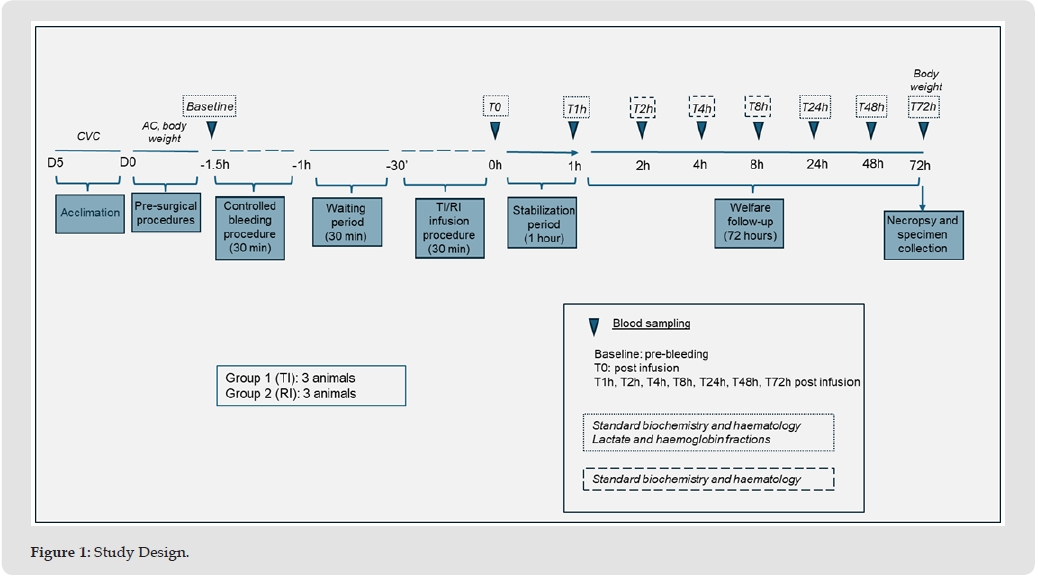
This study was designed to evaluate the efficacy and safety of SBX, the test item (TI), compared with Gelaspan® 40 mg/mL solution, the reference item (RI), in a controlled hemorrhage model using miniature pigs. A total of 6 animals (3 males and 3 females) were included in the study. The animals were randomly assigned to two experimental groups, with three animals per group. Group 1 (TI) consisted of 1 female and 2 males, whereas Group 2 (RI) included 2 females and 1 male. Hemorrhagic shock was induced by controlled withdrawal of blood corresponding to 30% of the estimated total blood volume over a 30-minute period. Following completion of the bleeding procedure, animals underwent a 30-minute stabilization phase. Subsequently, the TI or RI was administered intravenously at a volume corresponding to 25% of the estimated blood volume over a 30-minute infusion period. Vital signs were continuously monitored throughout the bleeding, stabilization, and infusion phases and were recorded approximately every 15 minutes until 1 hour after completion of the infusion. Animals were then allowed to recover and were monitored clinically for a total observation period of 72 hours.
Blood samples were collected at predefined time points for hematology, clinical biochemistry, lactate determination, and hemoglobin fraction analyses. At the end of the 72-hour observation period, all animals were euthanized and subjected to a complete necropsy. Selected tissues were collected for histopathological examination. The study design is summarized in Figure 1.
Figure 1
Study Objectives
The primary objectives of the study were:
• To evaluate the efficacy of SBX administration compared with Gelaspan in a controlled hemorrhage model in miniature pigs.
• To assess the safety profile of SBX following administration in study animals.
Study Endpoints and Assessments
Endpoint 1: Controlled Hemorrhage Procedure: The controlled hemorrhage model was established by withdrawing blood at a predefined rate corresponding to 30% of the estimated total blood volume over a period of 30 minutes.
Endpoint 2: Resuscitation Fluid Administration: The test item (SBX) or reference item (Gelaspan) was administered intravenously at a predefined volume corresponding to 25% of the estimated total blood volume over a 30-minute infusion period.
Endpoint 3: Efficacy Assessment: The efficacy of SBX was evaluated by monitoring markers of tissue perfusion and oxygen delivery, including:
• Blood lactate concentrations.
• Hemoglobin fraction profiles.
Assessments were performed at baseline, immediately before infusion (T0), and at 1, 24, 48, and 72 hours following completion of the infusion.
Endpoint 4: Safety Assessment: The safety profile of SBX was evaluated using the following parameters:
• Continuous monitoring of vital signs during and immediately after administration.
• Clinical pathology assessments, including hematology and serum biochemistry analyses, performed at baseline, T0, and at 1, 2, 4, 24, 48, and 72 hours post-infusion.
• Gross pathological examination at necropsy.
• Histopathological evaluation of the heart, liver, kidneys, spleen, and pancreas collected at terminal necropsy.
Test Article SBX
The investigational product was SBX, a PEGylated bovine hemoglobin formulation manufactured by SunBio, Inc. The product was stored frozen and thawed immediately prior to administration. After thawing, the solution was maintained under aseptic conditions until infusion. SBX was prepared from bovine hemoglobin, which was first stabilized by intramolecular crosslinking to prevent dissociation of the native tetramer into αβ dimers. Hemoglobin was reacted with 3-5 molar equivalents of bis(3,5-dibromosalicyl) fumarate in saline at pH 5-7 for 1-2 h at room temperature. The reagent primarily targets lysine residues, forming covalent bridges between subunits and producing a structurally stabilized hemoglobin with reduced dissociation. The crosslinked hemoglobin was then dissolved in 5 mM phosphate buffer (pH 8.5) at a concentration of 5%. PEGylation was performed sequentially using mPEG-SCM reagents of increasing molecular weight: 10 equivalents of 2 kDa PEG, followed by 4 equivalents of 5 kDa PEG, and 6 equivalents of 12 kDa PEG, each reacted for 30 min at room temperature. Sequential addition from smaller to larger PEG chains improved access to reactive sites and enhanced conjugation efficiency. Following PEGylation, the reaction mixture was subjected to purification steps, including microfiltration, ultrafiltration, and column chromatography, to remove unreacted PEG and low-molecular- weight impurities. The purified hetero-PEGylated hemoglobin (hetero-SBX) was then concentrated to a final hemoglobin concentration of 4.2%.
The degree of PEGylation was quantified using the TNBS (2,4,6-trinitrobenzene sulfonic acid) assay, which measures residual free amine groups on the protein [11]. Analysis performed after each conjugation step indicated that, on average, each hemoglobin molecule was modified with approximately 5 PEG chains of 2 kDa, 3 chains of 5 kDa, and 4 chains of 12 kDa. Based on this distribution, the total molecular weight of the SBX was estimated to be approximately 138 kDa. Previous PEG-hemoglobin products have generally been developed using PEG conjugation strategies in which all attached PEG molecules possessed the same molecular weight. Examples include Sangart’s MP4 [12], a human hemoglobin conjugated with 5 kDa PEG, and Prolong Pharmaceuticals’ Sanguinate [13], a bovine hemoglobin conjugated exclusively with 5 kDa PEG. In contrast, SBX is a bovine hemoglobin conjugated with a heterogeneous mixture of PEG molecular weights. This composition is specifically designed to maintain a total PEG load per hemoglobin molecule comparable to that of conventional PEG-hemoglobin products while achieving a larger overall hydrodynamic radius. The increased hydrodynamic size results from the incorporation of a limited number of higher-molecular-weight PEG chains, such as 12 kDa PEG. These larger PEG moieties play a critical role in prolonging intravascular retention by reducing renal filtration and clearance, thereby extending the circulation half-life of the SBX complex [unpublished data].
The hydrodynamic radius and polydispersity index (PDI) were determined using dynamic light scattering (DLS; Zetasizer, Malvern Instruments). The oxygen affinity of SBX was evaluated by measuring its p50 value, defined as the partial pressure of oxygen (pO₂, mmHg) at which 50% of the hemoglobin is oxygenated, using a Hemox Analyzer. SBX exhibited a p50 value of 8.2 mmHg, substantially lower than that of native blood hemoglobin (approximately 26 mmHg). This low p50 indicates a higher oxygen affinity, suggesting that SBX preferentially retains oxygen under normoxic conditions and may facilitate oxygen release primarily in hypoxic tissues (Table 1).
Table 1: Compositions of SBX.

Reference article used was Gelaspan® 40 mg/mL solution (B. Braun Medical S.A).
Ethics and Animal Care
The current experimental study was conducted at VERSA Biomedical S.L. (Autovia de Castelldefels C-31, Km 190.5, El Prat de Llobregat, Barcelona, Spain) and was reviewed and approved by its Ethics Committee for Animal Experimentation. Animals were housed under controlled environmental conditions with free access to food and water. All efforts were made to minimize animal suffering and reduce the number of animals used in the study.
Surgical Preparation
Animals (piglets, 6-8 months of age, 22-30 kg body weight at Day 0) were sedated using a combination of dexmedetomidine, midazolam, and methadone. General anesthesia was induced with propofol and maintained with inhaled isoflurane. Mechanical ventilation and physiological support were provided throughout the procedure. Body temperature was maintained within the normal physiological range using external warming devices. A central venous catheter was inserted, preferably through the jugular vein, to facilitate blood withdrawal, blood sampling, administration of the test article, and delivery of supportive medications. An arterial catheter was also placed, preferably in the femoral artery, to allow continuous monitoring of arterial blood pressure.
Pre-operative Procedures
On D0, the animals were sedated, intubated, anaesthetized, mechanically ventilated, and provided with maintenance support. The animals were continuously monitored throughout the entire surgical procedure. An arterial catheter was placed in the femoral artery for continuous blood pressure monitoring during the surgical procedure. And a central venous catheter (CVC) was inserted, in the jugular vein, for blood withdrawal (controlled bleeding and blood sampling), TI/RI infusion, and drug administration. Bleeding at the catheterisation site was closely monitored and tightly controlled when placing the catheters. The animal’s body temperature was maintained between 36,0 °C and 39,0 °C from the start of the bleeding procedure until 1-hour post infusion using a heating blanket.
Controlled Hemorrhage Procedure
The total blood volume of each animal was estimated using the standard calculation of 70 mL per kilogram of body weight. Controlled hemorrhage was then performed by withdrawing 30% of the estimated total blood volume through the central venous catheter over a period of 30 minutes. Blood withdrawal was performed at a constant rate using an infusion pump configured to provide controlled removal of blood. Following completion of the hemorrhage procedure, animals underwent a stabilization period of approximately 30 minutes before administration of the test article.
SBX Administration
Following the stabilization period, SBX was administered intravenously through the central venous catheter. The infusion volume corresponded to 25% of the animal’s estimated total blood volume and was delivered over a period of 30 minutes at a constant infusion rate. All infusion-related parameters, including total volume administered, infusion rate, and timing of administration, were recorded throughout the procedure.
Physiological Monitoring
Physiological parameters were continuously monitored during the hemorrhage procedure, the infusion period, and the subsequent observation phase. Parameters included heart rate, respiratory rate, oxygen saturation, capnography, body temperature, and arterial blood pressure. Measurements were recorded approximately every fifteen minutes beginning at the onset of blood withdrawal and continuing until two hours after completion of the infusion.
Post-operative Procedures
Following completion of the infusion procedure, animals remained under anaesthesia for 1 hour, during which vital parameters were monitored and recorded. After the observation period, the animals were recovered from anaesthesia.
In-Life measurements, Observations, and Procedures Once the animals were fully recovered from the anaesthesia, a veterinary animal welfare supervision was performed on D0, and daily until D3. Animals were provided with a heating lamp from D0 to D1. Feed intake was assessed starting on D1 until D3. Additional supervisions were conducted at the veterinarian’s discretion. Additionally, arterial blood pressure (MAP, SAP, and DAP) and heart rate was measured daily and recorded.
Blood Sample Collection
Samples were collected for blood analyses (standard biochemistry and hematology, lactate, and haemoglobin fractions) at the time points specified. Blood for haemoglobin fraction analysis was collected using oximetry syringes to prevent exposure to air. Venous blood at all time points was collected via the catheter placed in the jugular vein. Arterial blood was collected via the catheter placed in the femoral artery at baseline, T0, and T1h. For the remaining time points, arterial blood was obtained by ultrasound-guided direct puncture of the femoral artery.
Clinical Pathology
Blood samples were collected at three predefined time points. Baseline samples were obtained before induction of hemorrhage. Additional samples were collected immediately following completion of the SBX infusion and again twenty-four hours after administration. Hematological analyses included measurement of white blood cell count, differential leukocyte counts, red blood cell count, hemoglobin concentration, hematocrit, erythrocyte indices, platelet count, and mean platelet volume. Biochemical analyses included determination of albumin, glucose, total protein, gamma-glutamyl transferase, urea, and creatinine concentrations.
Welfare Assessment and Necropsy
Following recovery from anesthesia, animals underwent veterinary welfare evaluations that assessed feed intake, behavior, respiratory status, and oxygen saturation. Welfare assessments were repeated on the day following treatment. Animals showing evidence of severe distress would have been euthanized according to predefined humane endpoint criteria. At the completion of the study, all animals were euthanized by anesthetic overdose. A gross necropsy examination was subsequently performed to identify any visible abnormalities or lesions.
RBC, HGB, and HCT levels
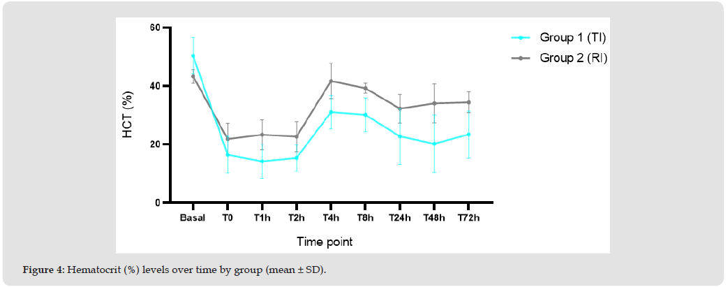
RBC, HGB, and HCT levels decreased markedly immediately after bleeding and infusion procedures (Figures 2-4). Recovery was observed by T4h for the three parameters and remained stable until the last time point (T72h). After the administration, the levels of RBC, HGB, and HCT were consistently lower for TI group compared to RI group. This was considered to be due to the oncotic effect of SBX.
Figure 2
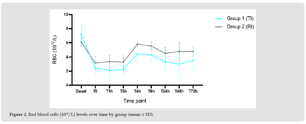
Figure 3
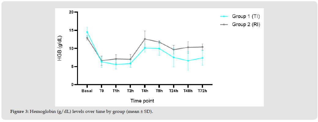
Figure 4
Platelets, White Blood Cells and Bilirubin Levels
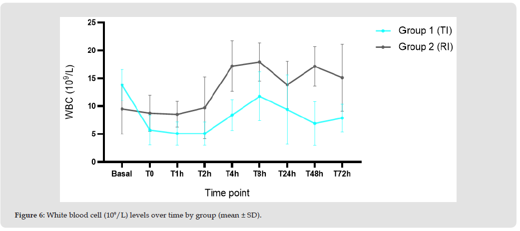
After the administration, consistently decreased level in platelet and white blood cells were observed in TI group compare to RI group, and this was considered to be the effect of oncotic nature of SBX (Figures 5 and 6).
Figure 5
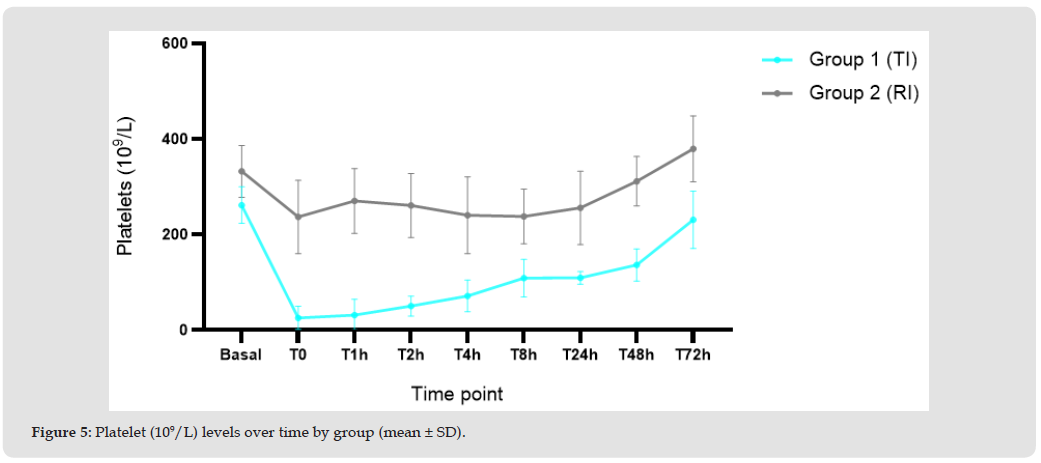
Figure 6
Biochemical Parameters
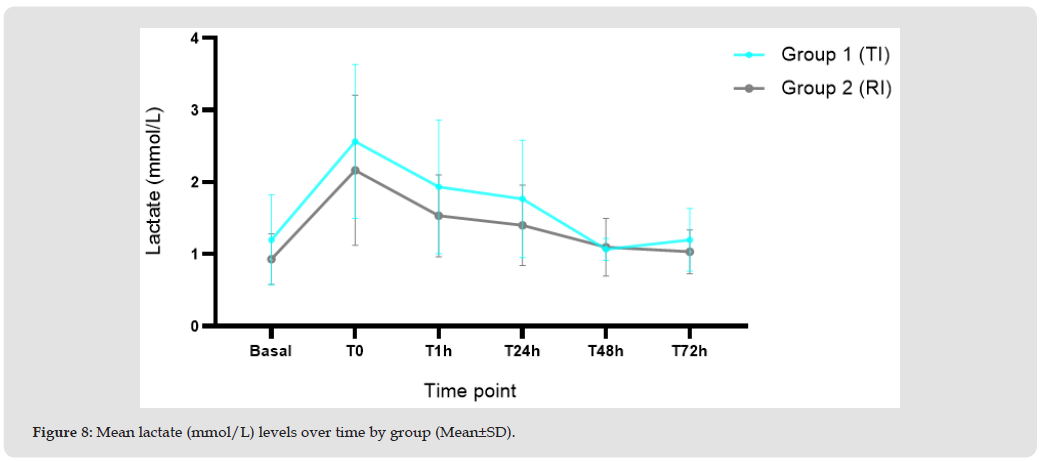
Regarding biochemical parameters, bilirubin levels increased after bleeding and infusion procedures in Group 1 (TI) and gradually decreased thereafter (Figure 7). This increased did not occur in Group 2 (RI). This finding was attributed to degradation of the haemoglobin haem group present in the TI. The remaining biochemical and haematological parameters showed no remarkable differences between groups, and any observed changes were considered procedure related. The results obtained from lactate analyses showed an increase following bleeding and infusion procedures and a progressive decrease at subsequent time points (Figure 8). Overall, results were comparable between groups.
Figure 7
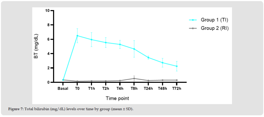
Figure 8
Hemoglobin Fractions
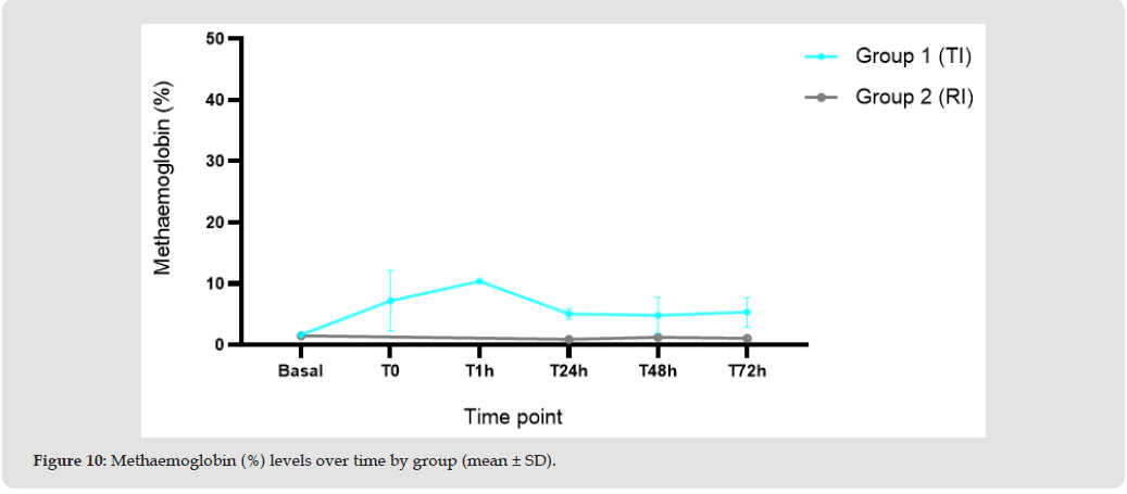
Oxyhaemoglobin levels (Figure 9) decreased following bleeding and infusion procedures and started to recover by T1h. From T24h to T72h, levels showed a slight decreasing tendency. Results for Group 2 (RI) at T0 and T1h were not available due to faulty sample handling. Results were very similar between groups. Methaemoglobin levels (Figure 10) showed a transient increase in Group 1 (TI) following bleeding and infusion procedures, peaking at T1h and then decreasing from T24h onwards. Group 2 (RI) showed consistently lower MetHb levels at the available time points. Group 2 (RI) results at T0 and T1h were not available.
Figure 9
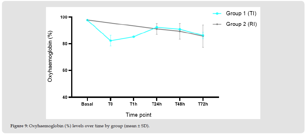
Figure 10
Heart Rate, DAP, MAP, SAP
Heart rate, systolic arterial pressure, mean arterial pressure, and diastolic arterial pressure were measured daily from Day 1 through Day 3. Continuous monitoring could not be performed because of equipment limitations. Variations in these parameters were observed during the follow-up period but were considered related to the experimental procedures. No clinically relevant differences in heart rate or arterial blood pressure were identified between the test-item and reference-item groups.
Post-surgical Recovery
Post-surgical recovery was satisfactory in both treatment groups. All animals were able to stand and walk, exhibited normal breathing, and showed no signs of pain. Recovery from the surgical procedure was therefore confirmed in all animals.
Body Weight
The animals were weighed on Day 0 before the bleeding procedure and again on Day 3. Body weights remained stable throughout the study, with no clinically significant changes observed in either treatment group.
Unscheduled Euthanasia and Unexpected Deaths
No animals required unscheduled euthanasia, and no unexpected deaths occurred during the study.
Necropsy
Necropsy examinations were performed on all study animals, and no clinically significant abnormalities were observed. A slight hematoma was noted at the arterial catheter insertion site in one animal from the test-item group and was considered procedure-related. Dilatation of the right renal pelvis and ureter was observed in one animal from the reference-item group; according to the study veterinarian, this finding was not considered clinically relevant. No other macroscopic abnormalities were identified in either group.
Histology and Microscopic Evaluation
Histopathological evaluation of the examined organs revealed overall minimal to moderate findings, most of which were limited in incidence and severity and showed no consistent pattern indicative of adverse effects. Most of the observed changes were of low severity, which is consistent with previously reported background lesions in minipigs. Table 10 shows the histopathological findings in heart, liver, kidney, pancreas, and spleen, for each animal. Based on the histopathological evaluation, no consistent or treatment-related microscopic findings were identified in the examined organs of animals receiving the TI compared to the RI. Observed changes were generally minimal to moderate in severity, limited in incidence, and distributed across both groups without a clear pattern. Findings in individual organs were considered compatible with the experimental model, known background changes in minipigs, or isolated occurrences. Under the conditions of this study, the TI did not induce any morphologically detectable adverse effects in the evaluated organs.
Survival
All animals survived throughout the 72-hour observation period without requiring unscheduled euthanasia or experiencing unexpected adverse events.
This pilot study evaluated the preliminary safety, physiological effects, and feasibility of SBX as an oxygen-carrying resuscitation fluid in miniature pigs subjected to controlled withdrawal of 30% of estimated blood volume. SBX was well tolerated, administration was successfully completed in all animals, and all animals survived the three-day observation period without adverse events or unscheduled euthanasia. Overall, its safety profile was comparable to that of Gelaspan, a clinically used plasma volume expander. The hemorrhage procedure produced the anticipated reductions in red blood cell count, hemoglobin concentration, and hematocrit in both treatment groups, confirming successful induction of acute blood loss. Subsequent changes reflected the combined effects of hemorrhage and hemodilution following fluid administration. Recovery patterns were broadly similar between the SBX and Gelaspan groups, indicating that SBX did not adversely affect hematological recovery. Lactate concentrations increased transiently following hemorrhage and infusion and subsequently declined toward baseline in both groups. Although SBX did not demonstrate a clear advantage over Gelaspan, normalization of lactate suggests that it provided sufficient physiological support to facilitate recovery from the hemorrhagic insult. The relatively mild hemorrhage model and small sample size may have limited the ability to detect differences in oxygen delivery between treatments.
Hemoglobin fraction analyses demonstrated the expected pharmacodynamic behavior of an administered hemoglobin-based oxygen carrier. SBX produced transient increases in methemoglobin, with corresponding changes in oxyhemoglobin fraction. These findings are consistent with the metabolism and redox cycling of cell-free hemoglobin. The changes progressively returned toward baseline and were not associated with adverse clinical outcomes. SBX administration was also associated with a more pronounced transient reduction in platelet counts, changes in leukocyte profiles, and an increase in bilirubin. The bilirubin elevation was expected from the metabolism of hemoglobin-derived heme and has been reported with other HBOCs. Platelet and leukocyte values showed recovery during the study, and these laboratory changes were not accompanied by clinically significant hepatic, renal, or systemic toxicity. Most other biochemical changes were comparable between groups and were considered related primarily to hemorrhage and perioperative management. Nitric oxide scavenging by cell-free hemoglobin, which may cause vasoconstriction and hypertension, has historically been an important concern in HBOC development [14-16]. Transient hypertension was observed during SBX infusion, while hypotension during blood withdrawal occurred in both groups. No persistent cardiovascular abnormalities or clinically meaningful between-group differences in daily heart rate or arterial blood pressure were subsequently identified.
Thus, the vasoactive effect of SBX appeared limited in magnitude and duration under the conditions examined.
Gross necropsy and histopathological examinations provided further evidence of acceptable tolerability. Findings were generally mild, occurred in both groups, and were consistent with background lesions in miniature pigs or consequences of the experimental procedures. No consistent pattern of SBX-related injury was detected in the liver, kidneys, heart, pancreas, spleen, or other examined organs. Interpretation of efficacy remains limited by the inclusion of only six animals, substantial inter-animal variability, the relatively moderate 30% hemorrhage, and incomplete hemoglobin-fraction data. In addition, Gelaspan provides plasma volume expansion but does not restore oxygen-carrying capacity and therefore cannot fully represent whole blood or blood-component therapy. Consequently, this study established preliminary safety and feasibility but was not sufficiently powered or designed to demonstrate superiority in oxygen delivery. In conclusion, SBX demonstrated an acceptable preliminary safety profile, predictable biological behavior, and physiological performance comparable to Gelaspan in this controlled hemorrhage model. Although a definitive efficacy advantage was not established, the recovery of lactate and other physiological parameters, together with the absence of clinically significant organ toxicity, supports continued development of SBX.
Future studies should use larger group sizes, more severe hemorrhagic shock models, longer observation periods, and direct measurements of tissue oxygenation, perfusion, and organ function. Comparisons with blood products or other oxygen-carrying resuscitation strategies will be important to define the potential role of SBX when conventional transfusion is unavailable, delayed, or impractical.
The authors affirm that there is no conflict of interest.
K. Nho conceived the project and designed the experiments. All authors participated in analyses and confirmed data.

International Association of Landscape Archaeology, Czech Glass Society, Czech Republic

Department of Chemistry, Semenov Institute of Chemical Physics, USSR Academy of Sciences, Moscow, Russia

Neurology, LA BioMed Research Institute, USA

Associate Professor at Department of Breast and Thyorid Surgey, Chongqing General Hospital, China

Professor of Nuclear Medicine, Faculty of Medicine and Surgery, University of Milan, Milan, Italy





 Abstract
 PDF