info@biomedres.us
+1 (502) 904-2126
One Westbrook Corporate Center, Suite 300, Westchester, IL 60154, USA
Site Map
 ISSN: 2574 -1241
Received: August 25, 2026; Published: October 09, 2026
*Corresponding author: Octavio Gamaliel Aztatzi-Aguilar, Instituto Nacional de Enfermedades Respiratorias Ismael Cosío Villegas, Departamento de Investigación en Toxicología y Medicina Ambiental, CDMX, México.
DOI: 10.26717/BJSTR.2026.67.010432
The cystic fibrosis transmembrane conductance regulator (CFTR) is implicated in epithelial ion transport and mucus secretion; dysfunction or lower expression is related with physiological dysfunction of organs such as lungs. It has been reported that cigarrete smoke and diesel particles affect CFTR expression in lung epithelium models, but not urban particles. The goal of the present brief communication is to present evidence of urban particles affect CFTR in an in vitro and in vivo experimental model affecting also non-respiratory tissues. In A549 cell line after 24h exposure to PM10 and PM2.5 from subway and outdoor environments evaluate the CFTR expression and intracellular chloride concentrations. On the other hand, in male rats evaluated CFTR expression after acute, and sub-chronic exposure to PM2.5 and filtered air (FA) in lung and kidney cortex tissues; serum- and urine-chloride concentrations were evaluated. In vitro, assays demonstrated that PM10 and PM2.5 can modulate CFTR expression and increased intracellular chloride concentrations. In vivo results showed that PM2.5 exposure reduced CFTR protein in lungs and kidney cortex, additionally, serum and urine chloride concentrations exhibited time-dependent changes. CFTR is a toxicological target of urban PM, which affects expression, and local and systemic chloride ion concentrations changes.
Keywords: Particulate matter, CFTR, lung tissue, non-respiratory tissues, kidney.
Air pollution by particulate matter (PM) is associated with the development of cardio-respiratory diseases [1,2] and there is evidence that air quality also contributes to neurological [3], and renal diseases [4]. The PM mechanism related with its toxicity is unknown and unexplained to the many diseases associated with air pollution by PM. Additionally, chronic diseases worsen under polluted ambient air conditions, such as metabolic diseases [5]. Some molecular targets have been suggested which mediate PM toxicity, that include Transient Receptor Potential (TRP) family channels [6], which are able to interact with other co-modulators channels such cystic fibrosis transmembrane conductance regulator (CFTR) [7].The CFTR gene codifies for an integral membrane protein, which is a member of the ABC transporter family and is implicated in chloride and bicarbonate ion transport in many epithelial secretions. CFTR also mediates anionic glutathione flux, which is the most important extracellular non-enzymatic antioxidant in the lungs [8,9]. The absence or malfunctioning of CFTR is implicated in Cystic fibrosis (CF), a hereditary recessive autosomal disorder caused by a mutation in CFTR gene [10]. The different CFTR mutations in the CF patients are related with diverse effects including airways, pancreas, male genital system, intestine, liver, bone, and kidney. Besides the fat malabsorption, CF causes chronic pulmonary infections that ultimately drives the development of bronchiectasis and progressive lung damage [11]. In CF, lungs become chronically infected by pathogens such Pseudomonas aeruginosa, suggested by the altered anionic composition of airway surface fluid of CF patients which affect the activity of innate immune response proteins called defensins. For the above reason CFTR it has been suggested as a pleiotropic protein [12].
PM exposure has been associated with the increment of respiratory infections. Fine particle exposure has been related with initial Pseudomona aeruginosa infection in the human alveolar epithelial cell line A549, and corroborate through epidemiological studies, where it was reported a risk augment (23-26%) in the P. aeruginosa acquisition by each 10 μg/ m3 augment in PM2.5 in children with a median age of 15 months (25th- 75th percentile, 9-25 mo) [13]. Chloride concentration has been used as a biomarker CFTR function in CF patients. It has been reported the CFTR participation in pneumocyte in chloride and fluid transport, it suggests the CFTR function in the pneumocyte layer could participate in the resolution of pulmonary edema [14]. Which is a pathological state, when increase permeability of the alveolar capillary barrier without changes in hydrostatic pressure, recurrent in burning smoke intoxication where many toxic products are formed including hydrogen chloride, chlorine, oxides, aldehydes, and particulate matter [15]. CFTR expression in rat isolated alveolar pneumocyte type-II [16], in alveoli from adult healthy human and alveoli from distal airway disease [17] and basal expression of CFTR in the lung adenocarcinoma cell line A549 (https://www.proteinatlas.org/ ENSG00000001626-CFTR/cell) have been reported [18]. In addition, extensive analysis of punctual mutation on CFTR gene sequence has allowed importance in tissues and organs dysfunction, however, the gene-environment interaction on CFTR transcript and protein continues being subject of study.
In this sense, CFTR might be a molecular target of air pollution, the changes in CFTR could associate an aggravating factor susceptibility of alveolar epithelium with the further development of pulmonary disease or compromise physio-pathological condition of other illnesses. Nguyen et al., (2021) reports the effect of Diesel exhaust particles and tobacco smoke extract in airway epithelial cells (Calu-3) on CFTR expression and dysfunction, but not by the exposure to urban particulate matter [19]. The present study evaluates whether urban PM exposure can modify CFTR mRNA and protein in the in vitro, A549 cell line, and in the lungs and non-respiratory tissues, such kidneys, of rats exposed to concentrated PM.
We investigated the effect of PM10 and PM2.5 collected in subway microenvironment and outdoor from Mexico City to explore whether CFTR is a molecular target of PM in the cell line A549. To corroborate the observations in vitro, we analyzed changes in CFTR mRNA and protein levels in lungs and in non-respiratory target of PM, kidney tissues, of rats exposed to whole body-chamber through a particulate concentrator.
In vitro Exposure
Particles were provided by Dr. Andrea de Vizcaya CINVESTAV-IPN. Particles were collected in September-October 2009 from subway microenvironment and outdoor of Azcapotzalco subway station. PM10 and PM2.5 were obtained with a HiVol sampler, nitrocellulose filters were scraped off in order to obtain the particles [20]. Human lung adenocarcinoma cells, A549, were obtained from ATCC. The A549 cell line was subcultured in F-12/Ham culture media (Pyruvate 0.05 mg/mL, 2 mM L-glutamine, penicillin/streptomycin 50 IU/mL and /50g/mL, from Sigma Aldrich, USA) with 10% inactivated FBS (GIBCO, Thermo Fisher Scientific, USA). Cells were maintained at 37°C in a 5% CO2 incubator. Cells were then plated at a density of 45 x 103 cells/cm2 24 h before PM exposure. Exposure media consisted of F-12 Ham supplemented with 0.5% FBS and PM solutions. The A549 cell line was exposed 20 and 40 μg/cm2 of PM10 and PM2.5 for 24 h, which are amount equal or below annual average stablished by the official Mexican standard [21]. LDH assay was performed to corroborate cytotoxicity (Sigma Aldrich, USA). All assays were performed in triplicate with at least three independent experiments.
In vivo Exposure
Sprague Dawley male rats (250 ± 40 g) were acutely (5 hours per day for 3 days) and subchronically (5 hours per day, 5 days per week, for 8 weeks) exposed to PM2.5 using a particulate concentrator at CINVESTAV- IPN in the North of Mexico City during months pf April-July 2013. Animal care and use were carried out according to the “Principles of Laboratory Animal Care” [22] and Mexican guidelines (NOM- 062-ZOO-1999) [23]. All procedures were approved by the institutional ethics committee for the care and use of animals. Concentration in chambers by particle concentrator was 445 and 375 μg/m3 in acute and subchronic exposure, respectively. After each exposure, urine form exposed animals was harvested 12 hours after being collected from a metabolic cage, followed by 15 min centrifugation at 1,000 rpm, and preserved at -80ºC for analysis. Twenty-four hours after the last exposure, anesthetized animals (40 mg/kg sodic pentobarbital) were sacrificed by exsanguination. Blood was obtained by abdominal aorta puncture and collected with a gel activator tube, lungs and kidney cortex tissue were dissected. Serum was separated after centrifugation at 1,500 rpm for 15 min and then aliquoted and preserved at -80ºC until their analysis.
Chloride Determination
Chloride concentrations in cell lysates obtained by osmotic shock with ultrapure MilliQ-water, serum, and urine were analyzed spectrophotometrically at 500 nm by the Mercury (II)-Thiocyanate method using a commercial kit (CL1645, Randox, UK) and read in a TECAN Infinite 200 PRO reader.
CFTR mRNA Expression
Total RNA was isolated by TRIzol reagent (Invitrogen, Thermo Fisher Scientific, USA) from cell cultures and lung and kidney cortex tissues. We then subjected 10 μg of total RNA to DNAse treatment (Ambion, Turbo DNAse-freeTM, Life Technologies, USA). cDNA synthesis was performed with 3 μg of total-RNA using Super Script II (Invitrogen, Thermo Fisher Scientific, USA). End point PCR was performed with the following CFTR oligonucleotides: forward 5’-TTCTGCCTCTTACCATATTTGACT-3’ and reverse 5’-GCCTTCAGATTCCAGTTGTTTG- 3’, and we used actin and GADPH as housekeeping genes; GADPH forward 5’-ACCACAGTCCATGCCATCAC-3’ and GADPH reverse 5’-TGCCAGTGAGCTTCCCGTT-3’; and Actin forward 5’-ACTATCGGCAATGAGCGGTTCC- 3’ and Actin reverse 5’-AGCACTGTGTTGGCATAGAGGTC- 3’.
CFTR Immunodetection
Total protein from A549 cells exposed to 0 and 20 μg/cm2 of the different PMs was obtained with Nonidet-P40 buffer (150 mM NaCl, 1 % NP-40, 50 mM Tris-HCl pH 8.0 and protease inhibitors) and lysed using an insulin syringe with an ultrafine 6 mm needle, centrifuged at 10,000 rpm and 4 °C, and the supernatant was collected and stored at -70 °C until use. Lung and kidney cortex tissues were homogenized with Nonidet-P40 buffer and centrifuged at 8,000 rpm and 4 °C. Protein concentrations were determined by Bradford protein assay. The total protein (20 μg) per sample was loaded in SDS-polyacrylamide gels and transferred in wet transfer units to a PVDF membrane at a constant voltage (25 V) and kept overnight (12 hours). Membranes were then blocked for 1 h with 5% non-fat milk and incubated overnight with the primary antibodies Anti-CFTR (1:400, ProSci, inc. 7717) and GADPH (1:1000, Santa Cruz Biotechnology SC-32233). HRP secondary antibodies Goat Anti-Rabbit (BioRad, 1706515) and Goat Anti-Mouse (BioRad, 1706516) were incubated for 1 h at a dilution of 1:10,000 to detect CFTR and GADPH, respectively. Immunoreactivity was detected using Luminata forte western blotting detection reagent (Millipore, WBLUF0500, Germany). Bands were visualized with a UVP EC3 imaging system (UVP Inc, USA). Finally, a semiquantitative analysis was performed with ImageJ free online software and expressed as arbitrary units.
Statistical Analysis
The normality of all data was assessed with a Kolmogorov-Smirnov test. We then performed one-way analyses of variance (ANOVA) or Kruskall-Wallis tests followed by Holm-Sidak or Dunn’s multiple comparisons post-hoc tests, respectively, to compare treatment groups with respect to the control group. All statistical analyses were carried out in GraphPad Prism version 6.00 for Windows (GraphPad Software, La Jolla California USA).
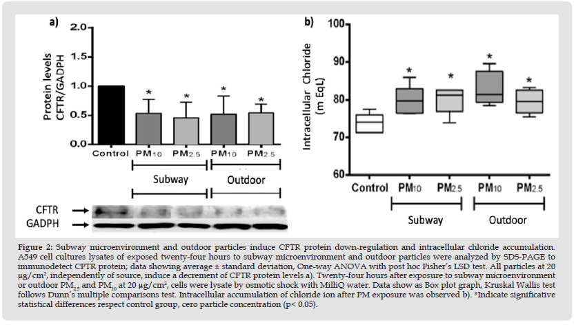
Effect of Ambient Particles on CFTR Expression in vitro After 24 hours of exposure to subway microenvironment and outdoor PM2.5 and PM10 particles, we observed cytotoxicity below 10% with respect to the control group (Figure 1a). Subway microenvironment PM2.5 at 20 and 40 μg/cm2 decreased CFTR expression, and a similar but less pronounced effect was observed with outdoor PM2.5 (Figure 1b). Subway PM10 at 40 μg/cm2 induce an increment statistical significative on CFTR mRNA, however, outdoor PM10 showed a statistical significative decrease at 20 μg/cm2, but not at 40 μg/cm2 (Figure 1b).To corroborate the effect of the different particles on CFTR expression, we immunodetected CFTR on the total protein of A549 cells. The results show that CFTR protein levels decreased with respect to the control group 24 hours after exposure to 20 μg/cm2, independently of the origin and size of the particle (Figure 2a). One proper function of CFTR is maintaining the balance between the intra and extracellular chloride ions concentration. Thus, we evaluated intracellular chloride ion concentrations in cell lysates obtained by osmotic shock. Chloride concentration increased statistically significantly into the cell lysates of cultures exposed to PM at 20 μg/cm2 in all particles tested, respect to the control cell group (Figure 2b).
Figure 1
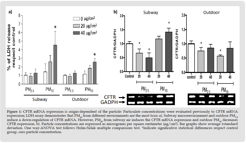
Figure 2
Effect of PM2.5 Exposure on CFTR Expression in vivo
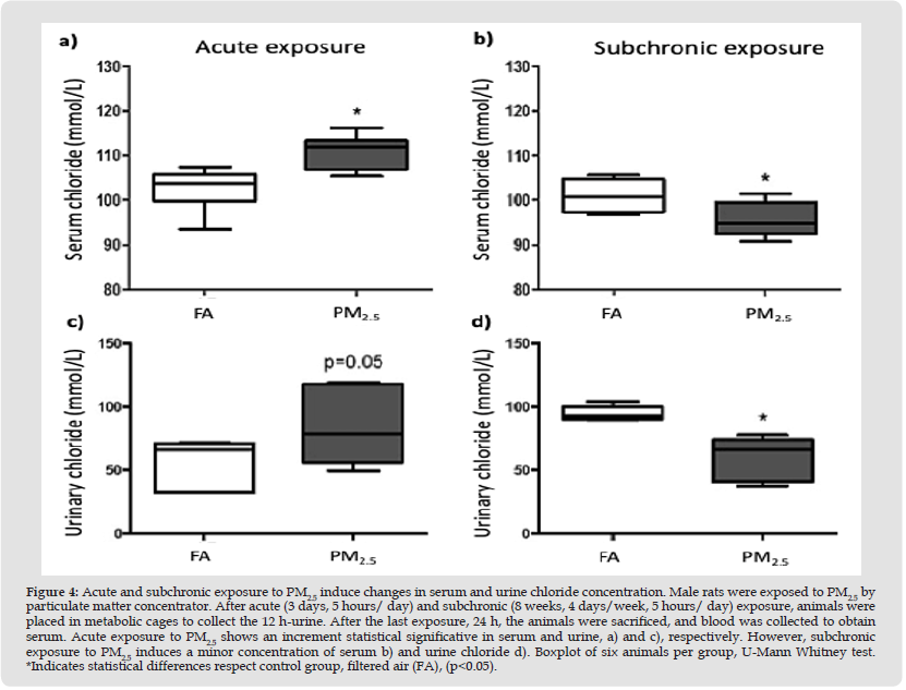
To provide evidence that CFTR is a molecular target of particle toxicity, we evaluated CFTR mRNA levels in lung tissue after acute (3 days, 5 hour/day) and subchronic (8 weeks, 5 days/week, 5 hour/ day) exposure. We observed a time-dependent response of CFTR mRNA levels (Figure 3). Acute exposure resulted in a statistically significant two-fold increment in CFTR mRNA (p<0.05; Figure 3a); however, subchronic exposure to PM2.5 reduced statistically significant CFTR mRNA to half with respect to the control group (p<0.05; Figure 3b). CFTR also has a function in other types of tissues, such as the pancreas and kidneys [24]. Therefore, we evaluated CFTR mRNA in kidney cortex tissue only in subchronic exposure to PM2.5 and observed a statistically significant reduction in CFTR mRNA in the particle-exposed group (p<0.05, Figure 3c), suggesting that a repeated exposure to PM2.5 during long periods can affect CFTR expression in non-respiratory tissues such as the kidneys. However, we also observed a statistically significant reduction (p<0.05) in CFTR protein expression in lung tissue after acute and subchronic exposure to PM2.5 (Figure 3d, 3e, respectively), as well as in kidney tissue (Figure 3f). CFTR protein levels were modified by PM2.5 exposure, suggesting changes in ionic transport such chloride anion. We observed significant statistical differences (p<0.05) in serum and urine chloride concentrations in both exposure periods (Figure 4). Our results show an increase in chloride ion concentrations in serum (Figure 4a) and urine (Figure 4c) after acute exposure to PM2.5; however, subchronic exposure decreased chloride ion concentrations in both fluids (Figure 4b and 4d).
Figure 3
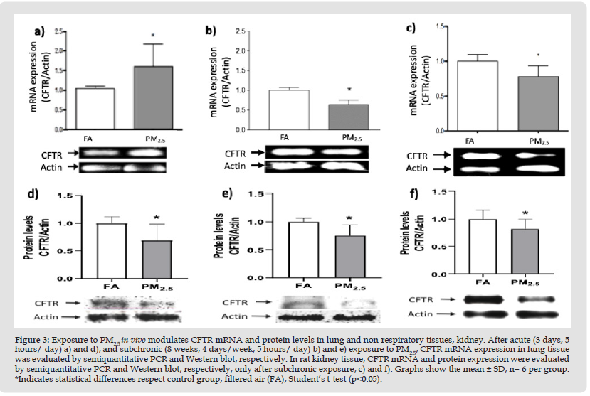
Figure 4
CFTR has been studied at different molecular levels, such as genome, transcript, proteomic, and cellular function. However, the effect of ambient factors, such as atmospheric PM, on CFTR expression was recently reported with effects on CFTR expression and function in Calu-3 cell line exposed to tobacco smoke extract and diesel exhaust particles. On the other hand, our study exposed A549 cell line and a rodent model we complement the findings reported by Nguyen, et al. [19], we observed that urban particles can impact CFTR expression on lungs and in extrapulmonary tissues in time-dependent course. The in vitro results provide evidence that CFTR mRNA levels respond to PM10 and PM2.5 from subway and outdoor environments. Particle size is related to the source, and chemical composition; however, independently of that factors marginal or statistical significative differences were observed in the cells exposed to PM10 and PM2.5 on CFTR mRNA. Bargon, et al. [25] has been reported that divalent cations as calcium and magnesium can down regulate CFTR mRNA in human bronchial cells [25]. Differences between PM10 and PM2.5 on CFTR mRNA could be influenced by the variances in the presence and concentrations of anions and cations by type and the particle origin. Regardless of the effect of particles on CFTR mRNA, all particles reduce statistical significative CFTR protein expression, concomitant with the retention of intracellular chloride ions under non-cytotoxic concentrations, cell viability minor to 10% through LDH assay. Chloride transport has been demonstrated in human and rat pneumocyte type-II alveolar epithelial. Apical pharmacological treatment of pneumocyte type-II monolayers with forskolin induce Cl- influx, which is inhibit by the CFTRinh-172, when high apical and low basal chloride concentrations are disposed in transwell assays [14]. Besides, it has been demonstrated that CFTR in pneumocyte type-II, through patchclamp studies, can regulate epithelial Na+ channel (ENaC), even when CFTR protein were minimal [26], as well other chloride channels of SLC26 family [27]. The pneumocyte exposition to PM affect CFTR expression contributing to change the chloride and fluid transport, because the presence of ions (e.g. chloride and sodium) as part of chemical PM composition are dissolved in the pulmonary lining fluid modulated apically to CFTR. This effect could be contributed to chloride influx or water resorption across the alveolar epithelium modified transcellular transport.
The CFTR expression reduction has been reported by cigarette smoke (CS) exposure, and the reduced chloride secretion by CS was reported since 80’s, before the description of CFTR gene, later was demonstrated the CS inhibit CFTR and induce its internalization, producing dehydration of liquid surface of airspace, a propound effect to development COPD [28]. Furthermore, the present results imply that CFTR is a molecular target of airborne PM and suggest that this effect occurs at different levels, such as mRNA, protein, and ion transport. CFTR regulation appears to be independent of particle size and chemical composition. The effect of PM2.5 on CFTR was confirmed in a rodent model. It is important to note that the effect was time-dependent, where repeated exposure chronically induced a down-regulation of CFTR mRNA. Additionally, we also observed a reduction on CFTR mRNA in non-respiratory tissues, the kidneys, which are affected in CF, and their function decline after PM2.5 exposure (4). Serum chloride concentrations corresponded to CFTR mRNA levels but not to CFTR protein expression, which suggests a difference in the temporality of the transcription, protein synthesis, and possibly channel activity.
Xu et al. [29] recently reported that the exposure to PM2.5 is a potential inductor of pulmonary fibrosis in in vitro and in vivo models by activating epithelial mesenchymal transition (EMT), and TGFβ1/ SMAD3 pathway, and increasing α-smooth muscle actin and Collagen type I [29]. Adding to this evidence, our results suggest that PM from different sources in non-cytotoxic concentrations can deregulate CFTR at different molecular levels, promoting an alteration in fluid secretion, ion transport, and it also likely contributes to EMT. It is significant to remark that the effect of PM on CFTR could contribute to the loss of glutathione transport, which is the most abundant antioxidant in lung epithelium lining fluid, and responsible for decreasing and attenuating oxidative stress and inflammation against toxic inhalants, such CS, [30] and which is reduced in CF patients [31].One of our most relevant results is the CFTR down-regulation in kidney cortex tissue after subchronic exposure to PM2.5, which suggests that PM has an impact on other types of tissues and physiological functions of CFTR. In this sense, the effect of PM on CFTR expression in non-pulmonary tissues might contribute to the development of other diseases. Metabolic pathologies such as Diabetes, the most common extrapulmonary complications of CF, since epidemiological studies have found an association between an increase in PM particles and the number of cases of DM [32,33].
Is well clinically documented Cystic Fibrosis-Related Diabetes (CFRD) as a frequent comorbidity of CF by CFTR impairment [34,35]. Further studies are needed to verify the effect of subchronic or chronic effect of PM on CFTR on pancreatic function. The impairment, severity, and lack of control of diseases caused by the exposure to air pollution has been reported in other pulmonary genetic diseases, such α1-antitrypsin deficiency, which predisposes to the development of chronic obstructive pulmonary disease (COPD); PM10 is associated with a decline in FEV1 and DLCO tests [36, 37]. Further epidemiological studies are necessary to demonstrate how the exposure to PM on CFTR can contribute to the impairment, increase in severity, and diseases control. The present experimental evidence demonstrates that CFTR is a toxicological target of PM in vitro and vivo models, affecting CFTR mRNA and protein expression, which could be related to chloride ion transport and probably with other physiological functions. These results imply a risk of developing diseases because of CFTR down-regulation by PM exposure.
The authors acknowledge to Toxicology Department from CINVESTAV- IPN for the equipment and infrastructure to carry out the present study.
Aztatzi-Aguilar OG performed the in vivo and in vitro exposure, analyzed the data and wrote the draft manuscript; Cruz-Guerrero M carried out the PCR analysis; Debray-García Y performed Western Blot and wrote the draft manuscript; Uribe-Ramírez TM participated in the in vivo and in vitro exposure; Narváez-Morales J participated in the metabolic cage and chloride quantification; Barbier O provided the resources to analyze samples by Western Blot and PCR.
The authors declare that no funds, grants, or other support were received during the preparation of this manuscript. The authors have no relevant financial or non-financial interests to disclose.

International Association of Landscape Archaeology, Czech Glass Society, Czech Republic

Department of Chemistry, Semenov Institute of Chemical Physics, USSR Academy of Sciences, Moscow, Russia

Neurology, LA BioMed Research Institute, USA

Associate Professor at Department of Breast and Thyorid Surgey, Chongqing General Hospital, China

Professor of Nuclear Medicine, Faculty of Medicine and Surgery, University of Milan, Milan, Italy





 Abstract
 PDF