info@biomedres.us
+1 (502) 904-2126
One Westbrook Corporate Center, Suite 300, Westchester, IL 60154, USA
Site Map
 ISSN: 2574 -1241
Received: July 17, 2026; Published: October 05, 2026
*Corresponding author: Zia Ul Hassan, Department of Chemistry, Government Post Graduate College Mansehra, Pakistan
DOI: 10.26717/BJSTR.2026.67.010428
Fungal infections, especially those which are caused by Candida species, are an increasing global health concern due to the rising resistance of pathogenic microorganisms to conventional antifungal and antibacterial drugs. In this work, a systematic investigation of Leptodermis virgata, which is traditionally used in Himalayan shrub that belongs to the family Rubiaceae, was carried out to analyze its antifungal activity, antibacterial activity, overall antimicrobial potential, and phytochemical composition. The ethanolic extract of L. virgata was prepared using the cold macera-tion technique, and its antimicrobial efficiency was evaluated using the agar well diffusion method against Candida albicans, Candida parapsilosis, Escherichia coli, Pseudomonas aeruginosa, and Staphylococcus aureus. The extract exhibited strong antifungal activity, with zones of inhibition of 13 mm against C. albicans and 19 mm against C. parapsilosis, along with notable antibacterial activity against E. coli, that produce an inhibition zone of 20 mm at a concentration of 25 µL. Phytochemical screening confirmed that the presence of major bioactive constituents, that includes flavonoids, tannins, terpenoids, saponins, glycosides, steroids, and anthraquinones. The results of this study is shown that L. virgata possesses significant antifungal, antibacterial, and antimicrobial properties, that highlights its potential as a promising natural source for the development of novel therapeutic agents.
Keywords: Phytochemistry; Antifungal Activity; Antibacterial Activity; Ethanolic Extract; Candida Species; Pathogenic Bacteria
Microbiology is the branch of biological sciences that investigates the microorganisms, that includes the archaea, viruses, protozoa, bacteria, and fungi, encompassing their structure, physiology, biochemistry, ecology, and clinical significance [1,2]. The foundations of microbiology were established in the seventeenth century by Antonie van Leeuwenhoek, whose pioneering microscopic observations first revealed the previous unseen microbial world [3]. The special study of fungi, which is known as mycology, and it focuses on a diverse group of eukaryotic organisms, and whose range is from unicellular yeasts to complex multicellular forms, that exhibits the sizes from a few micrometers to several centimeters [4]. Fungi play the vital roles in ecosystem functioning, specially in the nutrient cycling, organic matter decomposition, and the formation of mutualistic mycorrhizal associations, which support the growth and survival of over 90% of terrestrial plant species [5]. Although their ecological and economical importance, fungal biodiversity remains poorly characterized; it is estimated that only about 7-10% of fungal species have been described recently, with global diversity projected to range between 3.2 and 3.8 million species [6,7].
Fungi are classified as eukaryotic organisms, which is characterized by the presence of a true nucleus and exist as unicellular as well as multicellular forms. Based on morphological characteristics and modes of growth, fungi are categorized into yeasts, yeast-like fungi, molds, and dimorphic fungi [8,9]. Yeasts are unicellular, oval or spherical organisms that reproduce sexually through budding, with Cryptococcus neoformans serves as a clinically significant example [10]. Yeast-like fungi, such as Candida albicans, shows pseudohyphal growth, which shows the elongated cells that resemble true hyphae while retaining yeast-like characteristics [11]. In opposite, molds are multicellular fungi, that are composed of filamentous hyphae with diameters and it usually ranging from 2 to 10 µm, observed in genera such as Aspergillus and Penicillium [12]. Dimorphic fungi shows a medically important group which is capable of switching between yeast and mold forms in response of environmental conditions, with notable pathogens that includes the Blastomyces and Histoplasma species [13]. This morphological diversity underpins fungal adaptability and pathogenicity, emphasizes the clinical and biological importance of fungal classification systems.
Fungi play a very vital role and multifaceted role in human welfare, environmental sustainability, and industrial biotechnology. It is a very rich source of bioactive compounds, in which we have includes some of the important therapeutic agents that are used in modern medicine. The discovery of penicillin from Penicillium species is the initialization of the antibiotic era and revolutionized the treatment of bacterial infections [14,15]. Additionally, in the antibiotics, fungi make a wide range of secondary metabolites with anticancer, immunosuppressive, and cholesterol-lowering properties [16]. Many entomopathogenic fungi are used as biological control agents against insect pests, and antagonistic fungi such as Trichoderma species, to suppress plant pathogenic fungi, which show their importance in sustainable agriculture [17]. Additionally, fungi play a very important role in food and beverage production, with yeasts being a central component in baking, brewing, wine fermentation, and cheese manufacturing [18].
The Fungi are indispensable components of natural ecosystems due to their primary role as decomposers. It contribute to the degradation of complex organic materials, which includes the lignin, cellulose, pesticides, herbicides, and other xenobiotic compounds, and it’s facilitates the nutrient recycling and environmental detoxification [19]. The enzymatic activity, fungi mineralize is dead organic matter and releases the carbon dioxide and nitrogenous compounds that sustain primary method and maintain ecological balance [20]. Unlike their numerous benefits, fungi are also responsible for significant plant, animal, and human diseases, which is ranges from superficial infections to severe systemic mycoses such as candidiasis, cryptococcal meningitis, and plant blights [21]. The incidence of fungal infections and increasing resistance rises to exists antifungal drugs underscore the urgent need for novel antifungal agents, specially those derived from medicinal plants, which served as valuable sources of therapeutic compounds [22]. The use of plants as medicines to prehistoric times, although the exact origins of medicinal plant use remain unknown.
Archaeological evidence indicates that plants are used therapeutically as early as 60,000 years ago, which is shown by plant remains discovered in the grave of a Neanderthal in present-day Iraq, as well as medicinal plants that are found among the personal belongings of O¨ tzi the Iceman [23,24]. The earliest written medical records, dating back approximately 4,000–5,000 years, originate from ancient civilizations such as Mesopotamia, where Sumerian clay tablets documented herbal drug preparations is derived from over 250 plant species [25]. Traditional systems of medicine, spcially Ayurveda which is often regarded as the oldest organized healthcare system and emphasizes the therapeutic value of medicinal plants [26]. Similarly, ancient Chinese pharmacopoeias, notably the Pen T’Sao attributed to Emperor Shen Nung (circa 2500 BC), described hundreds of medicinal plants, many of which remain in use today [27].
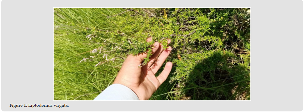
Leptodermis virgata belongs to the family Rubiaceae, one of the large angiosperm families, that comprises approx-imately 13,500 species distributed among about 620-630 genera with a cosmopolitan presence, spcially concentrated in tropical and subtropical regions [28,29]. Members of Rubiaceae are characterized by opposite or verticillate leaves with interpetiolar stipules, actinomorphic sympetalous flowers, and predominantly woody growth forms, that includes the shrubs and trees [30]. The genus Leptodermis Wall. is mainly distributed across the Himalayan region, China, and Japan, and derives its name from the Greek words leptos (thin) and derma (skin), that referres to its thin, mem-branous bractlets [31]. Species within this genus are shrubby, emit a characteristic odor when bruised, and possess opposite leaves with persistent stipules, although the genus remains taxonomically complex [32]. Leptodermis virgata is an erect, much-branched shrub typically 30–70 cm in height, bearing linear leaves, purplish flowers, and cylindrical capsules with fibrous seeds, features that distinguish it from related species [33]. Given the medicinal importance of several Rubiaceae members, including Coffea and Cinchona, the biological and pharmacological potential of L. virgata warrants systematic scientific investigation. Leptodermis virgata (Rubiaceae) is a shrubby species distributed predominantly in the Western Himalayas, that includes the regions of Pakistan, where it inhabits dry, stony soils up to elevations of approximately 2300 m. The family of Rubiaceae, one of the largest angiosperm families, is taxonomically and economically significant, encom-passes in medicinal, ornamental, and industrially very important plants such as Coffea and Cinchona. The members of this family are characterized by opposite leaves with interpetiolar stipules, sympetalous flowers, and predominantly woody growth forms, features that are well expressed in the genus Leptodermis. Now beyond its taxonomic relevance, it increases the scientific interest which has focused on the pharmacological potential of Rubiaceae taxa, specially their antifungal properties and rich phytochemical profiles. Antifungal agents, historically rooted in the concept of chemotherapy which is introduced by Paul Ehrlich, and it play a very critical role in combating fungal infections through selective inhibition of fungal cell wall or membrane synthesis. Concurrently, plant-derived phytochemicals such as flavonoids, alkaloids, terpenoids, tannins, and phenolic compounds have been bradly recognized for their anti-microbial and therapeutic efficacy. Standardized bioassays, that includes the agar diffusion (Kirby–Bauer) method, and it provides the reliable means to evaluate antifungal activity. In this method, the present study shows the botanical classification, antifungal relevance, and phytochemical significance of L. virgata, that supports its potential as a valuable source of bioactive compounds for pharmaceutical applications. Figure 1 shows the natural growth of Lip-todermis virgata, presenting its fine branching structure and dense green foliage. The image shows the characteristic leaf arrangement and morphology, which support botanical identification and ecological assessment.
Figure 1
Main Contributions
The main contributions of this study are summarized as follows: a. A comprehensive investigation of Leptodermis virgata, is used
in Himalayan shrub of the family Rubiaceae, is conducted to evaluate its antimicrobial potential.
b. The study systematically analyzed the antifungal activity, antibacterial activity, overall antimicrobial efficacy, and phytochemical composition of L. virgata in laboratry.
c. Ethanolic extracts of L. virgata are prepared using the cold maceration technique, and their efficacy is tested against multiple pathogenic microorganisms, that includes the Candida albicans, Candida parapsilosis, Es-cherichia coli, Pseudomonas aeruginosa, and Staphylococcus aureus.
d. The extract shows the strong antifungal activity with inhibition zones of 13 mm and 19 mm against C. albicans and C. parapsilosis, respectively, and notable antibacterial activity against E. coli with a 20 mm inhibition zone at 25 μL.
e. Phytochemical screening reveale the presence of key bioactive compounds, that includes the flavonoids, tannins, terpenoids, saponins, glycosides, steroids, and anthraquinones, which contribute to the observed antimicrobial effects.
f. This research highlights the potential of L. virgata as a natural source for the development of novel antifungal and antibacterial agents.
The use of medicinal plants is a cornerstone of traditional medicine, with knowledge passed through generations for the treatment of human and animal diseases. Ethnic groups worldwide have relied on plants as sources of natural remedies, primarily due to their production of antimicrobial secondary metabolites, and it offer relatively non-toxic and cost-effective method to synthetic drugs. Medicinal plants are evaluated based on their therapeutic, aromatic, or salty qualities, and are collected particularly for medicinal purposes and are recognized as true medicinal plants. The antimicrobial activity of these plants is linked to the presence of volatile oils and terpenes, and abundant in essential oils which is used in pharmaceuticals, cooking, and perfumery [34,35]. Plant extracts & essential oils are evaluated for their antifungal and antibacterial properties, and it shows their importance as natural bioactive compounds.
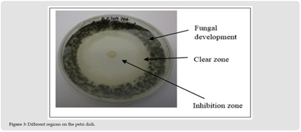
Many studies show the potent antimicrobial activity of plant extracts against both bacteria and fungi. Like, eleven ethanolic extracts from spices such as Melissa officinalis, Mentha piperita, Laurus nobilis, and Juniperus oxycedrus, and assayed against Gram-positive (Bacillus subtilis, Staphylococcus aureus, S. epidermidis) and Gram-negative (Escherichia coli, Pseudomonas aeruginosa) bacteria, as well as Candida albicans and Aspergillus niger. The minimum inhibitory concentration (MIC) of M. piperita, L. nobilis, and J. oxycedrus extracts is 5 mg/mL for all tested microorganisms, with P. aeruginosa being specially sensitive to P. nigrum and E. arborea extracts. The extracts of L. nobilis, D. coryophyllum, J. oxycedrus, and C. arborescens demonstrated higher antifungal activity than the standard drug nystatin [36]. Figure 2 shows the molecular structure of sulfanilamide, presenting a benzene ring which is substituted with amino and sulfonamide functional groups. The arrangement of these groups gives information about its chemical reactivity and underlies its antibacterial properties in pharmaceutical applications. Figure 3 shows the different regions on a petri dish, which includes the fungal development around the periphery and a distinct clear zone. The inhibition zone near the center shows the suppressed microbial growth due to the applied agent.
Figure 2
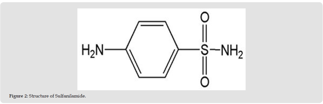
Figure 3
Similarly, six ethanolic extracts from spices including ginger (Zingiber officinale), cinnamon (Cinnamomum verum), black cumin (Nigella sativa), clove (Syzygium aromaticum), black pepper (Piper nigrum), and chamomile (Anthemis nobilis) were tested for antimicrobial activity against standard bacterial and fungal strains. Clove extract exhibited strong inhibitory activity against B. subtilis, P. aeruginosa, and C. albicans, whereas cinnamon was effective against B. subtilis and C. albicans only. Other extracts show either no activity or stimulatory effects. S. aureus and E. coli are resistant to most extracts, clove and cinnamon shown to potent antimicrobial effects at low MIC values and it ranges from 3.125 to 25 µg/mL [37].
Traditional Chinese medicine and ethnobotanical knowledge historically provided a rich source of empirical in-formation on the therapeutic value of plants. Before the emergence of modern medicine, herbalists accumulated significant knowledge about plant-based remedies through empirical observations of efficacy, failures, and successes. Modern pharmacology has built upon these traditional practices, that isolate active compounds from plant extracts for therapeutic purposes. Today, medicinal plants remain very important in combating infectious diseases; they are rich in secondary metabolites such as alkaloids, flavonoids, tannins, and terpenoids, which exhibit antibacterial, antifungal, and anticancer activities [38]. The study on plant extracts shown their selective antimicrobial and antifungal activities. Among 22 plant extracts evaluated, clove and ginger show the inhibitory activity against all tested fungal isolates, whereas garlic was effective against most fungi except Penicillium oxalicum. Extracts of cumin, turmeric, and cinnamon displayed activity against a limited number of fungi. Clove extract shows the highest mycelial growth inhibition (70-100%), which is followed by ginger extract (30-100%), and this suggests their potential as natural antimicrobial additives to improve food shelf-life.
The pharmacological potential of medicinal plants is also reflected in their chemical composition. Essential oils, flavonoids, terpenoids, saponins, steroids, and phenolic compounds contribute to the biological activity of plants. These phytochemicals not only confer the antimicrobial properties and also serve as antioxidants and nutraceuticals. The use of plant compounds like food preservatives is recognized due to consumer preference for natural products [39]. Research in different areas, like the Himalayas and Cameroon, highlights the nutritional and medicinal significance of indigenous plants. Pimpinella stewartii is identified as a rich source of phenolic compounds and antioxidants. And Cameroonian medicinal plants are reveals the presence of tannins, flavonoids, saponins, steroids, glycosides, and alkaloids, emphasizing their therapeutic potential. Certain plants, such as Morinda lucida and Spilanthes filicaulis, show the degrees of chemical diversity, which directly influence their efficacy in treating diseases [40-43].
Materials
Chemicals
a) For antifungal activity: The chemicals which are used for the evaluation of antifungal activity includes ethanol, distilled water, dimethyl sulfoxide (DMSO), and agar.
b) For phytochemical screening: The reagents which is employed for preliminary phytochemical analysis are ferric chloride (FeCl3), hydrochloric acid (HCl), magnesium ribbon, lead acetate, acetic acid (CH3COOH), sulphuric acid (H2SO4), Fehling’s solution, acetic anhydride, chloroform (CHCl3), and ammonia.
Apparatus
The apparatus are used in the present study comprises mortar and pestle, porcelain cloth, Whatman filter paper, beakers, volumetric flasks, micropipettes, aluminum foil, a magnetic stirrer, Eppendorf tubes, a cork borer, a digital balance, and sterilized Petri dishes.
Methodology
A systematic methodology is adopted to evaluate the antifungal activity of Leptodermis virgata. The experimental procedure involves the sequential steps, includes the collection and grinding of the plant material, preparation of crude extracts, preparation of a suitable culture medium, culturing of fungal strains with well formation, and assessment of antifungal activity of the plant extract.
Collection and Grinding of Plant Material
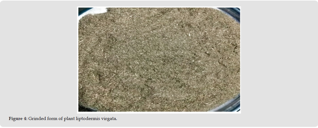
The plant, Leptodermis virgata, is collected from multiple locations in the Mansehra district. Initially, the samples are taken from Khait Sarash and Mang, Tehsil Balakot. Additional collections also collects from Tehsil Darband, Village Tarmang, and Tehsil Oghi, Village Chamial, and it is confirmed the presence of L. virgata in these regions. The collected plant material is shade dried at room temperature for a period of 65-70 days, in order to preserve its phytochemical constituents. After complete drying of plant, the whole plant, which includes the flowers, leaves, stems, bark, and branches, is finely ground into a uniform powder using a mortar and pestle. The powdered material is stored in clean, airtight containers for further experimental use. Figure 4 shows the powdered form of Liptodermis virgata after grinding, which presents a uniform and fibrous texture. This processed form is suitable for extraction and further phytochemical and biological analysis.
Figure 4
Preparation of Crude Extracts
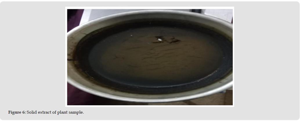
The ethanolic extract of Leptodermis virgata is prepared using the maceration technique. An estimated 4 kg of the dried and powdered plant material is soaked in 7.5 L of ethanol at room temperature. The mixture is intermittently stirred two to three times within a 24-hour period using a stainless-steel rod to guarantee the maximum extraction of bioactive constituents. After maceration, the extract is first filtered through a porcelain cloth and subsequently re-filtered using Whatman filter paper to obtain a clear filtrate. This extraction and filtration procedure is repeated three times to enhance this process. The combined filtrates are then left in the open air to allow complete evaporation of ethanol. This solvent evaporation process requires approximately two months, which show the results in a concentrated crude extract that is stored for further feature experimental use. Figure 5 shows the plant material soaked in ethanol, show the extraction stage where bioactive compounds are dissolved into the solvent. Figure 6 shows the solid plant extract and is obtained after solvent removal, it also represents the concentrated form which is also used for further experimental analysis.
Figure 5
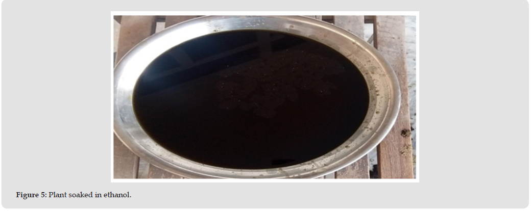
Figure 6
Preparation of Growth Medium
We evaluate the antifungal activity, and a suitable growth medium is prepared before microbial inoculation. We select nutrient agar as the culture medium due to its ability to support the growth of a wide range of microorganisms, and this includes fungi. The preparation method involves the dissolving of 20 g of nutrient agar powder in 500 mL of distilled water in a conical flask. The mixture is also heated with continuous stirring using a magnetic stirrer until the complete dissolution of the agar components is achieved. The flask is then covered with aluminum foil and autoclaved at 100◦C on a hot plate for one hour. After sterilization, the molten agar is allowed to cool without solidification and subsequently poured into sterile Petri dishes. The plates are then placed in a pressure steam sterilizer to guarantee the complete sterilization and left undisturbed until the agar solidifies.
Culturing of Fungi
Many techniques are available for fungal culturing, and the agar plate method is used in the present study. Fungi are ubiquitous organisms that exist as unicellular or complex multicellular forms and it play the essential roles in nutrient cycling within ecosystems. Some fungal species are pathogenic and responsible for diseases in plants, animals, and humans, which leads to significant agricultural and health-related losses. For the assessment of antifungal activity, two yeast species are selected as test organisms.
a) Candida albicans: Candida albicans is a pathogenic yeast that is commonly found as a commensal organism in the human gastrointestinal tract, but it may also exist outside the human body. It is also responsible for superficial infections such as oral and vaginal candidiasis, as well as it severe systemic infections that can be life-threatening, especially in immuno-compromised individuals.
b) Candida parapsilosis: Candida parapsilosis is the second or third most isolated fungal species from clinical specimens. It is a yeast associated with sepsis, wound infections, and tissue infections, especially in immunodeficient patients.
Assessment of Antifungal Activity
Antifungal activity is defined as the ability of a compound to selectively inhibit or kill fungal pathogens without causing damage to host tissues. Recently developed antimicrobial agents are chemically modified derivatives of natural compounds. The foundation of modern chemotherapy can be attributed to Paul Ehrlich (1910), who introduced the concept of chemotherapy using Salvarsan, an arsenic-based compound effective against syphilis. He also emphasized the selective toxicity of chemotherapeutic agents against pathogens. For antifungal evaluation, a solution of the crude plant extract is prepared using dimethyl sulfoxide (DMSO) as the solvent. Approximately 20 mg of crude extract is dissolved in 2 mL of DMSO in an Eppendorf tube and thoroughly shaken to gurantee the complete dissolution. Using a micropipette, 25 μL of the prepared solution is carefully dispensed into wells that is formed in the agar plates inoculated with fungal cultures. Only DMSO is used as a negative control and is added to a separate well. The same method is repeated for all Petri dishes. The inoculated plates are then incubated at appropriate conditions for 24 hours, after which antifungal activity is assessed by observation in a zones of inhibition around the wells.
Phytochemical Analysis
We also performed the phytochemical analysis is order to identify and evaluate the presence of various bioactive compounds in Leptodermis virgata. This screening is necessary for understanding the therapeutic potential of the plant and for correlating observed biological activities with specific classes of secondary metabolites. The phytochemical tests conducted in this study are follows:
• Test for Alkaloids: We have detected the nitrogen-containing
basic compounds, which are known for pharmaco-logical
activities.
• Test for Flavonoids: We identified the polyphenolic compounds
with antioxidant and antimicrobial properties.
• Test for Reducing Sugars: We also determined the presence
of monosaccharides or other reducing sugars.
• Test for Saponins: We detected the glycosidic compounds
that show the antifungal and surface-active properties.
• Test for Tannins: We identified phenolic compounds that are
capable of precipitating proteins and have contribution to the
antimicrobial activity.
• Test for Steroids: We also determined the presence of sterol-
based compounds with potential anti-inflammatory and
antimicrobial effects.
• Test for Terpenoids: We have detected the compounds which
is known for their diverse pharmacological activities, that includes
the antifungal and antibacterial effects.
These qualitative tests provide us with a preliminary insight into
the phytochemical composition of L. virgata, which contributes to its
observed antifungal and antibacterial activities.
Antifungal Activity
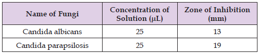
We evaluated the antifungal activity of the ethanolic extract of Leptodermis virgata using the agar well diffusion method. We also prepared the 25 µL solution of the extract in an Eppendorf tube and is subsequently added to the wells created using a cork borer on agar plates inoculated with the test fungi. We also determined the effectiveness of the extract which is measured the zones of inhibition around the wells after incubation. These results present that the Candida albicans gives us a zone of inhibition of 13 mm, while other Candida parapsilosis displays a larger zone of inhibition measuring 19 mm, which shows the stronger antifungal activity against C. parapsilosis. (Table 1) Figure 7 shows the antifungal activity of the plant extract against Candida albicans, which is evidenced by the visible inhibition of fungal growth. Figure 8 gives us a similar antifungal response against Candida parapsilosis, which indicates the broad-spectrum effectiveness of the extract. Figure 9 shows the inhibition zone, which is measured at a sample concentration of 25 microliter for two fungal strains. The extract shows the stronger antifungal activity against Candida parapsilosis and is compared to Candida albicans, which is indicated by the larger inhibition zone.
Figure 7
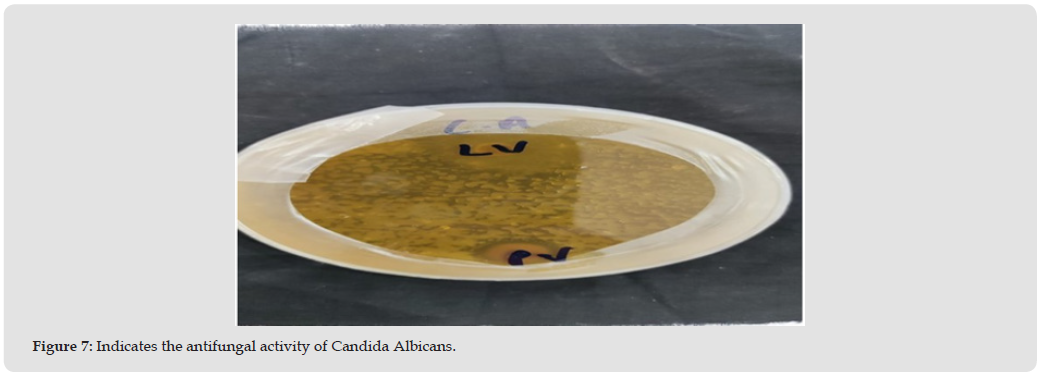
Figure 8
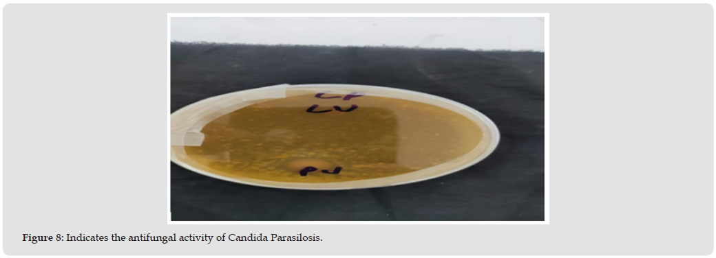
Figure 9
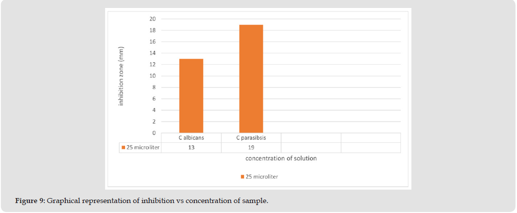
Table 1: Zone of Inhibition of Fungi by Leptodermis virgata Ethanolic Extract.
Antibacterial Activity
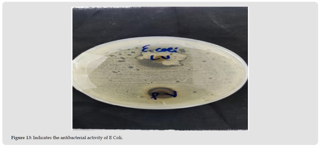
The antibacterial activity of the ethanolic extract of Leptodermis virgata is assessed using the agar well diffusion method. A 25 µL solution of the extract is added to wells in nutrient agar plates inoculated with the test bacterial strains. The zones of inhibition are measured after incubation to evaluate the effectiveness of the extract against each bacterial species. The results show that E. coli is the highest susceptibility with a zone of inhibition of 20 mm. Both Pseudomonas aeruginosa and Streptococcus aureus give us s smaller zones of inhibition, measuring the 6 mm each, suggesting comparatively lower antibacterial activity against these species. (Table 2) Figure 10 presents the zone of inhibition observed at a sample concentration of 25 microliter against different bacterial strains. The extract shows strong antibacterial activity against Escherichia coli, while moderate inhibition is observed for Pseudomonas aeruginosa and Staphylococcus aureus. Figures 11–13 shows the antibacterial activity of the plant which is extract against three clinically relevant bacterial strains.
Table 2: Zone of Inhibition of Bacteria by Leptodermis virgata Ethanolic Extract.
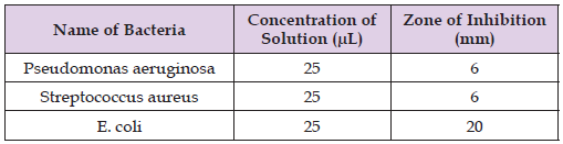
Figure 11 gives the information about the response of Pseudomonas aeruginosa, where a distinct inhibition zone confirms the sensitivity of this strain to the extract. Figure 12 demonstrates the antibacterial effect against Staphylococcus aureus, which indicates moderate growth suppression around the applied sample. Figure 13 shows the antibacterial activity against Escherichia coli, where a comparatively larger inhibition zone reflects stronger susceptibility. The variation in inhibition patterns shows the strain-dependent antibacterial potential of the plant extract. These observations suggest differences in cell wall structure and metabolic response among Gram positive and Gram-negative bacteria. The experimental results support the broad-spectrum antibacterial capability of the extracted compounds.
Figure 11
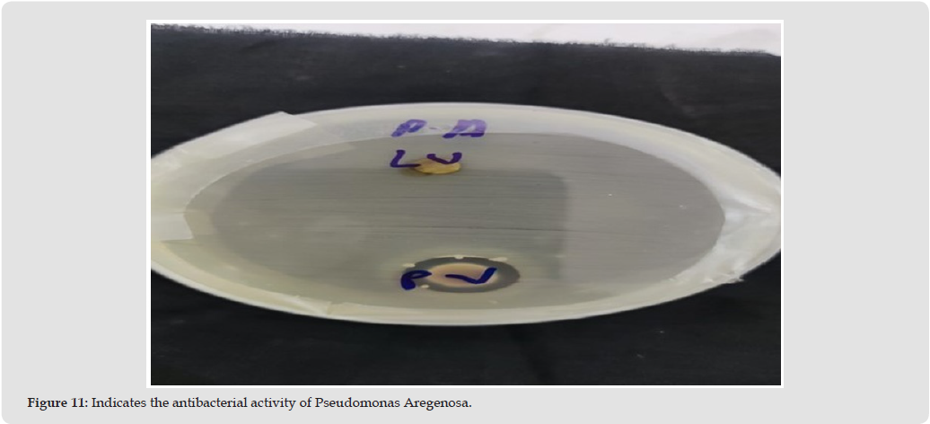
Figure 12
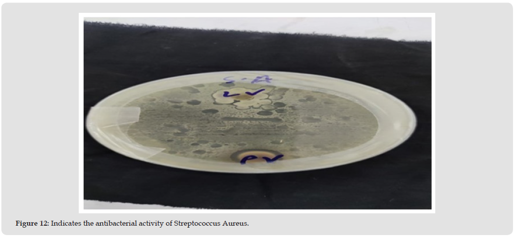
Figure 13
Phytochemical Analysis
Qualitative phytochemical analysis is performed to detect and localize bioactive compounds present in the ethanolic extract of Leptodermis virgata. Different standard tests are used to identify the various classes of phytochemicals, that are:
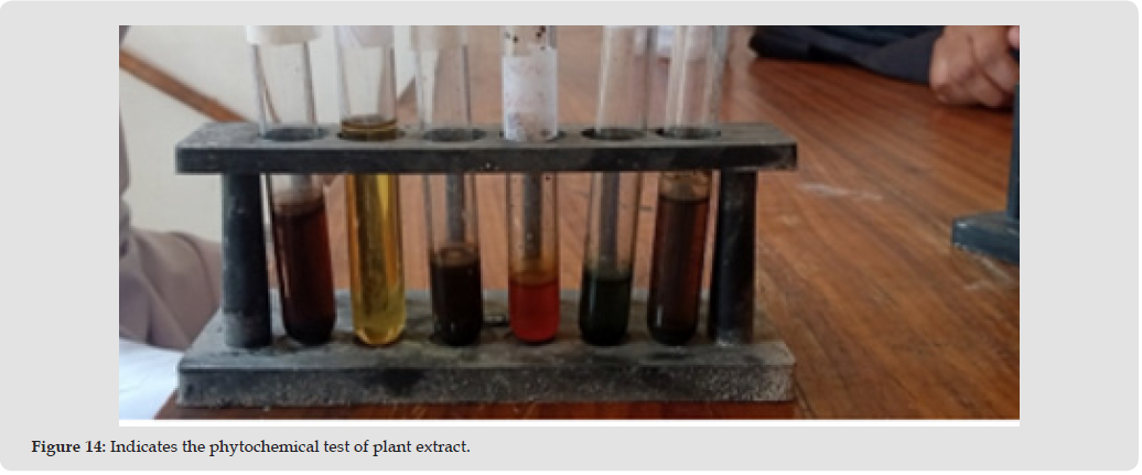
a) Flavonoids: The alkaline reagent test is used for flavonoids. Two milliliters of 2% NaOH solution are added to the crude extract. The appearance of a yellow color indicates the presence of flavonoids.
b) Saponins: The extract is mixed with 5 mL of distilled water in a test tube and warmed. The absence of significant change indicates the presence of saponins in the extract.
c) Alkaloids: Salkowski’s test is performed by mixing 2 mL of chloroform with the crude extract, which is followed by 2 mL of concentrated sulfuric acid. The development of a green color confirmed the presence of alkaloids.
d) Anthraquinones: We extract the two milliliters of plant and then mixed with 1 mL of 10% ammonia solution and shaken for 4–5 minutes. The formation of a deep red color shows the presence of anthraquinones.
e) Tannins: We also conduct the Ferric chloride test by stirring 2 mL of plant extract with distilled water and adding a few drops of FeCl3. The formation of a green precipitate shows the presence of condensed tannins.
f) Steroids: We also added the two milliliters of chloroform into 2 mL of plant extract, which is followed by 1 mL of concentrated sulfuric acid. The appearance of a blue-green color indicates the presence of steroids.
g) Terpenoids: We dissolve two milliliters of plant extract in 2 mL of chloroform, and 2 mL of concentrated sulfuric acid. The mixture is heated for 2 minutes, and the appearance of a greyish color confirmed the presence of terpenoids. Figure 14 shows the phytochemical screening of the plant extract using standard qualitative tests. Distinct color changes and precipitate formations show the presence of bioactive constituents such as alkaloids, flavonoids, tannins, and phenolic compounds. These phytochemicals are likely responsible for the observed antibacterial effects, and they validate the medicinal relevance of the plant extract.
Figure 14
Results of Phytochemical Tests
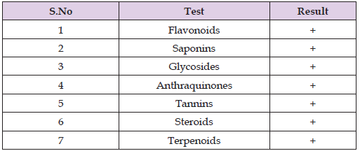
The qualitative phytochemical analysis of the ethanolic extract of Leptodermis virgata shows the presence of various bioactive compounds. The results are summarized in Table 3.
Table 3: Results of phytochemical tests for Leptodermis virgata ethanolic extract.
The active components of the plant Leptodermis virgata are extracted using ethanol and evaluate for their antibacterial and antifungal activities against some selected pathogenic microorganisms. The ethanolic extract also shows the activity against Pseudomonas aeruginosa, Streptococcus aureus, and E. coli for antibacterial testing, against Candida albicans and Candida parapsilosis for antifungal testing. We also observed the antimicrobial activity at room temper-ature and measured it through the zones of inhibition on agar plates. The results show the extract contains bioactive compounds such as flavonoids, terpenoids, glycosides, tannins, saponins, steroids, and anthraquinones, which contribute to the observed antimicrobial effects. These results give us the guarantee that L. virgata can be utilized for the treatment of gastroenteritis, urethritis, wound infections, and fungal diseases like sepsis.
The results of this study reveal that the ethanolic extract of Leptodermis virgata possesses antifungal, antibacterial, and overall antimicrobial activities. The extract shows the stronger antibacterial activity against Escherichia coli and as compared to Staphylococcus aureus and Pseudomonas aeruginosa. Moreover, both tested fungal strains, i.e. Candida albicans and Candida parapsilosis, show the marked susceptibility to the plant extract, show the antifungal activity of L. virgata is more pronounced and useful than its antibacterial activity. Phytochemical screening confirmed the presence of many major bioactive constituents, which includes flavonoids, tannins, terpenoids, saponins, glycosides, steroids, and anthraquinones, which are collectively contribute to the observed antimicrobial effects. These findings support the traditional use of L. virgata in the treatment of various infectious diseases, especially in fungal infections. Further studies will be used to investigate its efficacy against a broader spectrum of bacterial and fungal pathogens, as well as to isolate and characterize the active compounds responsible for its antimicrobial activity, that develop novel plant-based therapeutic agents.
No datasets were used or analyzed in this study.
The author is deeply grateful to his parents and uncle, especially Nazakat Hussain Shah, and one of his best friends, Muhammad Israr, for their unwavering sup-port and encouragement throughout his education and this research. The author also extends sincere thanks to the Department of Chemistry, Government Post Graduate College Mansehra, for their valuable support.
There is no conflict of interest among authors, and all authors did equal work.

International Association of Landscape Archaeology, Czech Glass Society, Czech Republic

Department of Chemistry, Semenov Institute of Chemical Physics, USSR Academy of Sciences, Moscow, Russia

Neurology, LA BioMed Research Institute, USA

Associate Professor at Department of Breast and Thyorid Surgey, Chongqing General Hospital, China
Professor of Nuclear Medicine, Faculty of Medicine and Surgery, University of Milan, Milan, Italy





 Abstract
 PDF