info@biomedres.us
+1 (502) 904-2126
One Westbrook Corporate Center, Suite 300, Westchester, IL 60154, USA
Site Map
 ISSN: 2574 -1241
Received: September 15, 2026; Published: September 30, 2026
*Corresponding author: Atsushi Imai, Department of Obstetrics and Gynecology, Matsunami General Hospital, 185-1 Dendai, Kasamatsu, Gifu 501-6062, Japan
DOI: 10.26717/BJSTR.2026.66.010419
The development and growth of uterine fibroids are primarily influenced by ovarian estrogen and progesterone. Following menopause, the decline in ovarian steroid hormone production generally induces spontaneous regression of uterine fibroids and improvement of associated symptoms. However, this regression is not necessarily complete, and some fibroids may persist or demonstrate limited reduction in size. For asymptomatic uterine fibroids or adenomyosis, routine imaging examinations, including magnetic resonance imaging (MRI), are generally unnecessary after menopause. However, if new symptoms such as genital bleeding, lower abdominal pain, or abdominal distension develop, prompt re-evaluation is essential to exclude malignant disease. Uterine fibroids are benign mesenchymal tumours that occur in approximately 30% of women over 30 years of age and in 40–50% of women over 40 [1-3]. Although approximately half of affected women are asymptomatic, many undergo periodic clinical surveillance or receive treatment for fibroids. The development and growth of uterine fibroids are strongly influenced by ovarian estrogen and progesterone. Many clinicians are concerned about the management of uterine fibroids after menopause. This article examines the appropriate management and follow-up of uterine fibroids during the menopausal transition and postmenopausal period.
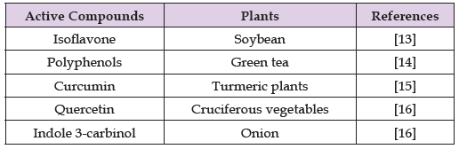
Following menopause, ovarian secretion of estrogen and progesterone declines substantially. Consequently, uterine fibroids generally undergo spontaneous regression, accompanied by improvement in associated symptoms, although the lesions themselves do not necessarily disappear completely. In a small proportion of women, however, fibroids may fail to demonstrate substantial regression after menopause. Possible explanations include peripheral estrogen production by adipose tissue, as well as the local production of growth factors, inflammatory mediators, or estrogen within the fibroid tissue itself [1-3]. This phenomenon may be particularly evident in women with obesity and a substantial amount of adipose tissue. Furthermore, calcification and degenerative changes within fibroids may limit or prevent appreciable reductions in lesion size. Women receiving estrogen- containing preparations [4-6] or supplements containing phytoestrogens [7-9], such as soy isoflavones (Table 1) -which are commonly used to alleviate menopausal symptoms or reduce the risk of osteoporosis-may likewise experience limited regression of uterine fibroids after menopause. Information regarding dietary compounds that may influence uterine fibroids is available at: https://www.mdpi. com/article/10.3390/nu14040734/s1.
Table 1: The active compounds from plants influencing uterine fibroids.
Adenomyosis, a condition sharing certain clinical and pathological characteristics with uterine fibroids [10,11], is similarly influenced by estrogen with respect to lesion growth and symptom progression. Accordingly, both lesion size and associated symptoms generally decrease after menopause. Although adenomyosis often produces more severe symptoms than uterine fibroids, it typically exhibits a more pronounced response to the hormonal changes associated with menopause. An extremely rare condition, occurring in less than 1% of cases, is endometrial carcinoma arising within adenomyosis [12]. This malignancy is thought to result from the malignant transformation of ectopic endometrial tissue within an adenomyotic lesion and is referred to as endometrial carcinoma arising in adenomyosis (EC-AIA). Although EC-AIA is considered to occur predominantly after menopause, no specific diagnostic imaging findings have yet been established. In many cases, uterine fibroids identified after menopause do not require routine surveillance with periodic imaging examinations. However, the development of new symptoms after menopause-such as genital bleeding, lower abdominal pain, or abdominal distension, including a sensation of increased abdominal fullness or tightness of clothing or undergarments-warrants further evaluation to exclude the possibility of malignancy.
In addition, very large fibroids may be associated with age-related complications, including an increased risk of deep vein thrombosis or renal impairment. Surgical intervention may also be considered when fibroid-related mass effects and compressive symptoms, such as urinary frequency or constipation, become clinically significant. For asymptomatic uterine fibroids or adenomyosis, routine imaging examinations, including MRI, are generally unnecessary after menopause. However, the emergence of new symptoms, such as genital bleeding, lower abdominal pain, or abdominal distension, warrants prompt clinical re-evaluation to exclude malignant disease [13-16]. The menopausal transition is a period during which a variety of physical symptoms and health concerns may arise as a consequence of hormonal fluctuations, particularly alterations in estrogen levels. Careful attention to changes in one’s physical condition and timely consultation with a healthcare professional when concerns arise are therefore important. Maintaining regular gynaecological examinations remains an essential component of preventive healthcare and contributes to the preservation of health and well-being throughout later life.
The authors declare no conflict of interest.

International Association of Landscape Archaeology, Czech Glass Society, Czech Republic

Department of Chemistry, Semenov Institute of Chemical Physics, USSR Academy of Sciences, Moscow, Russia

Department of Educational Sciences, Research Centre Qualities of Teachers, School of Education – HAN, Nijmegen, Netherlands
Associate Professor at Department of Breast and Thyorid Surgey, Chongqing General Hospital, China

Neurology, LA BioMed Research Institute, USA





 Abstract
 PDF