info@biomedres.us
+1 (502) 904-2126
One Westbrook Corporate Center, Suite 300, Westchester, IL 60154, USA
Site Map
 ISSN: 2574 -1241
Received: September 15, 2026; Published: September 25, 2026
*Corresponding author: Konstantinos Fousekis, Physiotherapy Department, School of Health Rehabilitation Sciences, University of Patras, 26504 Patras, Greece
DOI: 10.26717/BJSTR.2026.66.010406
Background: It is possible that instrument-assisted soft tissue mobilisation (IASTM) can affect neuromuscular
activity in areas that lie beyond the region that is directly treated, even though the spatial range and direction
of these effects are still not well understood. Passive movement offers a useful alternative for investigating
neuromuscular responses when the tissue is mechanically stimulated without any voluntary contraction of the
muscles. In this study local-to-remote surface electromyographic (sEMG) responses along the superficial back
myofascial line (SBL) were examined both during passive ankle movement and after ERGON® IASTM had been
applied to the plantar surface of the foot.
Methods: Thirty healthy adults (19 men and 11 women; average age 25 years; average BMI 22.9 kg/m²) took
part in the study. Surface electromyography was obtained from eight sites on the right side along the SBL: the
lateral gastrocnemius, the long head of the biceps femoris, the semimembranosus, the area above the iliac crest,
T12, T6, T1, and the upper trapezius. This surface electromyography was recorded while the ankle was being
passively plantarflexed and dorsiflexed. In the ERGON® condition, the participants were given a 10-minute treatment
on the plantar surface and on the longitudinal arches of the foot. After a 2-minute rest, their resting sEMG
was checked again.
Results: The sEMG activity was mainly increased by passive ankle movement, showing significant responses in
the lateral gastrocnemius, the long head of the biceps femoris, the semimembranosus, the area above the iliac
crest, and the upper trapezius. After undergoing ERGON® IASTM, the resting sEMG showed a heterogeneous
response that varied from site to site. In the lateral gastrocnemius, activity decreased significantly (from a median
of 0.945 to 0.890; p<0.001), and significant changes were also found in the long head of the biceps femoris
(p=0.021) and in the region above the iliac crest (p=0.045). The post-ERGON® responses were found to differ
significantly among the various recording sites (χ²=17.597, df=7, p=0.014).
Conclusion: There was a clear difference in the neuromuscular response patterns between passive ankle movement
and the use of the ERGON® IASTM. While passive movement mainly led to an increase in sEMG activity
when mechanical stimulation was applied, the ERGON® IASTM was associated with a non-uniform, site-dependent
adjustment of the resting muscle activity. These results suggest that further research should be carried out
into the stimulus-specific local-to-remote neuromuscular interactions that occur after IASTM.
Keywords: ERGON® IASTM; Superficial Back Line; Surface Electromyography; Passive Ankle Movement; Neuromuscular Response; Myofascial Continuity
Fascia is a continuous three-dimensional network of connective tissue which surrounds, links, and integrates the muscles and other parts of the musculoskeletal system [1,2]. Instead of acting as entirely separate anatomical units, the muscles are connected via both intramuscular and extramuscular connective tissue, thus providing a structural foundation for mechanical interactions between regions that are anatomically distant [3]. Since fascia also has sensory nerve endings and mechanosensitive structures, it indicates that its function goes beyond simple mechanical support to include proprioceptive and sensorimotor processes [4,5]. In this framework, myofascial chains have been suggested as longitudinal anatomical continuities that link muscles and connective tissues in different parts of the body. The superficial back line (SBL) is one of the most thoroughly studied of these, running from the bottom of the foot via the posterior part of the lower limb and the trunk to the upper region. A systematic review of anatomical studies has given evidence in support of several of the structural connections proposed within the myofascial chains, especially those relating to the SBL [6]. It is important to note that anatomical continuity does not mean that there is a uniform transmission of force functionally. However, experimental work on cadavers has shown that there is tensile transmission through the fascial structures, thus providing a possible mechanical basis for the interaction between different muscular areas [3].
There is an ever-increasing amount of evidence indicating that stimulating one area mechanically can be linked to functional changes at sites that are anatomically distant. Cases of remote responses have been documented after stretching [7,8], following exercise of the plantar muscles [9], as a result of self-myofascial release [10], and in relation to other interventions that involve mechanically connected regions. Together, these findings have led to a growing interest in the functional significance of myofascial connectivity [11]. Yet the mechanisms responsible for these responses are still unclear and could include mechanical interactions between tissues, changes in afferent input, neuromuscular regulation, or a combination of these processes. Instrument-assisted soft tissue mobilization (IASTM) consists of applying controlled mechanical stimulation to soft tissues by means of special instruments. It has now been increasingly included in musculoskeletal and sports rehabilitation, and both mechanical and neurophysiological mechanisms have been proposed together with reported effects on range of motion, pain, and function [12,13]. The acute changes in flexibility and range of motion that occur after myofascial interventions which are based on mechanical action also indicate that soft-tissue stimulation might quickly alter musculoskeletal behaviour [14]. ERGON® IASTM is a form of instrument-assisted therapy which includes the directed mobilisation of myofascial tissues. It is worth noting that the effects of ERGON® treatment can go beyond the area that is directly treated. Fousekis et al. [15] showed that when ERGON® IASTM was applied to either the proximal or distal part of the SBL, hamstring flexibility was significantly improved, indicating that treating one anatomical area may result in functional changes in other areas along the same myofascial continuum.
This earlier study therefore provides an important foundation for the current investigation. It cannot be determined from improved flexibility whether changes in neuromuscular activity occur as a result of remote responses. Surface electromyography (sEMG) offers a means of examining the neuromuscular manifestation of such responses. Weisman et al. [16] looked at sEMG activity during both passive and active movements involving various parts of the posterior myofascial kinematic chain, thus further supporting the need to investigate distributed neuromuscular responses when mechanical stimulation is applied. Surface EMG has also been established as a practical technique for assessing muscle activity under functional conditions [17]. It is especially relevant to consider passive movement since it offers mechanical and sensory input without requiring much voluntary motor activity. On the other hand, passive movement and IASTM are different types of mechanical stimulation; while it is possible to study neuromuscular activity during passive movement as the mechanical stimulus is taking place, resting sEMG taken after IASTM can be used to find out if neuromuscular modulation is still detectable once the interaction between the instrument and the tissue has stopped.
The main objective of the present study was to examine the local- to-remote changes in resting sEMG activity after a 10-minute ERGON ® IASTM intervention had been carried out on the plantar surface and the longitudinal arches of the foot. A second objective was to describe how the sEMG responses were distributed along the same myofascial line during passive ankle plantarflexion–dorsiflexion. We assumed that passive ankle movement would cause detectable sEMG responses in areas beyond the region that was mechanically moved, while ERGON® IASTM would result in a distinct modulation of resting sEMG activity at the proximal and more remote sites along the SBL.
Study Design
An experimental study using a randomized within-subject repeated- measures design was carried out to examine the neuromuscular responses associated with two experimentally different conditions involving the distal part of the SBL: passive ankle movement and ERGON® IASTM. The participants took part in both conditions, the order in which they did so being determined at random in order to minimize any possible order effects. While carrying out passive ankle movements, we simultaneously recorded the sEMG activity together with the mechanical stimulus. In the case of the ERGON® condition, the resting sEMG was measured both before and after the intervention had been completed. Passive movement therefore served to show the neuromuscular response during non-voluntary mechanical stimulation, while the ERGON® IASTM looked at the immediate change in resting neuromuscular activity after the intervention. There was a recovery period between the different conditions and the next condition only started once the sEMG activity had returned to baseline levels in order to reduce any possible carry-over effects.
Participants
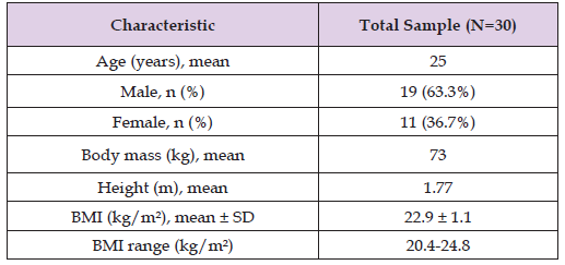
Thirty healthy adults (19 of them men and 11 women) took part in the study. Their average age was 25 years, their average body mass 73 kg, their average height 1.77 m, and their average BMI 22.9 kg/m². To be included in the study, participants had to be between the ages of 18 and 30, have a body mass index within the normal range (18.5 to 24.9 kg/m²), have reported experiencing no pain over the previous 3 months and no musculoskeletal injury in the past year, have no record of having undergone surgery, and not be on any pharmacological treatment. It was told to the participants that they should avoid strenuous physical activity in the 24 hours before the testing took place. The participants were made aware of the experimental procedures and gave their written informed consent before taking part.
The participants were placed on the treatment table in the prone position with the lower limbs at their distal end left unsupported so that they could carry out unrestricted plantarflexion and dorsiflexion of the ankle. Before electrode placement, the corresponding skin areas were shaved when necessary. Resting sEMG was recorded immediately before each experimental condition and was used as the condition-specific baseline. After completion of the first condition, participants remained at rest until resting sEMG activity returned to baseline before the second condition began. Each sEMG measurement took 10 seconds and was carried out three times; a rest period of 1 to 2 minutes was given between the repeated measurements and the highest value obtained during the three repetitions was used for the statistical analysis.
Surface Electromyography
The surface electromyographic activity was recorded with the use of a Noraxon surface EMG system.
Eight electrodes were positioned along the right side of the SBL at the following sites:
1. Lateral head of gastrocnemius;
2. Long head of biceps femoris;
3. Semimembranosus;
4. Region immediately superior to the iliac crest;
5. Paraspinal region at T12;
6. Paraspinal region at T6;
7. Paraspinal region at T1; and
8. Upper trapezius.
The anatomical sites were found by feeling them with the hands. Before the electrodes were placed, the lateral gastrocnemius, the long head of the biceps femoris, and the semimembranosus were identified while the subject was contracting voluntarily. T12 was located in relation to the lowest ribs, T6 was then found more towards the front, and T1 was identified in relation to C7. The electrode for the upper trapezius was placed at a point that was about one-third of the way from C7 to the acromioclavicular joint. The electrodes were placed on the right side of the body in order that the sEMG signal might be less likely to be contaminated by cardiac electrical activity.
Passive Ankle Movement
The participants kept a relaxed position with their bodies lying face down while the ankle was moved externally in both plantarflexion and dorsiflexion without any deliberate muscular involvement. Surface EMG activity was recorded at all eight sites at the same time as the passive ankle movement took place. The condition was thus an example of externally applied mechanical stimulation and served to show how the neuromuscular activity was distributed during the time the stimulus was active. Each recording took 10 seconds and was repeated three times, with a rest of 1 to 2 minutes between the repetitions.
ERGON® IASTM Intervention
The ERGON® treatment consisted of a single 10-min IASTM intervention applied to the plantar aspect of the foot. The Wave, Razor, and Globe strokes were used over the entire plantar surface, while the Excav techniques were applied in a targeted manner to the medial and lateral longitudinal arches. Surface EMG was not recorded during ERGON® application. After treatment, participants remained relaxed for 2 min, after which resting sEMG activity was reassessed at the same eight anatomical sites. As a result, the ERGON® outcome showed the change in resting neuromuscular activity immediately after the mechanical stimulus had stopped, not the muscle activation that occurred during the use of the instrument.
Statistical Analysis
The Shapiro-Wilk test and visual examination of the normal Q-Q plots, the detrended Q-Q plots, and the box plots were carried out to assess the data distribution. Since the normality assumptions were not met, non-parametric methods were employed. The continuous sEMG variables were described using medians and interquartile ranges. The differences within each site between the resting baseline and passive ankle movement were evaluated with the Wilcoxon signedrank test; the differences between pre- and post-ERGON® resting sEMG values were also assessed using the same test. Gain scores were calculated for each condition and for each anatomical site. The variations in response magnitude across the eight sites were investigated using the Friedman test, and where appropriate, pairwise comparisons were made. The statistical analyses were conducted using SPSS version 25 and a significance level of p<0.05 was used.
Thirty participants carried out both experimental conditions and were therefore included in the analysis. The demographic and anthropometric characteristics of these participants are given in Table 1.
Table 1: Demographic and Anthropometric Characteristics of Participants.
Note: BMI: body mass index.
Passive Ankle Movement
Compared to the resting baseline, passive ankle movement was mainly linked with higher sEMG activity, with significant increases noted in the lateral gastrocnemius (p=0.011), the long head of the biceps femoris (p=0.031), the semimembranosus (p=0.023), the area above the iliac crest (p=0.001), and the upper trapezius (p=0.008). At T12 (p=0.559), at T6 (p=0.632), and at T1 (p=0.105) no significant changes were found. The magnitude of response did not differ significantly across the eight recording sites (Friedman χ²=8.893, df=7, p=0.260).
ERGON® IASTM
After the application of ERGON® IASTM, the resting sEMG showed a heterogeneous response that varied from site to site. The most notable change was found in the lateral gastrocnemius, the median resting sEMG falling from 0.945 at baseline to 0.890 after the intervention (p<0.001). Significant changes after the intervention were also seen in the long head of the biceps femoris (p=0.021) and in the area superior to the iliac crest (p=0.045). No significant changes, however, were detected in the semimembranosus, at T12, T6, T1, or in the upper trapezius. The extent of the post-ERGON® response varied significantly among the different recording sites (Friedman χ²=17.597, df=7, p=0.014). A post-hoc analysis revealed a significant difference between the lateral gastrocnemius and the area superior to the iliac crest (p=0.025) (Table 2).
Table 2: Surface Electromyographic Responses During Passive Ankle Movement and Following ERGON® IASTM.
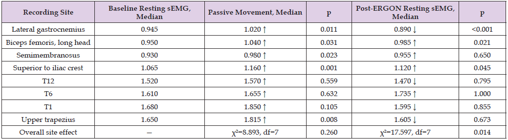
Note: The following values refer to the sEMG recordings made when the ankle is passively plantarflexed and dorsiflexed. The values obtained after the ERGON® procedure, following a 10-minute intervention and a further 2-minute rest, are known as the post-ERGON® values. The arrows show the direction of the median change compared with the baseline. Values in bold show that the result is significant at the p<0.05 level.
The main result of the current study was that passive ankle movement and the use of ERGON® IASTM were linked to quite different neuromuscular response patterns along the SBL. During passive ankle plantarflexion and dorsiflexion, sEMG activity increased mainly while the mechanical stimulus was being applied, with significant responses being found in the gastrocnemius, the hamstrings, the area above the iliac crest, and the upper trapezius. On the other hand, ERGON ® IASTM applied only to the plantar surface and the longitudinal arches was followed by a heterogeneous, site-dependent modification of resting sEMG; the most noticeable effect was a significant decrease in the resting activity of the lateral gastrocnemius, together with significant changes also being observed in the long head of the biceps femoris and in the area above the iliac crest.
Neuromuscular Responses During Passive Movement
It is noteworthy that there was a mainly increased sEMG activity during passive ankle movement since the movement was imposed from the outside without any intentional muscle contraction. At five of the eight recording sites there were significant responses and these extended considerably beyond the area of the mechanically moved ankle. This result is generally in agreement with that of Weisman et al. [16], who looked at the sEMG responses linked with both passive and active movement along the posterior myofascial kinematic chain. Yet the current findings must not be seen as direct proof that electrical activation was mechanically ‘transmitted’ through the fascia. Surface EMG refers to the electrical activity linked with motor-unit recruitment [17]. It follows that the remote sEMG responses might be due to a number of interacting mechanisms. Sensory input can be produced by the passive deformation of muscles, tendons, fascia, and the tissues surrounding the joints and this sensory input can in turn affect motor output. The fact that sensory nerves have been shown to innervate fascial tissues [4,5] provides a reasonable neurophysiological basis for these responses. At the same time, a mechanical factor must not be ruled out. Anatomical studies show there to be connective-tissue continuity in certain parts of the SBL [2,6], and experimental evidence obtained from cadavers proves that tensile loading can be transmitted through the fascial structures [3].
Hence, mechanical and neurophysiological mechanisms should not necessarily be seen as opposing explanations; a local mechanical disturbance may deform the interconnected tissues while at the same time altering afferent input and neuromuscular output. Interestingly, even though passive movement of the ankle mainly led to an increase in sEMG activity, there was no significant difference in the response among the eight sites (p=0.260). Moreover, T12, T6, and T1 also showed no significant changes. Hence, the pattern does not support a simple progression of activation from the distal to the proximal regions.
Neuromuscular Response Following ERGON® IASTM
The ERGON® condition resulted in a distinct pattern. In contrast to passive movement, sEMG was not recorded when the instrument was acting on the tissues. Resting sEMG was reexamined after the 10-minute intervention was completed and then again after a 2-minute rest period. The changes that were observed therefore reflect post-stimulus neuromuscular modulation and not activation which took place during mechanical stimulation.
The most notable result was a decrease in the resting sEMG of the lateral gastrocnemius from a median value of 0.945 to 0.890 (p<0.001). This is rather interesting since the treatment was applied to the plantar surface and the longitudinal arches rather than being given directly to the gastrocnemius. Significant changes were also seen in the long head of the biceps femoris (p=0.021) and above the iliac crest (p=0.045). The results of this study go beyond what has already been found concerning remote effects after ERGON® IASTM. Fousekis et al. [15] showed that there was an improvement in hamstring flexibility after ERGON® treatment was given to various areas of the SBL. The current study contributes a new aspect by showing that changes resulting from applying ERGON® distally can also be detected electrophysiologically at proximal sites which had not received treatment. More generally, remote functional responses have been shown to result from stretching [7,8], plantar muscle exercises [9], and self-myofascial release [10].
These findings lend support to the general idea that interventions given to one area can be linked with changes in other parts of an interconnected kinetic or myofascial system [11]. They do not, nevertheless, prove that there is a single mechanism accounting for these responses. The ERGON® response should be given special attention since, while passive movement was associated with an increase in sEMG during stimulation, ERGON® IASTM was accompanied by a decrease in resting activity at certain sites, most notably in the lateral gastrocnemius. The difference might to some extent be due to the different time-related features of the two situations. While the sEMG was being recorded, both continuous mechanical deformation and sensory input were occurring during passive movement. On the other hand, the ERGON® measurements were taken after the mechanical stimulation had stopped. The idea that IASTM can affect tissues is based on a combination of mechanical and neurophysiological mechanisms [12,13]. Since the plantar tissues are densely innervated, mechanical stimulation of them might change the afferent input, and this could in turn lead to a modification of the motor output of the muscles situated more proximally. The sensory innervation of the fascia as described by Tesarz et al. [4] and Stecco et al. [5] offers a biological basis for this kind of mechanism.
It cannot indeed be regarded as direct proof of ‘muscle inhibition’, ‘relaxation’, or a decrease in muscle tone that the resting sEMG has decreased. Since spinal excitability, motor-neuron excitability, and mechanical tissue stiffness were not directly measured, the correct interpretation is that the ERGON® IASTM led to lower resting electromyographic activity at certain proximal sites, this showing a shortterm neuromuscular modulation although the mechanism of this modulation has not yet been established. A further important finding was that there was a significant difference in the size of the post-ERGON ® responses at the eight recording sites (Friedman χ²=17.597, df=7, p=0.014). The response was not therefore evenly distributed along the SBL. This agrees with a more complicated model of myofascial interaction. Anatomical evidence does show there being specific fascial continuities [1,2,6], and mechanical studies have shown that force can be transmitted between connected tissues [3]. Yet neither of these findings demands that a mechanical stimulus applied at one end of a myofascial line should result in an equal or progressively diminished response at each subsequent anatomical site. On the contrary, the extent of the response could be influenced by the regional anatomy, the stiffness of the tissue, the orientation of the connective-tissue fibres, the mechanical coupling, the density of the sensory receptors, and neural regulation. The SBL can therefore be better understood as an anatomical framework which allows for distributed mechanical and neurosensory interactions, not as a rigid cable that transmits a fixed quantity of mechanical energy.
Clinical Implications
The results obtained at present indicate that the effects linked to IASTM might go beyond the tissues which are directly touched by the instrument. This idea is in agreement with the earlier ERGON® findings showing improvements in flexibility at a distance [15] and also with general evidence of non-local responses after interventions involving myofascial structures [7-11,14]. This situation could become important whenever treatment of a symptomatic structure is not well tolerated, is contra-indicated, or is technically difficult. It could also be used to support rehabilitation approaches that take into account the anatomical and functional relationships between the site of treatment and more distant areas. However, such clinical extrapolation should remain cautious. The participants were healthy young adults, and we examined only immediate electrophysiological responses. The results do not demonstrate reductions in pain, improvements in function, or superior clinical outcomes following remote treatment.
Limitations
It is necessary to recognize a number of limitations. The sample included 30 healthy young adults who had relatively similar anthropometric features, which limits the external validity. Moreover, the convenience sampling method employed in the original study also hampers generalizability. There was neither a sham-IASTM nor an untreated control condition included. As a result, the post-ERGON® changes observed cannot be solely attributed to a particular therapeutic mechanism. The passive and ERGON® conditions also involve different temporal paradigms since the sEMG was recorded when passive movement was taking place but only after the ERGON® treatment had been administered; they must therefore be regarded as complementary experimental conditions not as directly equivalent treatment effects. Surface EMG cannot directly measure fascial strain, mechanical force transmission, sensory afferent activity, spinal excitability, or muscle stiffness. As a result, the mechanical and neurophysiological mechanisms mentioned above still remain as plausible explanations rather than having been demonstrated as causal pathways. In the end, a number of comparisons specific to each site were carried out; since this raises the risk of a type I error, one must exercise special care when interpreting borderline results such as the response of the superior iliac crest (p=0.045).
Passive ankle movement and ERGON® IASTM were associated with distinct neuromuscular response patterns along the SBL. Passive ankle movement predominantly increased sEMG activity at multiple local and remote sites while mechanical stimulation was ongoing. In contrast, plantar ERGON® IASTM was followed by heterogeneous, site-dependent modulation of resting sEMG, with the clearest response being reduced lateral gastrocnemius activity. It seems that when the distal SBL is mechanically stimulated, neuromuscular reactions occur in tissues other than those that were directly stimulated. Significantly, the response depends on both the type and the temporal features of the mechanical stimulus. Rather than demonstrating simple linear myofascial force transmission, the findings support further investigation of interactions among anatomical continuity, mechanical loading, sensory input, and neuromuscular regulation.

International Association of Landscape Archaeology, Czech Glass Society, Czech Republic

Department of Chemistry, Semenov Institute of Chemical Physics, USSR Academy of Sciences, Moscow, Russia

Department of Educational Sciences, Research Centre Qualities of Teachers, School of Education – HAN, Nijmegen, Netherlands

Associate Professor at Department of Breast and Thyorid Surgey, Chongqing General Hospital, China

Neurology, LA BioMed Research Institute, USA





 Abstract
 PDF