info@biomedres.us
+1 (502) 904-2126
One Westbrook Corporate Center, Suite 300, Westchester, IL 60154, USA
Site Map
 ISSN: 2574 -1241
ISSN: 2574 -1241
ISSN: 2574 -1241
Received: September 07, 2026; Published: September 21, 2026
*Corresponding author: George B Stefano, Mind-Cell LLC, Baltimore, MD, USA
DOI: 10.26717/BJSTR.2026.66.010394
Glucagon-like peptide-1 (GLP-1) receptor agonists have become revolutionary therapies against metabolic diseases, obesity, and even neurological disorders. In addition to its role as an incretin hormone, GLP-1 is involved in modulating mitochondria energetics and the cellular redox status through direct actions. The crucial mediator in these mechanisms is the coenzyme nicotinamide adenine dinucleotide (NAD⁺), which regulates oxidation phosphorylation processes, metabolic plasticity, and stress response. In this Opinion piece, it is proposed that the appetite-suppressing and metabolic effects of GLP-1 depend on the evolutionary conserved mechanism coordinating nutrient sensing and mitochondria NAD⁺-linked energy metabolism. Evolutionary benefits in this process include optimal ATP synthesis efficiency, reduced oxidative load, and prevention of excessive caloric intake during nutrient deficiency periods. By synthesizing information from several studies on semaglutide, mitochondria, and NAD⁺, the hypothesis for a unifying concept of GLP-1 receptor as a systemic mediator of the organism’s redox status and energy distribution is formulated. Importantly, this proposal is framed as a testable hypothesis rather than an established causal pathway: while GLP-1 signaling and NAD⁺-dependent mitochondrial function are each well characterized, direct evidence that GLP-1 receptor activation raises intracellular NAD⁺ in vivo remains limited, and this Opinion identifies the specific predictions by which the model can be confirmed or refuted.
Keywords: Glucagon-Like Peptide-1; Semaglutide; Mitochondria; NAD⁺ Metabolism; Bioenergetics; Appetite Regulation; Neurodegeneration; Gut Microbiota; Redox Biology; Energy Homeostasis
Abbreviations: Glucagon-Like Peptide-1; GLP-1R : Glucagon-Like Peptide-1 Receptor; GLP-1RAs : Glucagon- Like Peptide-1 Receptor Agonists; NAD⁺: Nicotinamide Adenine Dinucleotide; ATP: Adenosine Triphosphate; ROS: Reactive Oxygen Species; CNS: Central Nervous System; AMPK: AMP-Activated Protein Kinase; SIRT1: Sirtuin 1 (NAD⁺-Dependent Deacetylase); NRF2: Nuclear Factor Erythroid 2–Related Factor 2; AAK-2: AMP-Activated Kinase-2 (C. elegans ortholog of AMPK); SIR-2.1: Silent Information Regulator 2.1 (C. elegans ortholog of SIRT1); SKN-1: Skinhead-1 (C. elegans ortholog of NRF2); C. elegans: Caenorhabditis elegans; MC4R: Melanocortin 4 Receptor; GLP-1–NAD⁺–Mitochondrial–Microbiome Axis, Conceptual Integrated Framework Linking Endocrine Signaling, Redox Biology, Mitochondrial Energetics, and Host–Microbiome Interactions
GLP-1 (Glucagon-Like Peptide-1) receptor (GLP-1R; Glucagon- Like Peptide-1 Receptor) signaling has classically been viewed as being specifically involved in glucose-dependent insulin secretion, but its significance is more broadly encompassed by its involvement in mitochondrial function and cellular energetic competence [1-3]. An important part of this function is its interaction with intracellular redox balance, including the ratio of NAD⁺/NADH (nicotinamide adenine dinucleotide, oxidized/reduced forms). NAD⁺ (nicotinamide adenine dinucleotide) is a critical electron carrier in the electron transport chain of oxidative phosphorylation and is a key factor in the efficiency of the mitochondria (Figure 1) [4]. Improved mitochondrial respiration, ATP (adenosine triphosphate) generation, and reduced reactive oxygen species (ROS; reactive oxygen species) are all indicative of more efficient electron transport chain efficiency, or stability [2,3]. Moreover, the enzymes involved in cellular energetic competence, including sirtuins, are all dependent on NAD⁺ [4]. Improved mitochondrial competence via GLP-1 signaling is thereby directly related to the presence of adequate intracellular NAD⁺ [5]. This body of evidence thereby underscores the notion that GLP-1 signaling integrates nutrient availability with intracellular energetic competence, or the balance of systemic metabolic status with mitochondrial redox efficiency.
Figure 1
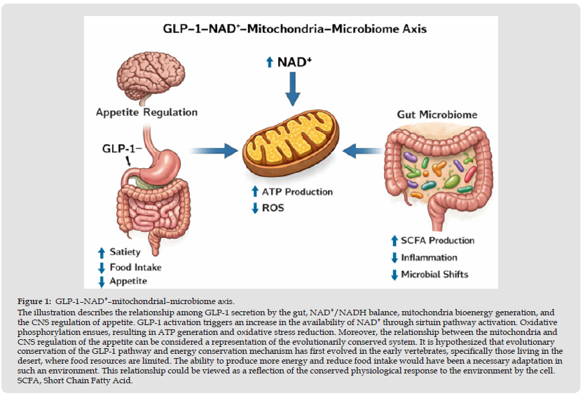
A plausible molecular substrate for this integration is the AMPK– SIRT1–PGC-1α axis: GLP-1 receptor signaling activates AMP-activated protein kinase (AMPK), while the NAD⁺-dependent deacetylase SIRT1 deacetylates and activates PGC-1α to drive mitochondrial biogenesis, thereby coupling receptor activation to the cellular NAD⁺ supply [6]. Consistent with this scheme, the GLP-1 receptor agonist liraglutide promotes mitochondrial function and beige-fat development in diet- induced obese mice partly through AMPK–SIRT1–PGC-1α signaling [7], and a recent systematic review reports GLP-1 receptor agonist– associated improvements in skeletal-muscle mitochondrial function, although the current evidence base remains largely preclinical [8].
One of the most striking effects of GLP-1 receptor agonism is its ability to suppress appetite, which has been demonstrated in clinical trials and feeding studies [9,10]. Semaglutide decreases energy, hunger, and food cravings and increases satiety. This suggests that there is physiological regulation of food intake [10]. The mechanisms of GLP-1 receptor agonism-induced appetite suppression can be viewed from different aspects of mitochondrial and redox biology and can be viewed from different perspectives, including central nervous system (CNS; Central Nervous System) mechanisms such as the hypothalamus and mesolimbic system [11]. GLP-1 receptors are densely expressed in hypothalamic and hindbrain circuits, including the area postrema and nucleus tractus solitarius, and current evidence attributes much of the anorectic action of GLP-1 receptor agonists to these central populations acting together with vagal afferent signaling rather than to peripheral bioenergetic status alone [12]. The NAD+/NADH ratio increases with improved metabolic efficiency and reduces the necessity for excessive energy intake [4]. This means that there is synchronization of central nervous system mechanisms that regulate appetite and peripheral bioenergetics status. When peripheral bioenergetics status is optimal, as reflected by improved ATP production through the mitochondrion, there is less physiological necessity for excessive energy intake. Furthermore, there is integration of peripheral metabolic status with central nervous system mechanisms that regulate appetite through GLP-1 receptor agonism-induced delay in gastric emptying and effects on the vagus nerve [1,2].
The physiological mechanisms underlying the increase of GLP-1 levels after nutrition is the universal physiological mechanism, which makes the digestion and the metabolism regulation processes interconnected [1]. From the evolutionary point of view, the coupling of the GLP-1 and NAD⁺-dependent mitochondrial function would provide a survival advantage for animals having intermittent nutrient availability [13]. Thus, an organism can get the maximal ATP amount due to the process of oxidative phosphorylation, keep the redox state inside the mitochondria in a suitable range through maintaining the NAD⁺/NADH ratio, and inhibit feeding behavior until the organism gets enough energy to meet the needs of its cells. The decrease of NAD⁺ content in the process of aging is currently associated with numerous modern pathologies [5]. Quantitative declines in tissue NAD⁺ during ageing have been directly linked to impaired mitochondrial and metabolic function across multiple organ systems, reinforcing the view that NAD⁺ availability is a limiting node in the energy-regulatory network proposed here [14].
Conservation of these metabolic pathways may be found in invertebrates, but no pharmacological activity of the classic GLP-1 receptor has been observed in invertebrates. However, the actions of GLP- 1 agonists have been studied in C. elegans, where the antioxidative and metabolic effects have been shown, indicating the involvement in interactions with conserved stress-response pathways [15]. Conserved metabolic pathways in C. elegans include AAK-2 (ortholog of the mammalian AMP-activated kinase, AMPK), SIR-2.1 (homologue of mammalian protein sirtuin 1, SIRT1), and SKN-1 (functional homologue of mammalian transcription factor NRF2, Nuclear Factor Erythroid 2–Related Factor 2) [16,17]. As seen in Figure 1, all these pathways are implicated in regulation of the mitochondrial functioning, lipid metabolism, and oxidative stress response. Thus, although there are no conserved receptors for GLP-1, metabolic effects of these pathways remain conserved throughout evolution, implying the conservation of metabolic architecture of the targeted metabolic pathways in spite of the lack of homology in GLP-1 receptors. All of the aforementioned observations support the hypothesis about the more extensive metabolic impact of GLP-1, mediated by the intracellular energy-regulatory systems. In conclusion, it is necessary to discuss the evolutionary history of the energy-regulatory systems as a series of evolutionary modules, which are more effective in comparison with their precursors.
It is natural to assume that there was selective pressure on such organisms that were able to efficiently use and store energy. The development of more advanced and complex brain structure required higher bioenergetic capacity of the organisms. Therefore, the pathways associated with GLP-1 can be considered as ancient bioenergetic regulatory pathways, which have been developed to meet the needs associated with brain development, especially regarding the evolution of the centralized nervous system.
A decrease in the concentration of NAD+ leads to impaired energy metabolism, mitochondrial dysfunction, and disturbance of redox homeostasis in the course of the disease development (Figure 1). In particular, NAD+-mediated energy metabolism abnormalities are characterized by the decreased production of ATP, oxidative stress, and reduced activity of cellular repair mechanisms [4]. In support of a causal contribution from NAD⁺ itself, pharmacological repletion with the precursor nicotinamide mononucleotide has been shown to increase muscle insulin sensitivity and improve metabolic function in humans, although the magnitude of clinical benefit reported to date has been modest [18]. Restoration of the cellular energy homeostasis is tightly connected to improving mitochondria functioning and normalizing NAD+-associated processes in neurons [3,19]. As the restoration of cellular energy metabolism ensures neuronal cell survival, maintenance of mitochondrial functioning and normalization of cellular redox status become important for neuroprotection mediated by GLP-1R agonists. It follows from the above information that energy homeostasis plays a systemic regulatory role in energy metabolism of cellular structures.
Due to lack of G protein-coupled receptors and GLP-1 receptors in prokaryotic cells, the impact of the GLP-1 receptor agonists on their energy homeostasis does not imply direct involvement. The impact of GLP-1 agonists on prokaryotic organisms occurs indirectly, being linked to physiological changes in the host body causing the reorganization of the GI tract environment [20,21]. Indeed, GLP-1RAs induce changes associated with the regulation of gastrointestinal mobility, delivery of nutrients and bile acids, as well as autonomic innervation of the GI tract. Hence, the composition of microflora and its metabolites, including short-chain fatty acids, change [20,21]. Conversely, microbially derived short-chain fatty acids can themselves stimulate secretion of endogenous GLP-1 through the free fatty acid receptor FFAR2, establishing a bidirectional host–microbiome loop in which GLP-1 both shapes and is shaped by the gut microbial community [22]. Based on the mechanism of action of GLP-1 receptor agonists, one can say that the elevation of norepinephrine concentrations caused by the activation of the sympathetic nervous system rapidly affects bacteria composition, including Escherichia coli [23].
The discovery can be taken as the evidence that GLP-1RAs can exert the impact on the prokaryotic energy homeostasis through the neurochemical signal mediated by the host organism. The above observations can indicate evolutionarily conserved interaction of the host’s organism, its nervous system and energy processes in prokaryotes (e.g., bacteria and viruses), and even mitochondria, which evolved out of bacteria-like organisms. This deep homology is consistent with the endosymbiotic origin of mitochondria from an ancestral α-proteobacterium, which provides an evolutionary rationale for why host-directed metabolic signals can influence bioenergetic processes across both prokaryotic and organellar compartments [24].
The integration of these observations supports a unified model in which GLP-1 signaling links systemic nutrient sensing with intracellular energy regulation and organismal behavior. Within this framework, GLP-1 signaling modulates systemic metabolic responses, NAD⁺/NADH balance governs intracellular redox state and metabolic efficiency, mitochondrial function determines ATP production and cellular viability, and central nervous system circuits regulate appetite and reward behaviors accordingly. At the same time, microbial ecosystems dynamically respond to host-driven changes in nutrient flux, bile acids, and neuroendocrine signals. Thus, GLP-1 receptor agonists can be understood as amplifiers of an endogenous evolutionary program that coordinates energy utilization across multiple biological scales, from mitochondrial bioenergetics to host–microbiome interactions [2,13].
Several limitations temper the model proposed here. First, much of the evidence linking GLP-1 signaling to NAD⁺ metabolism is associative or derives from preclinical systems, and a direct, quantitative demonstration that GLP-1 receptor activation elevates intracellular NAD⁺ in humans is still lacking [8]. Second, the anorectic and bodyweight effects of GLP-1 receptor agonists are substantially explained by central and vagal mechanisms and by delayed gastric emptying, so improvements in peripheral bioenergetics may in part be a consequence of reduced caloric intake and weight loss rather than a primary cause of appetite suppression [12]. Third, clinical trials of NAD⁺ precursors have so far produced modest and inconsistent metabolic benefits, indicating that raising NAD⁺ is not by itself sufficient to reproduce the effects of GLP-1 receptor agonists [18]. These caveats do not negate the proposed axis, but they define the experiments—incorporating tissue-specific NAD⁺/NADH measurement, loss-of-function models, and human pharmacodynamic studies—needed to distinguish correlation from causation.
The signaling of the GLP-1 receptor is a nodal point between nutrient sensing, mitochondrial bioenergetics, and redox signaling. The addition of NAD⁺ biology into this model offers a rationale for appetite suppression, metabolic efficiency, and neuroprotection. Evidence from invertebrate models offers support for the conservation of key metabolic pathways, whereas microbiome research offers evidence of the indirect modulation of prokaryotic bioenergetics via host-mediated effects. These data offer support for the concept of the GLP-1-NAD⁺- mitochondrial axis as an evolutionarily conserved energy management system of relevance to various diseases of metabolism and neurodegeneration with implications for healthy longevity [25].
Not applicable.
Not applicable.
This research received no external funding.
The authors declare no conflicts of interest.
The authors together are accountable for all aspects of the work.
Process
During the preparation of this work, the authors used LLM to organize information, copyedit the text, and an generate original figure. Following the use of this tool, the authors reviewed and edited the content as necessary and take full responsibility for the final publication.

International Association of Landscape Archaeology, Czech Glass Society, Czech Republic

Department of Chemistry, Semenov Institute of Chemical Physics, USSR Academy of Sciences, Moscow, Russia

Department of Educational Sciences, Research Centre Qualities of Teachers, School of Education – HAN, Nijmegen, Netherlands

Associate Professor at Department of Breast and Thyorid Surgey, Chongqing General Hospital, China

Neurology, LA BioMed Research Institute, USA





 Abstract
 PDF
PDF



























