info@biomedres.us
+1 (502) 904-2126
One Westbrook Corporate Center, Suite 300, Westchester, IL 60154, USA
Site Map
 ISSN: 2574 -1241
ISSN: 2574 -1241
ISSN: 2574 -1241
Received: September 03, 2026; Published: September 16, 2026
*Corresponding author: Susumu Tanaka, Department of Nutrition Science, Faculty of Nursing and Nutrition, University of Nagasaki, Siebold, 1-1-1 Manabino, Nagayo-cho, Nishi-Sonogi-gun, Nagasaki 851-2195, Japan
DOI: 10.26717/BJSTR.2026.66.010388
Decidualization occurs when endometrial stromal cells are influenced by progesterone. Decidualization promotes the spiral arteries remodeling, endometrial immune tolerance, and the invasion of fetal cells and placental formation—all of which are critical for embryo implantation. In this study, we found that the expression level of SLC6A6, a taurine transporter, increased during decidualization. Stimulation with low concentrations of taurine regulated the decidualization markers—HAND2, FOXO1, PRL, IGFBP1, and IL15 —to a constant level. Low concentrations of taurine induced appropriate decidualization by increasing GSR, which possesses antioxidant properties, thereby reducing the excessive oxidative stress associated with decidualization and suppressing the overreaction of decidualization marker genes caused by oxidative stress. In other words, the presence of taurine in the blood is thought to contribute to successful decidualization, subsequent successful embryo implantation, and maintenance of pregnancy. However, excessive taurine reversed the responses of many genes, including EF1A, which regulates the cell cycle, and its indiscriminate use should be avoided.
Keywords: Endometrium; Decidualization; Endometrial Stromal Cells; Taurine
Abbreviations: E₂: Estrogen; P₄: Progesterone; ENSCS: Endometrial Stromal Cells; IGFBP1: Insulin-Like Growth Factor Binding Protein 1; EVT: Extravillous Trophoblasts; Il15: Interleukin 15; CDO1: Cysteine Dioxygenase Type 1; CSAD: Cysteine Sulfinate Decarboxylase; SLC6A6: Solute Carrier Family 6 Member 6; FBS: Fetal Bovine Serum; Hand2: Heart and Neural Crest Derivatives Expressed 2; Foxo1: Forkhead Box Protein O1; EF1A: Elongation Factor 1-Alpha 1
The menstrual cycle of the human endometrium is approximately 28 days and repeats the menstrual, proliferative, and secretory phases. These three stages are regulated by estrogen (E₂) and progesterone (P₄). During the menstrual period through ovulation, increased secretion of E₂ from ovarian follicular cells induces re-epithelialization, stromal proliferation, and angiogenesis during the proliferative phase. At this time, endometrial stromal cells (EnSCs) proliferate as the concentration of E₂ in the blood increases, and the endometrium increases in thickness; this is called the proliferative phase. After ovulation, the endometrium responds to P₄ secreted by the corpus luteum and changes to a state suitable for implantation [1]. As the blood level of P₄ increases, endometrial epithelial cells increase mucus-like secretions; therefore, the endometrium during this period is called the secretory phase. In addition, EnSCs, which are the most abundant cells in the endometrium, express the P₄ receptor; therefore, they respond to P₄ and become rounded and differentiate into epithelial-like cells. This differentiation is known as decidualization [2]. The attenuation of P₄ signaling in the endometrium due to P₄ receptor deficiency causes dysregulation of downstream genes of this signaling pathway, resulting in various pregnancy disorders [3]. With normal progression of P₄ signaling, EnSCs undergo decidualization and initiate not only morphological changes but also the synthesis and secretion of decidualization markers listed below [3].
Prolactin (PRL) from decidualized EnSCs promotes angiogenesis by acting on vascular endothelial cells of the endometrium. Insulin- like growth factor binding protein 1 (IGFBP1) from decidualized EnSCs promotes migration of extravillous trophoblasts (EVT) and contributes to placentogenesis [3]. controls the Uterine natural killer (uNK) cells, which are regulated their proliferation and differentiation by Interleukin 15 (IL15) from decidualized EnSCs, promote remodeling of the spiral arteries together with EnSCs, which further supports decidualization of the endometrium [3]. In addition, uNK cells are important for immune tolerance and embryo receptivity [4,5]. Abnormal decidualization of EnSCs causes implantation disorders [6]; therefore, normal decidualization of EnSCs is essential for embryo implantation and fetal development in utero [7]. Taurine (2-aminoethane sulfonic acid) is an essential micronutrient and one of the most abundant amino acids in the human body. It is also well known as a major ingredient in energy drinks, but it is also abundant in the bodies of mammals and in various foods. It is present in high concentrations in excitable tissues such as the heart, eyes, brain, and muscles, reaching up to 1 g/kg of body weight [8,9]. It contributes to metabolic homeostasis in vivo by participating in various biological and physiological functions, including bile salt binding, osmoregulation, cell membrane stabilization, calcium regulation, antioxidant action, and immunomodulation [10].
The amount of taurine in human cells depends on biosynthesis via cysteine dioxygenase type 1 (CDO1) and cysteine sulfinate decarboxylase (CSAD), as well as taurine transport via solute carrier family 6 member 6 (SLC6A6) [9]. Taurine reduces mitochondrial redox stress depending on the amount transported into cells [9]. Conversely, taurine transport in the liver suggests the involvement of SLC6A13 rather than SLC6A6 [11,12]. Blood taurine levels decrease with age, and taurine deficiency is associated with age-related diseases due to reduced antioxidant capacity [13]. Antioxidant levels are significantly lower in women with luteal phase failure and recurrent miscarriages than in healthy women of the same age [14]. Moreover, expression of SLC6A6 has been reported in the endometrium [14]; therefore, we hypothesized that taurine-mediated regulation plays a role in endometrial function, particularly in decidualization. However, the effects of taurine on decidualization of EnSCs have not yet been examined. We investigated the taurine effects on human endometrial function, particularly during decidualization.
Culture of Human EnSC Line KC02-44D [15]
The human EnSC line KC02-44D was purchased from the American Type Culture Collection (Manassas, VA, USA). KC02-44D cells were cultured in DMEM (phenol red, Life Technologies, NY, USA) containing 10 mM HEPES (pH 7.4), 10% fetal bovine serum (FBS), and 1% antibiotics, and the medium was replaced every 3 days at 37°C and 5% CO₂ in a CO₂ incubator until confluence. After reaching confluence, cells were treated with trypsin-EDTA solution (0.5 g/L trypsin/0.53 mmol/L EDTA) (NACALAI TESQUE inc., Kyoto, Japan) and passaged into 24-well plates (Corning Incorporated COSTAR, NY, USA) for experiments.
Decidualization and Taurine Treatment to KC02-44D
As a control group, KC02-44D cells were cultured in phenol redfree DMEM containing 10% charcoal-stripped FBS, 10 mM HEPES (pH 7.4), 1% antibiotics, and 1% GlutaMAX for 6 days, with medium changes every 3 days. For decidualization treatment, KC02-44D cells were cultured in the above medium supplemented with 10⁻⁸ M estrogen (Sigma-Aldrich Corp., St. Louis, MO, USA), 10⁻⁶ M medroxyprogesterone acetate (Sigma-Aldrich Corp.), and 0.5 mM 8-bromo-cAMP (Kanto chemical Co., Inc., Tokyo, Japan) for 6 days, with medium changes every 3 days. To examine the effects of taurine, different concentrations of taurine were added to the medium during decidualization.
Total RNA Extraction and Reverse Transcription Reaction
After 6 days of treatment, cells were lysed by using Sepasol RNA I Super G (NACALAI TESQUE Inc.) for total RNA extraction. Reverse transcription was conducted by using ReverTra Ace™qPCR RT Master Mix with gDNA Remover (Toyobo Co., Osaka, Japan).
Quantitative Polymerase Chain Reaction (qPCR)
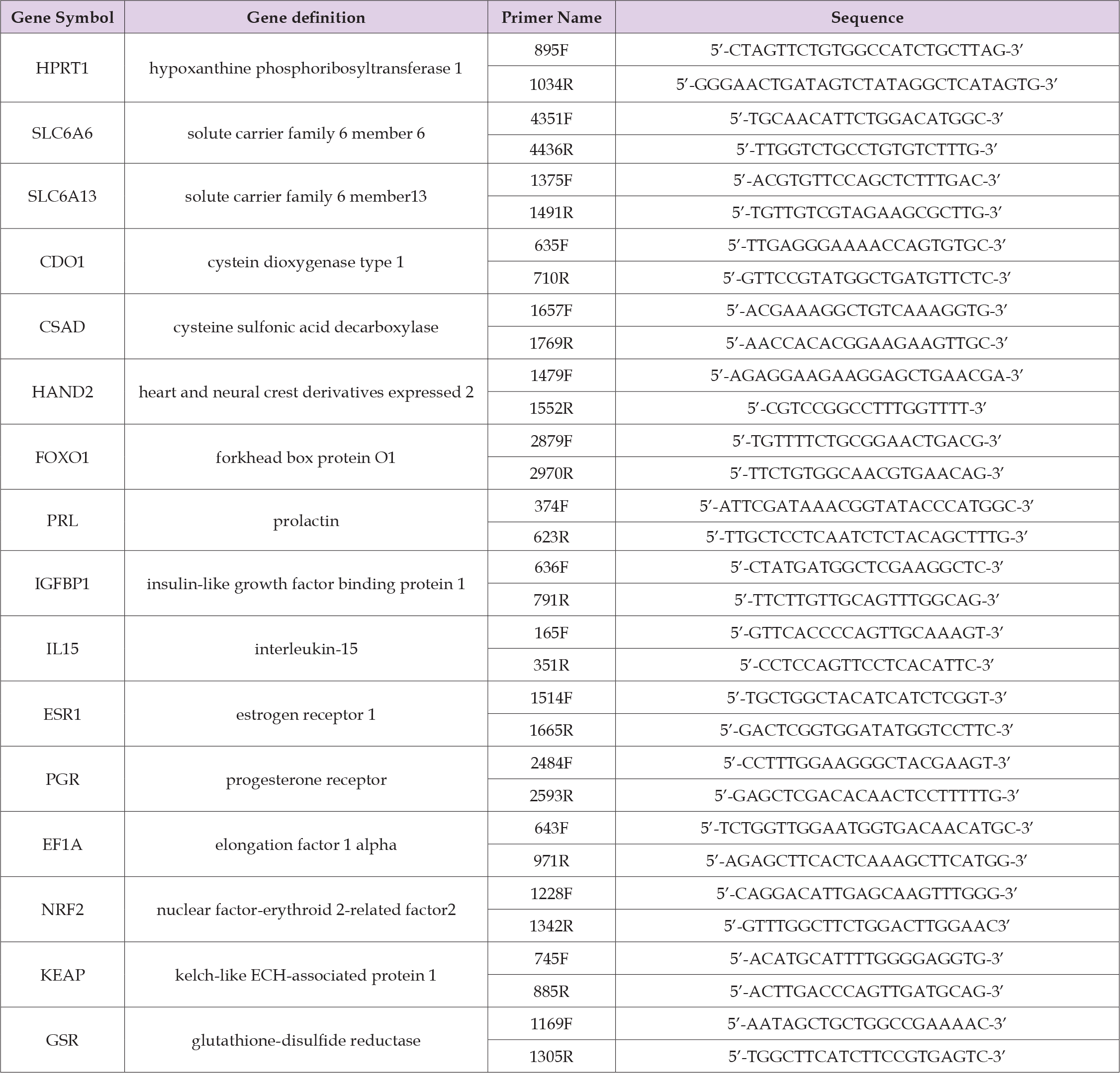
We performed qPCR to confirm decidualization and to examine the effects of taurine on decidualization. qPCR was performed using SYBR™ Green qPCR Master Mix (Thermo Fisher Scientific, MA, USA), primers, and a LightCycler 96 system (Roche Diagnostics K. K, Tokyo, Japan). The gene definitions and primer sequences are described in Table 1. Relative gene expressions were calculated by using the delta- delta-Ct method [16], with hypoxanthine phosphoribosyl transferase 1 (HPRT1) used as the internal control.
Table 1: Primer list.
Statistical Analysis
To assess the each group normality, we performed a Shapiro-Wilk normality test, followed by a two-tailed Welch’s t-test. Statistical significance was set at p < 0.05, and in cases of multiple comparisons, the Bonferroni correction was applied to adjust the significance threshold for each experiment. IBM SPSS Statistics (version 29.0; IBM Corp., Armonk, NY, USA) was used for statistical analyses.
The Role of Taurine in Decidualization
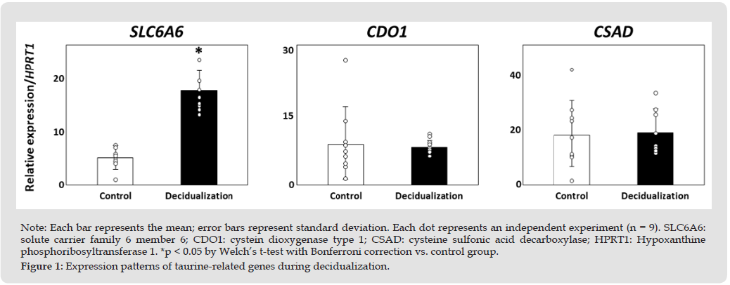
We investigated the requirement for taurine in decidual KC02- 44D cells by examining the expression of SLC6A6, SLC6A13, CDO1, and CSAD. SLC6A6 was detected in the control group and was significantly increased in the decidualization group compared with the control group (p < 0.05, Figure 1). SLC6A13 was not detected in either the control or decidualization groups. CDO1 and CSAD were detected in both control and decidualization groups, but there were no significant differences between the two groups (Figure 1). These results suggest that decidualized EnSCs require taurine and that taurine is likely supplied through uptake from outside the cell rather than through intracellular synthesis.
Figure 1
Effect of Taurine on Decidualization
Heart and neural crest derivatives expressed 2 (HAND2) and forkhead box protein O1 (FOXO1), transcription factors whose expression is initially increased as master regulators of decidualization, were significantly increased in the decidualization and decidualization + taurine groups compared to the control group (p < 0.05, Figure 2). HAND2 and FOXO1 expression was significantly decreased in the decidualization + 1 mM taurine treatment group compared with the decidualization- only group (p < 0.05, Figure 2). PRL, IGFBP1, and IL15 were significantly increased in the decidualization group compared with the control group (p < 0.05, Figure 2). Significant decreases in these genes were observed in the decidualization + 1 mM taurine treatment group compared with the decidualization-only group (p < 0.05, Figure 2). There were no significant differences in estrogen receptor 1 (ESR1) among all groups (Figure 3). The P4 receptor (PGR) was significantly increased in all decidualization groups compared with the control group (p < 0.05, Figure 3). The expression of elongation factor 1-alpha 1 (EF1A), which encodes a translation elongation protein, was significantly suppressed in the decidualization-only and decidualization + 1 mM taurine treatment groups compared with the control group (p < 0.05, Figure 3). As shown in Figure 1, SLC6A6 expression was significantly increased in all decidualization groups compared with the control group (p < 0.05, Figure 3).
Figure 2
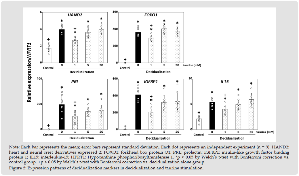
Figure 3
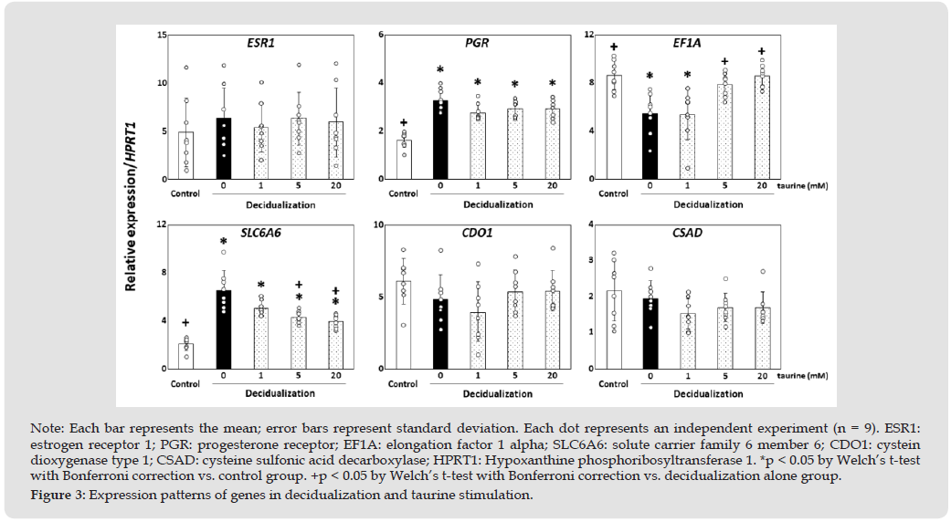
The decidualization + 20 mM taurine treatment group showed significantly reduced expression compared with the decidualization-only group, suggesting a negative feedback mechanism in response to excessive taurine (p < 0.05, Figure 3). There were no differences in CDO1 and CSAD among all groups (Figure 3). SLC6A13 was not detected under any of these conditions. Since taurine has a reducing effect on oxidative stress, we investigated changes in oxidative stress markers. Nuclear factor-erythroid 2-related factor 2 (NRF2) enhances the antioxidant capacity by becoming activated under oxidative stress [17], and NRF2 levels are controlled with Kelch-like ECH-associated protein 1 (KEAP1) [18]. NRF2 and KEAP1 showed a significant increase in the decidualization-only group compared with the control group (p < 0.05, Figure 4). NRF2 and KEAP1 showed a significant reduction in all decidualization + taurine groups compared with the control and decidualization-only groups (p < 0.05, Figure 4). Glutathione disulfide reductase (GSR) maintains intracellular glutathione concentration and reduces oxidative damage [19]. GSR was significantly increased in the decidualization + 1 mM taurine treatment group compared with the control and decidualization-only groups (p < 0.05, Figure 4).
Figure 4
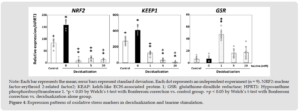
In this study, we found that the expression of the taurine transporter SLC6A6 increased when the EnSC line KC02-44D underwent decidualization, suggesting a requirement for taurine in decidualized EnSCs. Since there was no difference in the expression levels of taurine synthases, it was concluded that the increased taurine requirement of EnSCs during decidualization relies almost entirely on extracellular supply. We then examined the effects of taurine on human endometrial function, particularly on EnSCs during decidualization. As previously reported, HAND2 [1] and FOXO1 [20], known master regulatory transcription factors for decidualization, were elevated in the decidualization groups compared with the control group; however, the addition of a low concentration (1 mM) of taurine reduced their expression. Furthermore, PRL and IGFBP1 [3], known as decidualization markers, exhibited similar patterns. Interestingly, changes were also observed in the EF1A housekeeping gene. Conversely, treatment with a high concentration (20 mM) of taurine suppressed the expression of SLC6A6 itself. Given taurine’s antioxidant properties, these changes were considered to result from modulation of oxidative stress. Consequently, we examined changes in the expression of oxidative stress markers and found, as expected, that taurine likely alters the oxidative stress state in EnSCs, thereby influencing decidualization.
HAND2 is known to act as a master mediator that plays a central role in P₄ signaling during EnSC decidualization [1], and P₄ increases HAND2 expression in a concentration- and time-dependent manner, thereby closely regulating successful embryo implantation [21]. In this study, HAND2 expression increased significantly in response to decidualization stimulation. However, the addition of low-concentration taurine resulted in a significant decrease compared with decidualization stimulation alone, although expression remained higher than in the control group. Since HAND2 is involved in the regulation of various cellular functions, particularly the regulation of oxidative stress-resistant genes that are crucial for decidualization [22], the elevation of HAND2 during decidualization suggests that it is driven not only by P₄ stimulation but also by oxidative stress; low concentrations of taurine may therefore help maintain HAND2 levels within a physiological range. FOXO1 exhibited a similar response. FOXO1 is one of the key transcription factors involved in the decidualization of EnSCs and plays a role in the transcriptional regulation of factors that control numerous cellular processes, including oxidative stress resistance [23]. Oxidative stress refers to a pathophysiological state caused by the excessive cellular oxidizing capacity of reactive oxygen species (ROS) and affects reproductive processes, including pregnancy [24]. Therefore, it was hypothesized that taurine prevents excessive decidualization by reducing oxidative stress and regulating the expression of master transcription factors.
PRL, a marker of decidualization, is transcriptionally regulated by HAND2 [25], and IGFBP1 is regulated by FOXO1 [26]; therefore, it is believed that PRL and IGFBP1 levels also decrease as a result of the suppression of HAND2 and FOXO1 expression by taurine. However, even with taurine treatment, the expression levels of both genes remained significantly higher than those in the unstimulated group. PRL enhances the effects of P4 in the endometrium, contributes to the regulation of fetal cell proliferation and invasion, and supports immune tolerance, thereby playing a significant role in successful decidualization, embryo implantation, and maintenance of pregnancy [27].
Accordingly, hyperprolactinemia is associated with recurrent pregnancy loss [28]. Upon secretion from EnSCs, IGFBP1 promotes the migration of EVT and contributes to placental formation by regulating EVT invasion into the decidua [29,30]. Therefore, IGFBP1 dysregulation is associated with abnormal placental development and fetal growth restriction [31]. Therefore, it is believed that the regulation of transcription factors via taurine leads to appropriate decidualization and contributes to successful embryo implantation and subsequent pregnancy maintenance. Similarly, IL-15, which is transcriptionally regulated by HAND2 in EnSCs, increases during the secretory phase when decidualization occurs and shows a strong positive correlation [32].
EF1A has various functions, including [1] binding to guanine quadruplex sequences and regulating transcription via promoter activity, [2] acting as a component of the protein synthesis machinery, and [3] regulating the cell cycle through interactions with actin and microtubules [33,34]. Therefore, it is known as an “essential gene”—a gene required for an organism to survive under specific conditions—and is widely used as an internal control [35]. However, in this study, EF1A expression was significantly reduced in the decidualization-only group and in the group supplemented with 1 mM taurine, but recovered upon supplementation with 5 mM or higher concentrations of taurine. One possible explanation is that EF1A expression may be more unstable expression than that of other genes commonly used as internal controls [35]. Furthermore, since P4 stimulation causes En- SCs to arrest proliferation (cell cycle) and enter the G0 phase, leading to differentiation into decidualized cells, it is reasonable that EF1A, which is involved in cell cycle regulation, is downregulated. It may also be suggested that high concentrations of taurine inhibit this differentiation, leading to the resumption of cell proliferation. Therefore, treatment with high taurine concentrations is not desirable for decidualization. Elevated levels of the oxidative stress markers FOXO1, NRF2, and KEAP1 were observed following decidualization, suggesting increased oxidative stress.
In contrast, low concentrations of taurine increased the expression of GSR, which plays a crucial role in maintaining antioxidant function by reducing oxidized glutathione to reduced glutathione under oxidative stress [36]. As a result, intracellular oxidative stress was reduced, and the levels of oxidative stress markers NRF2 and KEAP1 decreased. However, since FOXO1 is not regulated solely by oxidative stress but is also influenced by multiple pathways induced by decidualization [20,37], the effects of low-concentration taurine were less pronounced, and the expression of IGFBP1, which is transcriptionally regulated by FOXO1, was not completely suppressed.
In other words, decidualization is accompanied by oxidative stress, which leads to increased expression of genes such as FOXO1 and IGFBP1; however, these increases appear excessive compared to normal physiological responses. It is believed that taurine, by reducing oxidative stress via GSR, induces appropriate decidualization that is partially independent of oxidative stress. In contrast, taurine treatment at concentrations exceeding 5 mM reversed the responses of several genes, including those encoding oxidative stress markers. Although no toxicity of taurine has been reported, and taurine supplementation is recommended from an anti-aging perspective [13], the potentially adverse effects observed in EnSCs in this study suggest that its effects should be re-evaluated—for example, by measuring taurine levels in the serum of patients with recurrent miscarriages.
In fact, stimulation with taurine concentrations of 5 mM or higher— which is considered excessive for EnSCs—reduced the expression of the taurine transporter SLC6A6 (i.e., decreased uptake capacity). It is possible that EnSCs protect themselves not only by downregulating SLC6A6, but also by modulating other metabolic enzymes and taurine efflux mechanisms.
Experimental Limitations in this Study
Although taurine toxicity in KC02-44D cells has been suggested, this was not examined in the present study; therefore, concentration- dependent toxicity assays for taurine are necessary in the future. In addition, for embryo implantation, not only decidualization but also proliferation of EnSCs during the proliferative phase and the resulting thickening of the endometrium are critically important; thus, it will be necessary to investigate the effects of taurine on cell proliferation in future studies.
In this study, we found that the expression level of SLC6A6, a taurine transporter, increased, and we further identified the necessity of taurine in decidualized EnSCs. Stimulation with low concentrations of taurine regulated the expression of the decidualization markers HAND2, FOXO1, PRL, IGFBP1, and IL15 to a constant level. Low concentrations of taurine are believed to induce appropriate decidualization by increasing GSR, which possesses antioxidant properties, thereby reducing the excessive oxidative stress associated with decidualization and suppressing the overexpression of decidualization marker genes caused by oxidative stress. In other words, the presence of taurine in the blood is thought to contribute to successful decidualization, subsequent successful embryo implantation, and maintenance of pregnancy. However, excessive taurine reverses the responses of many genes; therefore, its indiscriminate use should be avoided.
Not applicable.
This research was funded by the Takeda Science Foundation (2018) and the Yamaguchi Endocrine Research Foundation (2025) awarded to Susumu Tanaka; and JSPS KAKENHI grants awarded to Susumu Tanaka (grant number 25K14871).
The data presented in this study are available upon request from the corresponding author.
Conceptualization, S.T.; methodology, S.T.; validation, S.T., M.T.; formal analysis, S.T., M.T., and N.Y.; investigation, S.T., M.T., K.I., K.M., M.U., A.T., M.Y., and N.Y.; resources, S.T.; data curation, S.T., M.T.; writing— original draft preparation, S.T.; writing—review and editing, M.T., K.I., K.M., M.U., A.T., M.Y., and N.Y.; visualization, S.T., and M.T.; project administration, S.T.; funding acquisition, S.T. All authors have read and agreed to the published version of the manuscript
The authors declare no conflict of interest. The funders had no role in the study design; collection, analyses, or interpretation of data; writing of the manuscript; or decision to publish the results.
Not applicable.
Not applicable.
Not applicable.

International Association of Landscape Archaeology, Czech Glass Society, Czech Republic

Department of Chemistry, Semenov Institute of Chemical Physics, USSR Academy of Sciences, Moscow, Russia

Neurology, LA BioMed Research Institute, USA

Associate Professor at Department of Breast and Thyorid Surgey, Chongqing General Hospital, China

Professor of Nuclear Medicine, Faculty of Medicine and Surgery, University of Milan, Milan, Italy





 Abstract
 PDF
PDF



























