info@biomedres.us
+1 (502) 904-2126
One Westbrook Corporate Center, Suite 300, Westchester, IL 60154, USA
Site Map
 ISSN: 2574 -1241
Received: August 27, 2026; Published: September 04, 2026
*Corresponding author: Alparslan Dilsiz, Department of Periodontology, Faculty of Dentistry, Atatürk University, 25240-Erzurum/ Turkey
DOI: 10.26717/BJSTR.2026.66.010372
Background/Aim: Hereditary gingival fibromatosis (HGF) is a rare genetic disorder characterized by slowly progressive fibrous enlargement of the gingival tissues surrounding the teeth, which may lead to functional impairment as well as esthetic concerns. The condition is most commonly inherited in an autosomal dominant pattern and is generally classified into two clinical forms: syndromic and non-syndromic. Although HGF is an uncommon disorder, its clinical manifestations may substantially affect the oral health and quality of life of affected individuals and their families. Therefore, the present study aimed to describe the clinical and histopathological characteristics of five patients with the non-syndromic form of HGF, including three members of the same family, and to present the clinical management together with the long-term treatment outcomes.
Methods: This case series included five patients diagnosed with hereditary gingival fibromatosis (HGF), all of whom were clinically managed and monitored for a period of 5 years following periodontal treatment. Among the affected individuals were three members of the same family, consisting of a mother and her two children (one female and one male). The initial clinical diagnosis of HGF was confirmed by histopathological examination of the excised gingival tissue. Periodontal management comprised comprehensive oral hygiene instruction, mechanical periodontal debridement, and surgical treatment involving internal bevel gingivectomy performed in combination with open flap debridement.
Results: The clinical presentation of HGF varied among the patients and included poor oral hygiene, dense fibrotic gingival enlargement, delayed eruption of permanent teeth, tooth displacement, supernumerary teeth, delayed root resorption, and anterior open or deep bite. Clinical evaluation throughout the 5-year follow-up period revealed no evidence of recurrence after completion of periodontal treatment. Both the patients and the treating clinicians considered the therapeutic outcomes to be satisfactory.
Conclusion: Clinical evaluation over a 5-year follow-up period demonstrated successful long-term management of gingival overgrowth in all patients. A comprehensive treatment protocol combining periodontal therapy with surgical removal of the fibrotic gingival tissue proved to be an effective approach for the management of hereditary gingival fibromatosis. Clinicians should carefully evaluate patients for accompanying dental and developmental abnormalities, particularly delayed tooth eruption and the presence of supernumerary teeth. Furthermore, regular periodontal maintenance and long-term clinical surveillance are essential for preserving periodontal health, maintaining treatment outcomes, and minimizing the risk of recurrence.
Keywords: Gingival Overgrowth; Gingival Fibromatosis; Periodontal Debridement; Oral Surgical Procedures; Gingivectomy; Gingivoplasty; Case Report
Hereditary gingival fibromatosis (HGF), also referred to as familial gingival fibromatosis, idiopathic gingival fibromatosis, or elephantiasis gingivae, is a rare benign disorder characterized by slowly progressive fibrous enlargement of the gingival tissues, which may present as either localized or generalized overgrowth [1,2]. Since its first description by Gross in 1856, considerable progress has been made in understanding the clinical characteristics of HGF; however, its exact pathogenesis remains incompletely understood [3,4]. Current evidence suggests that gingival enlargement may result from alterations in connective tissue metabolism, including increased collagen synthesis by gingival fibroblasts and/or impaired collagen degradation [1,2]. HGF may occur as an isolated condition or in association with several inherited syndromes, including Cowden syndrome, Cross syndrome, and Laband syndrome. Depending on the underlying syndrome, additional clinical manifestations such as hypertrichosis, intellectual disability, pigmentary abnormalities, epilepsy, cherubism, corneal dystrophy, microphthalmia, developmental delay, syndactyly, and abnormalities of the fingers and toes may also be present [5-16]. The disorder affects males and females with similar frequency, with an estimated phenotypic prevalence of approximately 1:750,000, although the clinical severity and expression may differ considerably even among members of the same family [2,3].
Genetically, HGF is a heterogeneous condition and has been reported to follow autosomal dominant, autosomal recessive, and sporadic patterns of inheritance [1,3,6,9]. The clinical appearance of HGF is characterized by firm fibrotic gingival enlargement, with the gingiva typically exhibiting either a normal color or mild erythema [1-3]. Although the underlying alveolar bone is generally unaffected, excessive gingival overgrowth frequently results in pseudopocket formation and compromises plaque control, thereby increasing the risk of periodontal problems. Gingival enlargement may be localized or generalized, unilateral or symmetrical, depending on the extent of the disease. Interestingly, HGF has not been described in completely edentulous individuals, suggesting that the presence of teeth may be necessary for the development and persistence of gingival overgrowth [5,10]. Considerable variation in the clinical presentation has been reported, even among affected individuals from the same family [9-14]. As the disease progresses, the enlarged gingival tissues may interfere with tooth eruption, mastication, occlusion, speech, lip competence, and overall facial esthetics. The present study describes the clinical and histopathological characteristics of five patients diagnosed with the nonsyndromic form of hereditary gingival fibromatosis, including three members of the same family, and presents the clinical management together with the long-term treatment outcomes. This case series was prepared in accordance with the CaReL guidelines [17].
Description of Three Cases
A 15-year-old girl presented to the Department of Periodontology, Faculty of Dentistry, Atatürk University, Türkiye, with the chief complaint of generalized gingival enlargement accompanied by delayed eruption of the permanent dentition. Her medical history was unremarkable, with no known allergies or use of medications associated with gingival enlargement, including antiepileptic, antihypertensive, or immunosuppressive agents. The patient reported that the gingival enlargement had first become noticeable at approximately 5 years of age. The condition progressed gradually over time and eventually resulted in difficulties with mastication and speech, in addition to causing considerable esthetic concerns (Figure 1A1, 1A2 & 1A3). A positive family history was identified during the clinical interview. Similar gingival enlargement affecting both the maxillary and mandibular arches was observed in the patient’s 8-year-old brother (Figure 1B1, 1B2 & 1B3) and her 46-year-old mother (Figure 1C1, 1C2 & 1C3), although the severity of the enlargement differed among the affected family members. Extraoral examination demonstrated lip protrusion associated with facial disharmony. Intraoral examination revealed generalized moderate gingival enlargement involving both the maxillary and mandibular arches. The gingival tissues were pink, firm, and fibrotic, covering approximately one-half to two-thirds of the clinical crowns of the erupted teeth. No acute inflammatory findings, including spontaneous pain or bleeding on probing, were detected.
Figure 1
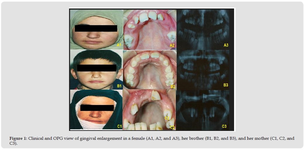
The patient also exhibited marked malocclusion characterized by tooth displacement, rotation, and generalized diastemas. Panoramic radiographic examination demonstrated normal alveolar bone morphology together with delayed root resorption, unerupted permanent teeth, and the presence of supernumerary teeth.
Case 4
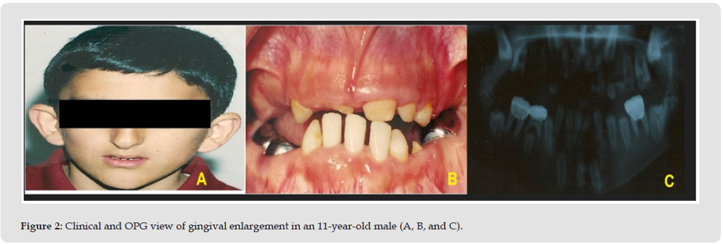
An 11-year-old boy was referred to our department because of generalized gingival enlargement associated with delayed eruption of the permanent dentition. The patient had no history of systemic disease or medication use known to induce gingival overgrowth, and no similar gingival enlargement was reported among his family members. Clinical and panoramic radiographic examinations demonstrated the characteristic findings of hereditary gingival fibromatosis (Figure 2A, 2B & 2C).
Figure 2
Case 5
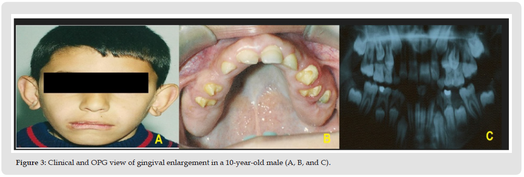
A 10-year-old boy attended our clinic with generalized gingival enlargement as his primary complaint. His general medical condition was good, and he was not receiving any medication associated with gingival enlargement. No positive family history was identified. Based on the clinical and radiographic findings, a diagnosis of hereditary gingival fibromatosis was established (Figure 3A, 3B & 3C).
Figure 3
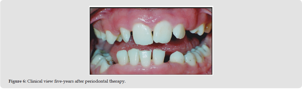
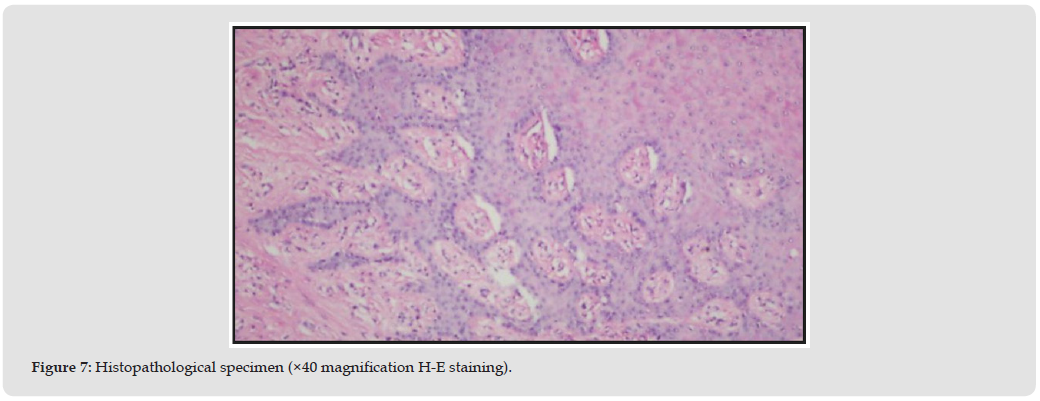
Before surgical intervention, all patients received initial periodontal therapy, including oral hygiene instruction, full-mouth scaling and root planing, and crown polishing. Extraction of retained deciduous teeth and restorative treatment of carious teeth were completed whenever indicated. Periodontal flap surgery was subsequently performed in all quadrants under local anesthesia using an internal bevel incision to excise the excessive fibrotic gingival tissue. The surgical flaps were repositioned and secured with 3-0 non-resorbable silk sutures (Figure 4). Following surgery, all patients were prescribed analgesic medication together with a chlorhexidine mouth rinse. The postoperative healing period was uneventful in every case. Orthodontic treatment was initiated in Case 4 to facilitate eruption of the impacted permanent teeth, which subsequently erupted into the occlusal plane. All patients were enrolled in a regular periodontal maintenance program and were monitored periodically for five years after treatment. Throughout the follow-up period, neither clinical nor radiographic evidence of recurrence was observed (Figures 5 & 6). Both the patients and the treating clinicians were satisfied with the long-term treatment outcomes. Histopathological examination of the excised gingival tissue demonstrated stratified squamous epithelium with elongated rete ridges extending deeply into the underlying connective tissue.
Figure 4
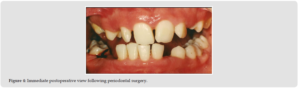
Figure 5
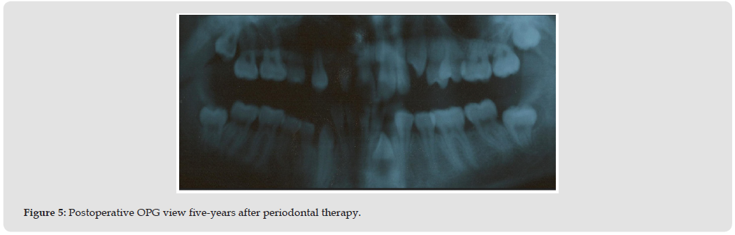
Figure 6
The connective tissue consisted predominantly of dense interwoven collagen bundles containing relatively few fibroblasts, sparse vascular structures, and mild chronic inflammatory cell infiltration, findings consistent with hereditary gingival fibromatosis (Figure 7). The present case series was conducted in accordance with the ethical principles of the Declaration of Helsinki. Written informed consent for publication of the clinical findings and photographic documentation was obtained from the parents or legal guardians of all participating patients.
Figure 7
In the present report, five patients with hereditary gingival fibromatosis (HGF) were evaluated clinically and followed after periodontal treatment. HGF is an uncommon disorder characterized by a slowly progressive fibrous enlargement of the gingival tissues [1-3]. The clinical severity of gingival enlargement may vary considerably among affected individuals with respect to both the thickness and vertical extension of the gingival tissues. Typically, the enlarged gingiva appears firm, fibrotic, non-hemorrhagic, and either normal in color or slightly erythematous. As the enlargement progresses, it may cover a substantial portion of the clinical crowns, thereby compromising speech, mastication, occlusion, and facial esthetics, findings that were also evident in the present cases [1-4]. Familial occurrence of isolated HGF has been well documented, and the disorder is most frequently inherited in an autosomal dominant pattern [9-12]. Although pathogenic variants of the SOS1 gene have been identified in some families, the marked genetic heterogeneity of HGF indicates that additional genetic alterations may also contribute to the development of the disease [18,19]. HGF may occur as an isolated condition or in association with several inherited syndromes, including Cowden, Cross, Laband, Murray–Puretic–Drescher, Ramon, Rutherford, and Schinzel–Giedion syndromes [1-4]. None of the patients included in the present case series demonstrated systemic abnormalities or clinical findings suggestive of syndromic HGF, supporting the diagnosis of the nonsyndromic form of the disease.
Because of its broad spectrum of clinical manifestations, HGF may occasionally be misdiagnosed during the initial clinical examination. Accurate diagnosis relies on careful evaluation of the patient’s clinical presentation, family history, and histopathological findings. The variability observed among affected members of the same family in the present series further illustrates the heterogeneous clinical expression of HGF [6-9]. In the familial cases, gingival enlargement first appeared during childhood, progressed gradually until puberty, and subsequently remained relatively stable during adulthood. In addition to its characteristic gingival enlargement, HGF frequently causes significant functional and esthetic impairment [7,9,13]. The patients described in this report exhibited several well-recognized clinical manifestations of the disease, including delayed eruption of permanent teeth, supernumerary teeth, and prolonged retention of deciduous teeth. Progressive eruption of the permanent dentition was observed during the postoperative period, particularly in the molar region, indicating that surgical removal of the excessive fibrotic gingival tissue facilitated normal tooth eruption, consistent with previous reports [2,3]. Histopathological examination of the excised tissue demonstrated the characteristic microscopic features of HGF and confirmed the clinical diagnosis in all patients.
Although the precise biological mechanism responsible for excessive gingival enlargement remains unclear, available evidence suggests that abnormalities in connective tissue metabolism, including altered synthesis and turnover of gingival extracellular matrix components, may contribute to the pathogenesis of the disease [1-5].
Although hereditary gingival fibromatosis cannot currently be cured, successful long-term control can be achieved with appropriate periodontal management. Surgical intervention is generally recommended after eruption of the permanent dentition because recurrence appears to be more common when treatment is performed at an earlier stage. The selection of treatment should be guided by the severity of gingival enlargement and the associated functional and esthetic impairment. Mild cases may be managed with meticulous plaque control and supportive periodontal therapy, whereas advanced enlargement generally requires surgical intervention [13-16]. In the present series, periodontal flap surgery using an internal bevel incision was selected because it allowed effective removal of the fibrotic tissue while promoting favorable wound healing. Throughout the 5-year follow-up period, no recurrence was observed, most likely owing to regular periodontal maintenance and satisfactory oral hygiene. These findings emphasize the importance of long-term supportive periodontal care and periodic recall visits for preserving treatment outcomes and minimizing the risk of recurrence [2,3].
Long-term clinical evaluation over a 5-year follow-up period demonstrated successful management of gingival overgrowth in all patients without evidence of recurrence. A comprehensive treatment strategy combining periodontal therapy with surgical removal of the excessive fibrotic gingival tissue proved to be an effective approach for the management of hereditary gingival fibromatosis. Dental clinicians should carefully assess affected individuals for associated dental and developmental abnormalities, particularly delayed eruption of the permanent dentition and the presence of supernumerary teeth. Furthermore, continuous periodontal maintenance together with regular long-term clinical follow-up plays an essential role in preserving periodontal health, maintaining treatment outcomes, and reducing the likelihood of disease recurrence.
The authors have nothing to report.
The manuscript is original and has not been published or submitted elsewhere.
The authors declare no conflicts of interest.
Alparslan Dilsiz, diagnosed and managed the patient, conceived the idea of the case report, drafted the manuscript, and critically revised the manuscript Alparslan Dilsiz, Fevziye Bekdas, Zeynep Tekin, and Aysegul Turksoy performed periodontal therapy and surgery procedure. All authors read and approved final version of text.

International Association of Landscape Archaeology, Czech Glass Society, Czech Republic

Department of Chemistry, Semenov Institute of Chemical Physics, USSR Academy of Sciences, Moscow, Russia

Department of Educational Sciences, Research Centre Qualities of Teachers, School of Education – HAN, Nijmegen, Netherlands

Associate Professor at Department of Breast and Thyorid Surgey, Chongqing General Hospital, China

Neurology, LA BioMed Research Institute, USA





 Abstract
 PDF