info@biomedres.us
+1 (502) 904-2126
One Westbrook Corporate Center, Suite 300, Westchester, IL 60154, USA
Site Map
 ISSN: 2574 -1241
ISSN: 2574 -1241
ISSN: 2574 -1241
Received: August 10, 2026; Published: August 19, 2026
*Corresponding author: Obot I P, Department of Zoology, Faculty of Life Sciences, Federal University of Lafia, Nigeria, Email: patienceobot36@gmail.com
DOI: 10.26717/BJSTR.2026.66.010346
Background: In sub-Saharan Africa, onchocerciasis (river blindness), which is caused by Onchocerca volvulus
and spread by Simulium blackflies, is still a neglected tropical illness. In two communities in Akwanga LGA,
Nasarawa State, Nigeria, this study assessed the effects of mass drug administration (MDA) on onchocerciasis
prevalence, transmission, and control.
Methodology: A cross-sectional study was carried out in the two communities between July and November
of 2024. Capillary blood samples were collected from 97 participants (Ungwan Zaria: n=53; Ungwan Habu:
n=44). Thick and thin blood smears were Giemsa-stained (10%) and examined microscopically for microfilariae.
Hematological parameters (hematocrit (HCT), white blood cell count (WBC), neutrophils, lymphocytes,
monocytes, eosinophils, basophils, red blood cell count (RBC), hemoglobin) were analyzed. OV16 ELISA
confirmed elimination status.
Results: Microfilariae were not detected in any of the samples (0.0% prevalence; 95% CI: 0.0-3.7%).
ELISA for OV-16 showed no O. volvulus antigens. Hematological analysis revealed significant inter-community
variations (p<0.05) with Ungwan Zaria having higher HCT (41%), WBC (3417±358.36 cells/μL) and hemoglobin
(13.24±0.901 g/dL) than Ungwan Habu (HCT: 32%; hemoglobin: 1.20±0.677 g/dL) indicating a high prevalence
of anemia. MDA adherence was 100% following five annual treatment rounds.
Conclusion: Sustained MDA effectively interrupted onchocerciasis transmission in both communities.
However, significant hematological disparities -particularly severe anemia in Ungwan Habu-warrant integrated
nutritional and immunological interventions. Post-elimination surveillance remains critical to prevent
reintroduction and ensure sustained elimination gains.
Keywords: Onchocerciasis; Onchocerca volvulus; River Blindness; OV-16 ELISA; Blackfly; Simulium Species; Mass Drug Administration; Microfilaria; Transmission Interruption; Post-Evaluation; Hematological Parameters; Biomarkers; Nasarawa State; Central Nigeria
Abbreviations: NTD: Neglected Tropical Diseases; MDA: Mass Drug Administration; WHO: World Health Organization; BCR: Blood Composite Ratios; LGA: Local Government Area; FCT: Federal Capital Territory; PHC: Primary Healthcare; DBS: Dried Blood Spot; ELISA: Enzyme-Linked Immunosorbent Assay; KAP: Knowledge, Attitude, And Practice; CDTi: Community Drug With Ivermectin; PCR: Polymerase Chain Reaction; WBC: White Blood Cell; NET: Neutrophil Extracellular Trap
Onchocerca volvulus is a filarial worm that is responsible for one of the neglected tropical diseases (NTDs) called onchocerciasis, which can also be referred to as river blindness. O. volvulus is transmitted by female blackflies of Simulium species, the female blackfly reproduces around swift-moving water bodies, especially in tropical and subtropical areas, and Simulium spp. bite humans to spread the disease [1,2]. Onchocerciasis prevails in Yemen, Latin America, and many regions of sub-Saharan Africa; the disease is most prevalent in Africa. Over the years, Nigeria, as an endemic country in Africa, has implemented a number of strategic measures to control and eliminate disease with the mass drug administration (MDA) campaign serving as a crucial intervention [3]. The primary health impact of onchocerciasis includes debilitating skin conditions, intense itching, skin depigmentation, and nodules under the skin, as well as severe ocular damage that can cause irreversible blindness. These effects not only cause physical suffering, but also impose a significant socioeconomic burden on affected communities, leading to decreased productivity and impaired quality of life [4].
Onchocerca volvulus infection is characterized by a type 2 immune response with alterations in lymphocytes, eosinophils, basophils, neutrophils, and mass cell distribution [5,6]. Onchocerciasis has been shown to stimulate both the innate and adaptive immune cells within the host [7,8]. Historically, the disease was rampant in many riverine communities, where all populations were at risk of blindness, resulting in economic decline and mass migration to less fertile upland areas to avoid exposure to blackfly bites [9]. The World Health Organization (WHO) launched extensive initiatives to control and eradicate onchocerciasis in response to the serious health and socioeconomic problems the disease posed, working with domestic and foreign partners [10]. Ivermectin, a medication used to treat onchocerciasis, was made available for free in 1987 through Merck & Co.’s Mectizan Donation Program. Since then, this medication has served as the mainstay of MDA initiatives [11] lowering the parasitic load in effected people. MDA campaigns have played a crucial role by providing ivermectin to help reduce burden or eliminate microfilariae (infective stage) that are responsible for most of disease symptoms in many endemic areas [4]. Millions of people in endemic states, including Nasarawa state, have received ivermectin an MDA application thanks to the National Onchocerciasis Control Program [12].
Onchocerciasis remains an issue nationwide, despite great efforts to achieve high coverage and compliance of treatment, the complete effectiveness of MDA programs has been hampered by elements such as limited public knowledge, sociocultural beliefs, logistical challenges in reaching rural populations, and inadequate health infrastructure [13]. Some communities in Akwanga LGA, Nasarawa state, have been said to be endemic due to the swift-moving rivers or streams that serve as ideal breeding sites for blackflies, resulting in high transmission rates seen in the past. However, to lower the prevalence and transmission of onchocerciasis, MDA campaigns have been carried out in these LGA’s, but post-MDA evaluation are essential in ascertaining the efficacy of past intervention and spot any gaps that would need to be filled in subsequent treatment cycles [14]. Onchocerciasis can activate both innate and adaptive immune system cells in the host, helminths infection can be characterized by the activation of inflammatory cells through strong inflammatory cells, and blood composite ratios (BCR) may be an independent discriminatory predictor of chronic disease. According to some research, the innate immune system kills most L3 infiltrants; this can be examined using a hematological profile [15]. This study is important and timely given that the World Health Organization wants to eradicate onchocerciasis as a public health issue in Africa by 2030, providing evidence-based suggestions to improve drug delivery tactics, strengthen control initiatives, and ensure that affected communities are actively involved in the process of eradication [16]. Therefore, this study evaluated the impact of MDA after treatment on the prevalence, transmission, and control of onchocerciasis, as well as hematologic biomarkers in two selected communities in Akwanga Local Government Area (LGA) of Nasarawa State, Nigeria.
Study Area
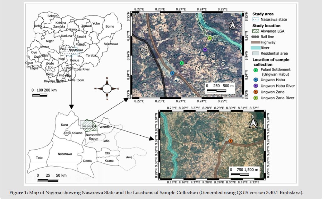
The study was conducted in two localities in Akwanga LGA, Nasarawa State, North Central Nigeria: Ungwan Habu (8.936767°N, 8.232139°E) and Ungwan Zaria (9.016064°N, 8.293720°E) (Figure 1). Positioned between Plateau State to the south and the Federal Capital Territory (FCT) to the west, Nasarawa State is the fifteenth largest state in Nigeria. The state lies mainly in the tropical Guinean forest savanna mosaic ecoregion, with an estimated population of 2.5 million [17].
Figure 1
Study Design
A cross-sectional study was carried out in the two communities.
Ethical Approval
Ethical approval was sought from the Ministry of Health of Nasarawa State, through the Human Research Ethics Committee Lafia, Nasarawa State. Permission was obtained from the Nasarawa Local Government Area Director of Primary Healthcare (PHC) with reference number NHREC Protocol No: 18/06/2017 and the Chief Medical Officer of the community clinic, while householders were contacted and informed on the nature of the research work and also asked for their consent.
Sample Size
The sample size was determined as described by Okoro et al. [18] in the formula below.

n = Sample size
Z = is the standard normal distribution at 95% confidence
interval = 1.96
p = is the anticipated population [19] = 0.295
1-p = q = 1-0.295 = 0.705
L= is the allowable error, which is taken as 5% = 0.05

n = 319.58
The expected sample size was 319.58 people, but only 97 subjects
consented for samples to be collected (Ungwan Habu: n = 44 and
Ungwan Zaria: n = 53).
Sampling
The target audience was not age specific, while the collection site was determined by previous research of lymphatic filariasis infection within the two communities of study [20]. Sampling and transportation of a high-quality sample was a critical aspect in ensuring accurate test results. Capillary blood was collected from the finger of a human host in which a portion was dried on an appropriate filter paper by air drying while the remaining was further processed wetwise. Wet and dried blood spot (DBS) samples were transported to the laboratory in a zip lock bag for testing. The following materials were used for blood collection: retractable sterile lancets with tips smaller than 2.4 mm for adults, 2.0 mm for children, and 1.5 mm for infants; scissors; alcohol wipes with a 70% isopropyl alcohol content; sterile gauze pads; disposable gloves made of latex, vinyl, and nitrile; safety goggles; a single-use, flammable waste container; filter paper; small, clean plastic bags to store individual filter papers; large plastic zipper-close bags; and a desiccant pack [21].
Laboratory Analysis of Blood Sample
Using 10% Giemsa staining at pH 7.2, blood slides were prepared and stained according to the standard protocols of the World Health Organization [16]. Using an aseptic technique, blood samples were taken by swabbing the fingers with 70% alcohol, letting them dry and then pricking each person with a sterile lancet. The thin blood smear was made by putting a drop of blood on a labelled, clean, grease-free glass slide. A 60° angled spreader was used to disseminate the blood and make a smooth tail. The smear was fixed with methanol for two minutes following air drying. After five seconds of water flushing, the smear was stained with 10% Giemsa stain for sixty minutes, rinsed, and allowed to air dry. Three droplets of blood were spread to a reasonable thickness on a grease-free slide in order to prepare the thick blood smear. Giemsa was used to stain the thick and thin film blood smears, and a 100x objective light microscope was used to apply oil immersion to the stained slides, Onchocerca volvulus microfilaria, and other blood parasites. Positive results indicate the presence of microfilaria, although the presence of white blood components such as eosinophil, basophil, neutrophil and lymphocytes with an abnormal range could serve as an indicator of the presence of onchocerciasis, and ring forms of Plasmodium trophozoites, schizonts, or gametocytes for the malaria parasite. The parasitological colour atlas was used for the identification of parasites in blood smears.
Parasite Identification
The stained smears were read at a magnification of 10x using a light microscope by trained technicians. Onchocerca volvulus was identified using the microfilarial identification keys of Amambo et al. [22].
ELISA-Based Detection of OV-16
Onchocerca volvulus specific antibodies (OV-16) were detected using an enzyme-linked immunosorbent assay (ELISA) according to standard protocols. The DBS samples were used for this assay. The process entailed a number of major phases, which include elution from filter paper samples, coating plate with antigen, washing plates, blocking plates, preparation of standard curve and blank, empty plate, adding samples, washing plates, adding conjugate, washing plates, adding streptavidin-AP, washing plates and Substrate.
Questionnaire Administration
Each subject screened for O. volvulus infection was administered a well-structured questionnaire in order to evaluate their knowledge, attitude, and practice (KAP) regarding onchocerciasis.
Statistical Analysis
The data obtained was analyzed using the Minitap Statistical Package version 21.1.3. Prevalence of O. volvulus infection was determined using simple percentage calculation. The hematological biomarkers of the subjects between the two study areas were compared using one-way analysis of variance (ANOVA). P value < 0.05 was considered statistically significant.
Prevalence of Onchocerciasis
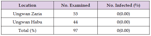
None of the 97 samples screened for onchocerciasis tested positive for O. volvulus infection (0%) in both Ungwan Zaria and Ungwan Habu communities (Table 1). Furthermore, ELISA analysis confirmed the complete absence of O. volvulus.
Table 1: Prevalence of Onchocerciasis in the Ungwan Habu and Ungwan Zaira Communities, Akwanga LGA, Nasarawa State, Nigeria.
Haematological Profile of the Subjects Screened for Onchocerca volvulus
The haematological parameters of the subjects tested for Onchocerca volvulus in this study are revealed in Table 2. The hematocrit (HCT) was higher in Ungwan Zaria with (41%) than in Ungwan Habu with (32%) compared to the normal range in men- 42-50% and women- 37-47%. The white blood cell count (WBC) had a high value of 3417±358.36 in Ungwan Zaria than in Ungwan Habu (2106.82±408.08) although both fell within the range when compared to the normal range of 1500-8000 cell/μL. The neutrophil record was higher in Ungwan Habu than in Ungwan Zaria (22.59±2.57 and 20.25±2.299, respectively) but both were low compared to the normal range of 55-70%. Ungwan Zaria had a high lymphocytes value of 58.98±4.52 than Ungwan Habu with 44.77±4.76, and both higher compared to the normal range of 20-40%. Regarding monophyls, Ungwan Habu had a high record of 3.45±0.785 than Ungwan Zaria with 0.98±0.45 as compared to the normal range of 2-8%. The eosin was high in Ungwan Habu with 1.81±0.572 than in Ungwan Zaria with a value of 0.57±0.348 compared to the normal range of 1-4%. Furthermore, Ungwan Habu had a high basophils value of 0.43±0.188 than Ungwan Zaria with a value of 0.19±0.101 compared to the normal range of 0.5-1%. Additionally, Ungwan Zaria recorded a high red blood cell count (RBC) value of 1.74±0.160 with Ungwan Habu recording a low value of 0.71±0.191 compared to the normal range of men - 4.35-5.65 mil/mcl and women- 3.92-5.13 mil/mcl. The hemoglobin (HGB) was high in Ungwan Zaria with a value of 13.24±0.901 while Ungwan Habu recorded a low (1.20±0.677) and both were lower compared to the normal range in men 14-18 g/dl and women- 12-16 g/dl. The HCT, WBC, lymph, monocyte, RBC and HGB values between the two communities varied significantly (P < 0.05) while the neutrophil, eosin, and basophil were not significantly different (P > 0.05) between the two areas.
Table 2: Haematological Profile of Subjects Evaluated for Onchocerca volvulus Infection across the Two Selected Communities in Akwanga LGA, Nasarawa State, Nigeria.
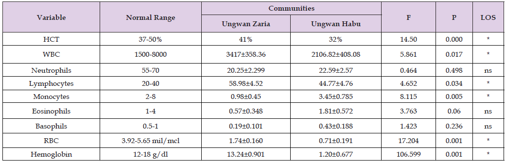
Note: * = Significant at p < 0.05, ns = Not significant, LOS = Level of significance.
Effectiveness of the Mass Drug Administration Program in Reducing Onchocerciasis Infections
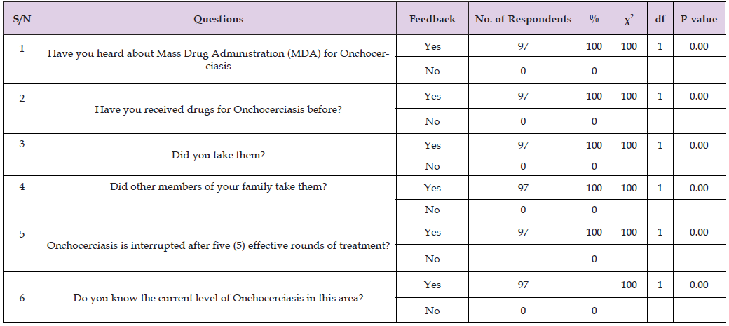
Table 3 revealed the level of onchocerciasis after five (5) rounds of MDA from 97 respondents in the two study areas. The six (6) questions had yes responses from all 97 (100%) respondents. Statistically, there was a significant difference (P<0.05) in all the questions with respect to the level of knowledge of the onchocerciasis of the patient after five (5) rounds of MDA.
Table 3: Effectiveness of the Mass Drug Administration Program in Reducing Onchocerciasis Infections.
An important turning point in the battle against onchocerciasis in this study reveals a zero prevalence (0.00%) of onchocerciasis, indicating that transmission has been successfully stopped in Ungwan Zaria and Ungwan Habu communities. This result is consistent with that of Dauda et al. [23] who similarly found no onchocerciasis prevalence, suggesting that there is no active transmission in the southwest Nigerian waterfalls of Arinta and Erin-ijesha. This result is also consistent with WHO [24] recommendations for disease elimination tactics, which emphasize the value of long-term initiatives such as the Mass Drug Administration (MDA). The low frequency of 3.5% reported by Aniaku et al. [25] in the Igbo-Eze North Local Government Area, Enugu State, Nigeria, is in contrast to this study. The public awareness campaign and local knowledge of onchocerciasis may have contributed to the study’s lower prevalence. Additionally, the mass drug administration previously documented in the study area may have contributed to the low prevalence; however, Okoye et al. [26] re-assessment study of onchocerciasis following a 10-year mass mectizan chemotherapeutic intervention found that onchocerciasis prevalence remained high even after the drug was freely distributed in the study area for ten years. The lack of active transmission of O. volvulus has also been reported in populations like Northern focus and Oaxaca focus in northern Mexico after several years of distribution of community drug with ivermectin (CDTi) [27]. Polymerase chain reaction (PCR) testing and other additional diagnostic techniques are still essential, even though microscopy and the results of the OV-16 ELISA indicate that the disease may have been eradicated. Microscopy can sometimes miss very light or hidden infections, but the OV-16 ELISA test is a reliable means that can detect even low-level cases. This is in support of the view of Ajero et al. [28] who proposed the use of different diagnostic tools together to ensure elimination. The success seen in this study area is not only encouraging but provides useful evidence for health workers and policy makers in Nigeria and other affected regions. This highlights the need for robust surveillance to prevent re-emergence of the disease either from undetected cases or from neighboring communities [29].
The haematological results revealed significant immune differences between Ungwan Habu and Ungwan Zaria. The differences in immune activation between the two communities is reflected in the changes in white blood cell (WBC) counts. Ungwan Zaria had the higher WBC counts, which suggest more immune responses, probably due to current or previous exposure to infection or environmental factors. This pattern of immune stimulation is further supported by the higher level of lymphocytes in Ungwan Zaria (58.98+4.52) than in Ungwan Habu (44.77+4.76) as observed by Ajero et al. [28] where such lymphocyte- driven activity usually reflects chronic antigen exposure and persistent immune activation. In both communities, lower-than-expected neutrophil counts in both communities may suggest compromised innate immune responses and an increased vulnerability to infections. This finding is consistent with WHO [24] that reported the weakening of innate responses may increase a population’s susceptibility to reinfection.In addition to the above observations, some blood parameters are also increasingly being recognized as useful indicators of the risk of onchocerciasis. Higher eosinophil counts and eosinophil-based ratios such as ENR, EMR, EBR and ELR have been found to be significantly associated with skin microfilaria suggesting eosinophil profiles might be a marker of active infection and parasitic load. Similarly, activity of Wolbachia, the endosymbiotic bacteria of O. volvulus, is highly associated with neutrophil responses. Wolbachia- driven neutrophil activation and neutrophil extracellular trap (NET) release are central to disease pathology, particularly ocular complications. The general pattern in this study provides important information on immune status of the studied populations. Differences in WBC, lymphocytes, neutrophils and eosinophils do not reflect past or present exposure to O. volvulus, but also biological factors that may affect susceptibility, persistence of infection and community-level risk of transmission.
The findings of this study provide strong evidence of the effectiveness of MDA in mass drug administration to control onchocerciasis. There was a marked reduction in disease prevalence and communities adhered to treatment, this finding confirms that sustained MDA cycles are critical to stopping transmission. The WHO [24] and Ajero et al. [28] have stressed the importance of community participation in the reception of ivermectin, which is essential for the success of a public health program in the elimination of onchocerciasis.
The zero prevalence (0%) of onchocerciasis recorded in this study indicates that the disease has been effectively controlled in the two communities studied. This finding underscores the impact of long-term interventions, especially the regular application of MDA in preventing recrudescence and sustaining eradication efforts. Meanwhile, deviations noted in the haematological profile of participants suggest a possible underlying immune related factor that may affect individual health status and vulnerability, the broader health context of affected population still requires close attention, and also identifying and addressing risk factors that may contribute to the reoccurrence of infection transmission is recommended.
The authors declare no conflict of interest.

International Association of Landscape Archaeology, Czech Glass Society, Czech Republic

Department of Chemistry, Semenov Institute of Chemical Physics, USSR Academy of Sciences, Moscow, Russia

Neurology, LA BioMed Research Institute, USA

Associate Professor at Department of Breast and Thyorid Surgey, Chongqing General Hospital, China

Professor of Nuclear Medicine, Faculty of Medicine and Surgery, University of Milan, Milan, Italy





 Abstract
 PDF
PDF



























