info@biomedres.us
+1 (502) 904-2126
One Westbrook Corporate Center, Suite 300, Westchester, IL 60154, USA
Site Map
 ISSN: 2574 -1241
Received: July 27, 2026; Published: August 14, 2026
*Corresponding author: Anwarul Islam, M.D, Ph.D, FRCPath, FACP, Clinical Associate Professor of Medicine, Division of Hematology/ Oncology, Department of Medicine, Buffalo General Medical Center, Buffalo, New York 14203, USA
DOI: 10.26717/BJSTR.2026.66.010344
We used low-dose prednisolone to treat an elderly patient with myelodysplastic syndrome (MDS) associated with marrow fibrosis and a complex mutational profile. Our findings suggest that this treatment approach is feasible, safe, and effective, even in patients with MDS complicated by marrow fibrosis and adverse molecular features.
Abbreviations: CTC: Common Toxicity Criteria; HMAs: Hypomethylating Agents; MDS: Myelodysplastic Syndrome; G-CSF: Granulocyte Colony-Stimulating Factor; AML: Acute Myeloid Leukemia; HMA: Hypomethylating Agent; IPSS: International Prognostic Scoring System; NCCN: National Comprehensive Cancer Network; NGS: Next-Generation Sequencing; PT: Prothrombin Time; PTT: Partial Thromboplastin Time
Myelodysplastic Syndromes are a heterogeneous group of hematologic disorders affecting the blood and bone marrow [1]. They encompass a wide spectrum of clinical presentations and prognostic implications. Bone marrow fibrosis associated with MDS is relatively uncommon and is generally considered to confer a poorer prognosis, placing affected patients in a higher-risk category [2]. Unfortunately, bone marrow fibrosis is not incorporated into conventional prognostic scoring systems and therefore may not always be adequately considered when treatment decisions are made.
Various therapeutic approaches have been explored in MDS; however, no established effective treatment currently exists for patients with MDS associated with marrow fibrosis. At present, most patients with high-risk MDS are treated with low-intensity regimens based on hypomethylating agents (HMAs), such as Azacitidine or Decitabine [3]. However, these agents are associated with significant toxicity. Across studies involving azacitidine and decitabine, approximately 50% of patients develop Common Toxicity Criteria (CTC) grade 3 or 4 cytopenias [4,5], particularly during the first treatment cycle. This often necessitates intensive transfusion support and close clinical monitoring, while also increasing the risk of febrile neutropenia and early mortality. We treated a newly diagnosed elderly patient with MDS associated with marrow fibrosis and adverse molecular features using low-dose Prednisone alone. The rationale for this approach was to avoid the undesirable toxicities associated with chemotherapy while potentially prolonging survival and improving quality of life.
Our patient continued to demonstrate positive and satisfactory results 12 months after therapy was initiated. He achieved and maintained a stable, continuous partial remission. The circulating neutrophil count increased and generally remained within normal limits, while the hemoglobin level was maintained at approximately 8 g/dL without the need for blood transfusions. The platelet count remained low (approximately 40,000/μL) but stable, and no platelet transfusions were required. Occasional decreases in the neutrophil count (to approximately 2,000/μL) were managed successfully by increasing the dose of prednisolone. Peripheral blood smears continued to show occasional blasts and blast-like cells, along with morphologic features of myelodysplasia involving all three hematopoietic cell lines. Although the patient’s response was initially slow, it was sustained over an extended period. Unfortunately, the patient later suffered a fall, developed a subarachnoid hemorrhage, and died shortly thereafter.
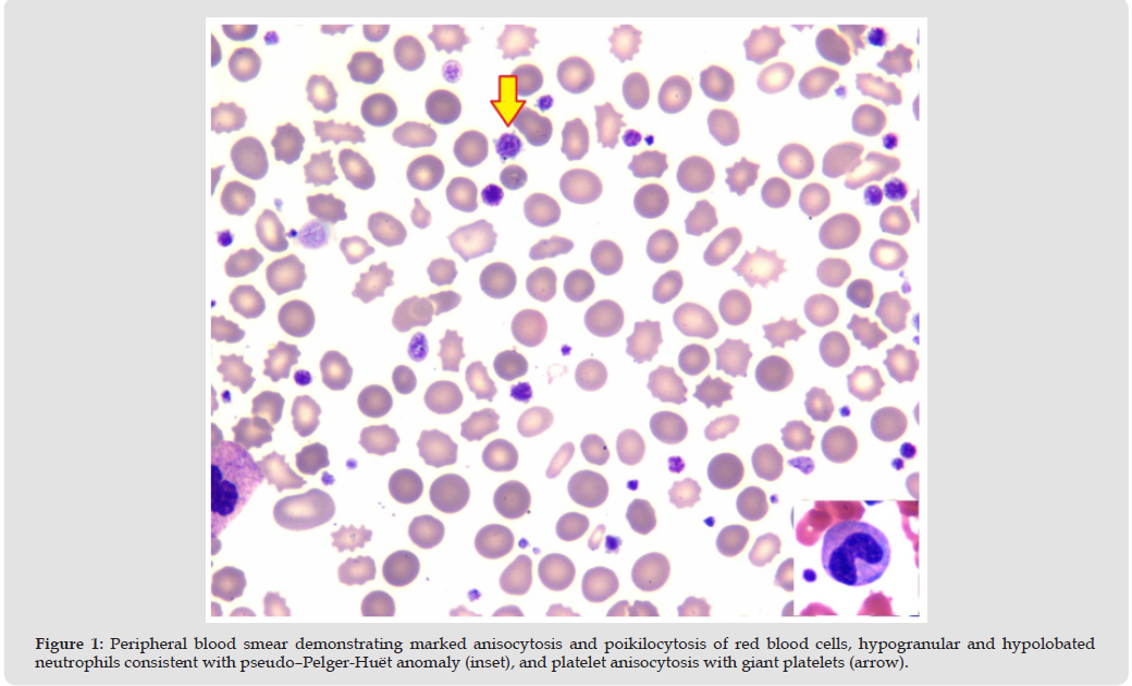
The patient was an 81-year-old White male with a past medical history significant for hypertension, hypercholesterolemia, hyperlipidemia, degenerative disc disease, and prior myocardial infarction. He had been scheduled to undergo a dental implant procedure on April 17, 2025. As part of the preoperative surgical clearance, his primary care physician ordered routine blood tests, which revealed pancytopenia, including a white blood cell count of 1.3 × 10⁹/L, hemoglobin of 8.7 g/dL, and platelet count of 34,000/μL. He was subsequently advised to present to the emergency department for further evaluation and management. At presentation, the patient reported intermittent shortness of breath and fatigue. He denied fever, chills, dyspnea, chest pain, chest tightness, palpitations, nausea, vomiting, diarrhea, constipation, abdominal pain, headaches, visual changes, lightheadedness, or syncope. On physical examination, the patient appeared anemic but was not in acute distress. There was no jaundice, cyanosis, or peripheral edema. The abdomen was soft and non-tender, with normal bowel sounds. The liver, spleen, and kidneys were not palpable, and there was no palpable lymphadenopathy. Cardiac examination revealed normal S1 and S2 heart sounds with a soft ejection systolic murmur. Chest examination was clear to auscultation bilaterally. Vital signs were stable, with blood pressure of 125/70 mm Hg, pulse rate of 72 beats/min, respiratory rate of 18 breaths/min, and temperature of 97.6°F. Laboratory investigations revealed a white blood cell count of 1.7 × 10⁹/L, hemoglobin of 8.1 g/dL with normal MCV and MCH, and a platelet count of 23 × 10⁹/L. Manual differential count of the peripheral blood smear showed 53% neutrophils, 40% lymphocytes, 3% monocytes, 2% bands, 1% myelocytes, and 1% metamyelocytes. The peripheral blood smear demonstrated marked anisocytosis and poikilocytosis of red blood cells (Figure 1), hypogranular and hypolobated neutrophils consistent with pseudo–Pelger-Huët anomaly (Figure 1, inset), and platelet anisocytosis with giant platelets (Figure 1, arrow).
Figure 1
Serum iron level was normal at 94 μg/dL (reference range 45–182 μg/dL), and total iron-binding capacity was also within normal limits at 374 μg/dL (reference range 261–478 μg/dL). Percent iron saturation was slightly decreased at 19% (reference range 20–50%), while ferritin was markedly elevated at 2521 ng/mL. Reticulocyte count was normal at 0.5% (reference range 0.5–1.5%). Haptoglobin was elevated at 344.7 mg/dL. Vitamin B12 level was normal at 315 pg/ mL, and folic acid level was normal at 9.8 ng/mL. Prothrombin time (PT) and INR were mildly prolonged at 20.2 seconds (reference range, 9.3–13.5 seconds) and 1.7 (reference range, 0.9–1.2), respectively. The activated partial thromboplastin time (aPTT) was within the normal range at 32.8 seconds (reference range, 25.9–40.5 seconds). The comprehensive metabolic panel was largely unremarkable. Viral studies including influenza, COVID-19, CMV, and EBV were negative. JAK2 mutation analysis and routine urinalysis were also negative. Because of anemia, severe leukopenia, thrombocytopenia, and abnormal cytologic findings on the peripheral blood smear, a hematologic malignancy was suspected, and the patient was admitted for further evaluation, diagnosis, and management. Shortly after admission, the patient underwent a bone marrow examination.
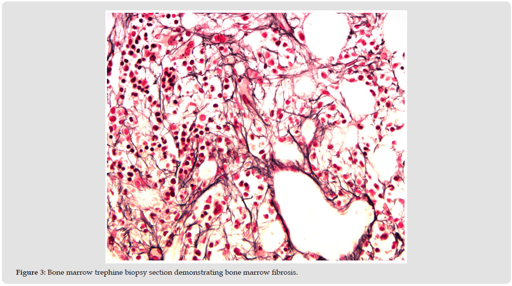
Bone marrow aspirate, touch imprints, clot sections, and core biopsy revealed a hypocellular marrow with marked dysmyelopoiesis (Figure 2), mildly increased blasts, and increased reticulin fibrosis (MF 1–2+) (Figure 3), consistent with a myeloid neoplasm associated with myelofibrosis. There was insufficient material for flow cytometry or cytogenetic studies. Next-generation sequencing (NGS) demonstrated mutations in IDH2, TET2, TP53, and ASXL1. Overall, the findings were most consistent with a diagnosis of myelodysplastic neoplasm (MDS) with myelofibrosis. Because of the patient’s advanced age (81 years), the diagnosis of MDS with marrow fibrosis, and the complex mutational profile identified by NGS studies, he was considered to have higher-risk disease (IPSS-R score: 3). The patient was referred to a local cancer center, where treatment with Inqovi (decitabine and cedazuridine) was initiated.
Figure 2
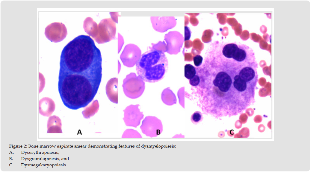
Figure 3
However, the therapy was poorly tolerated and resulted in severe clinical deterioration. Consequently, the patient’s family declined further treatment at that institution and returned to our care. At that point, the patient was started on low-dose prednisone at 20 mg orally daily. The prednisone dose was subsequently adjusted according to the patient’s blood counts, with gradual tapering to as low as 5 mg daily or escalation up to 30 mg daily when clinically indicated. Within two weeks of initiating this treatment, the patient’s circulating neutrophil count began to improve. Although the hemoglobin concentration and platelet count remained low (hemoglobin approximately 8 g/dL; platelet count approximately 40–60 × 10³/μL), they remained stable and did not require red blood cell or platelet transfusions. Peripheral blood smears showed no blasts or blast-like cells, and there was no overt evidence of leukemic transformation. As noted earlier, the patient subsequently sustained a fall at home resulting in a subarachnoid hemorrhage, and he expired shortly thereafter.
Treatment strategies for patients with MDS rely heavily on prognostic scoring systems such as the revised International Prognostic Scoring System (IPSS-R) [6]. Bone marrow fibrosis has been identified as an independent adverse prognostic factor associated with reduced survival in patients with MDS, regardless of the IPSS-R risk category [2]. The IPSS-R incorporates cytogenetic abnormalities, bone marrow blast percentage, and laboratory parameters including hemoglobin level, platelet count, and absolute neutrophil count (ANC) at diagnosis. However, the degree of bone marrow fibrosis is not included in the IPSS-R risk stratification model or prognostic assessment [6]. The clinical significance of bone marrow fibrosis was not recognized in the 2008 WHO classification [7], whereas the 2016 WHO classification acknowledged MDS with marrow fibrosis as an unclassified subtype of MDS [8]. Overall survival is generally considered inferior in patients with de novo MDS associated with moderate to severe bone marrow fibrosis.
The ideal therapeutic approach for older patients (≥65 years) with MDS, particularly those with higher-risk disease, remains uncertain. Although allogeneic hematopoietic stem-cell transplantation can induce long-term remission in patients with MDS [9-12], this option is not feasible for most patients because the median age at diagnosis is typically over 70 years [13], as in the present case. In general, elderly patients with higher-risk MDS are treated with hypomethylating agents such as 5-azacitidine or decitabine following studies demonstrating that these agents achieve superior remission rates compared with supportive care and may delay progression to acute leukemia [13]. In a randomized trial comparing azacitidine with conventional care in patients with high-risk MDS, median survival was 24.5 months in the azacitidine group compared with 15.0 months in the conventional care group [4]. However, treatment with hypomethylating agents in elderly patients with high-risk MDS is associated with significant toxicity, and treatment-related mortality may be substantial [4,14]. Moreover, prognosis becomes particularly poor once these agents fail. Subsequent therapeutic options are limited, and median survival after treatment failure is generally less than six months [15,16].
MDS has traditionally been regarded as a malignant hematologic disorder, and treatment has therefore relied primarily on chemotherapy- based approaches, except in a limited number of studies in which granulocyte colony-stimulating factor (G-CSF) and erythropoietin were used in patients with low-risk disease and minimal transfusion dependence [17,18]. Because our patient did not tolerate chemotherapy, we adopted a different and less intensive therapeutic strategy, with the goal of prolonging survival while preserving quality of life. Accordingly, the patient was treated with prednisolone rather than chemotherapy. Corticosteroid therapy has been reported to improve blood counts in approximately 50% of cases. To our knowledge, this is the first reported case of MDS with marrow fibrosis and multiple genetic alterations successfully managed with prednisolone alone, resulting in favorable clinical outcomes. We believe this observation merits further evaluation in larger studies and prospective clinical trials.
As we have previously reported [19], the diagnosis of MDS is not entirely uniform, and considerable heterogeneity exists both among patients and among diagnosticians. This variability may partly explain the marked differences in disease course and treatment response observed in patients with MDS. Consequently, therapeutic approaches should be individualized according to symptom burden, risk of progression to acute myeloid leukemia (AML), and associated comorbidities [20]. Hypomethylating agent (HMA) therapy is approved in Europe primarily for patients with higher-risk MDS according to the International Prognostic Scoring System (IPSS), whereas in the United States HMAs are approved for the treatment of all patients with MDS [21]. Current National Comprehensive Cancer Network (NCCN) guidelines recommend HMA therapy mainly for patients with intermediate- or high-risk MDS who are not candidates for intensive treatment, are unlikely to benefit from alternative therapies, or require bridging therapy before allogeneic stem cell transplantation [21].
This disease, Myelodysplastic Syndromes, is frequently misclassified, as evidenced by the fact that more than 50% of patients in certain registries were categorized as “MDS-unclassifiable” [14,22]. When bone marrow biopsies are not evaluated in conjunction with bone marrow aspirates during the diagnostic workup of MDS, important findings such as abnormal localization of blast cells [19] and bone marrow fibrosis may be overlooked. Consequently, high-grade MDS may be incorrectly classified as low- or intermediate-risk disease, thereby compromising accurate risk stratification and adversely affecting prognostic assessment and clinical management.

International Association of Landscape Archaeology, Czech Glass Society, Czech Republic

Department of Chemistry, Semenov Institute of Chemical Physics, USSR Academy of Sciences, Moscow, Russia

Department of Educational Sciences, Research Centre Qualities of Teachers, School of Education – HAN, Nijmegen, Netherlands

Associate Professor at Department of Breast and Thyorid Surgey, Chongqing General Hospital, China

Neurology, LA BioMed Research Institute, USA





 Abstract
 PDF