info@biomedres.us
+1 (502) 904-2126
One Westbrook Corporate Center, Suite 300, Westchester, IL 60154, USA
Site Map
 ISSN: 2574 -1241
ISSN: 2574 -1241
ISSN: 2574 -1241
Received: July 13, 2026; Published: July 28, 2026
*Corresponding author: Andrew J Fishman, Department of Otolaryngology–Head and Neck Surgery, University of Missouri School of Medicine, Columbia, Missouri, USA
DOI: 10.26717/BJSTR.2026.66.010326
Background: Patients with chronic craniofacial pain frequently experience prolonged diagnostic delays because clinically important information becomes fragmented across multiple clinical encounters, healthcare systems, and medical specialties. Although artificial intelligence (AI) has demonstrated considerable promise in image interpretation, pattern recognition, and diagnostic support, few systems have been designed to assist physicians with the longitudinal evolution of clinical reasoning. To address this gap, we developed the Comprehensive Patient Assessment and Symptom System (COMPASS), a physician-directed framework that integrates structured Patient Self-Assessments, serial physician evaluations, diagnostic interventions, imaging studies, therapeutic response, and AI-assisted evidence synthesis into a continuous diagnostic process (Committee on Diagnostic Error in Health Care, et al. [1-5])
Case Presentation: A 27-year-old man presented with a six-year history of refractory unilateral deep otalgia despite extensive investigation and multiple unsuccessful treatments. Sequential Patient Self-Assessments within the COMPASS framework documented progressive evolution of the clinical phenotype from presumed musculoskeletal pain to glossopharyngeal nerve localization. Partial response to oxcarbazepine unmasked previously unrecognized tongue-base pain, prompting focused evaluation for glossopharyngeal pathology. High-resolution magnetic resonance imaging demonstrated vascular contact without convincing neurovascular compression. Continued physician-directed analysis, supplemented by AI-assisted literature review, suggested an atypical presentation of Eagle syndrome despite a styloid process measuring 2.9 cm on preoperative CT, below the commonly used 3.0 cm threshold for elongation. Dedicated high-resolution computed tomography with multiplanar and three-dimensional reconstruction demonstrated abnormal medial angulation and marked narrowing of the styloid-C1 interval. Minimally invasive transcervical styloidectomy resulted in near-complete resolution of symptoms, providing surgical confirmation of the diagnostic pathway (Badhey, et al. [6-10]).
Conclusions: This surgically validated proof-of-concept case demonstrates the feasibility of a physician-directed longitudinal framework that combines adaptive Patient Self-Assessments, sequential physician reasoning, and AI-assisted evidence synthesis to support complex diagnostic decision making. Rather than replacing physician judgment, COMPASS preserves the temporal evolution of clinical information and facilitates progressive refinement of diagnostic hypotheses throughout extended patient care. This proof-of-concept study establishes the conceptual foundation for prospective evaluation of the physician-directed COMPASS framework and provides a model for investigating how artificial intelligence can strengthen longitudinal clinical reasoning without replacing physician judgment (Fishman, et al. [2,5,11]).
Keywords: Artificial Intelligence; Compass; Eagle Syndrome; Chronic Otalgia; Craniofacial Pain; Glossopharyngeal Neuralgia; Clinical Decision Support; Longitudinal Diagnosis; Neurotology
Diagnosis is inherently a longitudinal process, yet modern healthcare is largely organized around isolated clinical encounters. Patients with uncommon disorders frequently undergo years of consultations, repeated diagnostic testing, empiric treatments, and multiple specialist referrals before a definitive diagnosis is established. In many cases, the primary limitation is not the absence of clinical information but the inability to integrate observations that accumulate gradually over time into a coherent diagnostic narrative. This challenge is particularly evident in neurotology and craniofacial pain, where overlapping symptom patterns, evolving clinical phenotypes, and low disease prevalence often obscure uncommon pathological processes (Committee on Diagnostic Error in Health Care et al. [1,4,12,13]). The Comprehensive Patient Assessment and Symptom System (COMPASS) was developed to address this limitation. COMPASS is a physician-directed longitudinal clinical framework built upon three integrated components:
1. Adaptive Patient Self-Assessments that evolve as the diagnostic process progresses;
2. Sequential physician-directed clinical reasoning that continuously refines the differential diagnosis; and
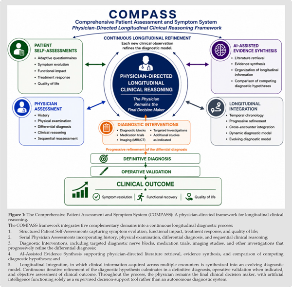
3. Artificial intelligence-assisted evidence synthesis that supports organization of clinical information, literature retrieval, and comparison of competing diagnostic hypotheses. Rather than functioning as a diagnostic algorithm, COMPASS preserves evolving clinical information and supports continuous refinement of diagnostic reasoning as clinical evidence accumulates over time. The overall COMPASS framework is illustrated in Figure 1.
Existing symptom questionnaires and patient-reported outcome instruments have important roles in clinical practice but are generally designed to assess disease severity, treatment response, or quality of life at discrete points in time. Most are static, disease-specific instruments that do not adapt as new diagnostic information emerges. Consequently, they are not intended to support iterative diagnostic reasoning across prolonged clinical journeys. COMPASS was developed to complement these established instruments by preserving the temporal evolution of symptoms and allowing patient-reported information to evolve together with physician-directed diagnostic reasoning (Cella, et al. [14,15]). Within COMPASS, artificial intelligence serves exclusively as a physician-supervised clinical decision-support tool rather than an autonomous diagnostic system. AI assists with organization of longitudinal clinical information, identification of diagnostic inconsistencies, retrieval of relevant medical literature, comparison of competing diagnostic hypotheses, and synthesis of complex clinical data.
Clinical interpretation, diagnostic decision making, selection of investigations, and patient management remain entirely under physician supervision (Fishman, et al. [5,11,16]). This report presents the first clinical validation of the COMPASS framework through the evaluation of a patient with chronic unilateral otalgia whose diagnosis evolved through multiple stages of investigation before definitive surgical confirmation. The case also illustrates an unexpected application of publicly available artificial intelligence in healthcare navigation. After conventional referral pathways failed to establish an appropriate diagnosis, the patient independently used AI-assisted searches to identify a fellowship-trained neurotologist with expertise in complex craniofacial pain, ultimately leading to definitive treatment.
The objective of this report is to introduce COMPASS as a physician- directed framework for longitudinal clinical reasoning and to demonstrate its potential value through a surgically validated proofof- concept case. To our knowledge, this is the first report describing a longitudinal diagnostic framework that integrates adaptive Patient Self-Assessments, sequential physician reasoning, and AI-assisted evidence synthesis into a continuously evolving diagnostic model. The emphasis is not on autonomous AI diagnosis, but on illustrating how structured integration of evolving clinical information, supported by artificial intelligence and directed by physician expertise, may facilitate diagnosis of uncommon disorders over extended periods.
Figure 1
Study Design
This report describes the prospective development and first clinical validation of the Comprehensive Patient Assessment and Symptom System (COMPASS) during the evaluation of a patient with chronic unilateral craniofacial pain. The patient’s diagnostic course was documented longitudinally from initial consultation through definitive diagnosis, operative treatment, and postoperative follow-up. Following surgical confirmation of the diagnosis, the longitudinal clinical course and diagnostic methodology were retrospectively analyzed to characterize the COMPASS framework. Because this work represents the initial clinical application of a novel physician-directed diagnostic framework, the primary objective was to evaluate the feasibility of integrating structured Patient Self-Assessments, serial physician evaluations, diagnostic interventions, imaging studies, therapeutic response, and AI-assisted evidence synthesis into a unified longitudinal model of clinical reasoning.
The COMPASS Framework
COMPASS is a physician-directed longitudinal clinical assessment framework designed to preserve, organize, and continuously refine evolving clinical information throughout a patient’s diagnostic journey. The framework integrates structured Patient Self-Assessments with serial physician examinations, diagnostic procedures, medication response, imaging studies, operative findings, postoperative outcomes, and AI-assisted evidence synthesis into a continuously evolving clinical record. Unlike traditional symptom questionnaires, Patient Self-Assessments within COMPASS are adaptive. Information obtained during one stage of evaluation informs the design of subsequent assessments, allowing progressively more focused investigation as the differential diagnosis evolves.
Rather than functioning as static surveys, these assessments become active components of the clinical reasoning process, documenting changes in symptom distribution, provoking factors, treatment response, and diagnostic confidence over time (Cella, et al. [14,15]). The framework is intended to complement conventional history-taking rather than replace it. By preserving the chronology of symptom evolution and integrating information acquired across multiple encounters, COMPASS supports physician-directed clinical reasoning while maintaining the longitudinal context that is frequently lost during fragmented healthcare delivery (Committee on Diagnostic Error in Health Care et al. [1,4])
Role of Artificial Intelligence
Artificial intelligence functioned exclusively as a physician-supervised clinical support system throughout the diagnostic process. At no point did AI independently diagnose the patient, interpret radiologic studies, recommend operative intervention, or direct patient management.
Within COMPASS, AI served six principal functions:
• Organization of longitudinal clinical information; • Structured summarization of serial patient encounters; • Refinement of differential diagnostic hypotheses; • Retrieval of relevant medical literature; • Comparison of competing diagnostic possibilities; and • Synthesis of emerging clinical evidence.
Every AI-generated observation underwent independent physician review before incorporation into clinical decision making. Clinical interpretation, diagnostic reasoning, selection of investigations, operative planning, and treatment decisions remained entirely under physician supervision (Fishman AJ [2,5,16]). COMPASS is intentionally platform independent. The framework does not depend upon any specific artificial intelligence model, software platform, or commercial product. Any AI system capable of organizing longitudinal clinical information, retrieving relevant literature, synthesizing evidence, and supporting physician-directed reasoning may be incorporated into the framework, provided that all outputs remain subject to independent physician review and clinical judgment. This design allows COMPASS to evolve alongside future advances in artificial intelligence without requiring modification of its underlying clinical methodology.
Longitudinal Diagnostic Strategy
Evaluation followed a sequential, hypothesis-driven approach in which each diagnostic investigation was selected according to information obtained during the preceding stage of assessment rather than according to a predetermined algorithm. Initial evaluation focused on common causes of chronic unilateral otalgia, including temporomandibular dysfunction, myofascial pain syndrome, and other recognized sources of referred otalgia (Earwood, et al. [12,13]). Following unsuccessful conservative management, serial Patient Self-Assessments together with repeated physician evaluations suggested a progressively stronger neuropathic component. Sequential peripheral nerve blocks, pharmacologic testing with oxcarbazepine, focused magnetic resonance imaging of the lower cranial nerves, disease-specific symptom assessment, AI-assisted literature review, and dedicated high-resolution computed tomography with multiplanar and three-dimensional reconstruction were subsequently incorporated into the diagnostic pathway (Hiwatashi, et al. [7-10,17,18]).
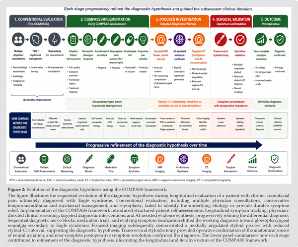
Importantly, each diagnostic intervention informed the subsequent stage of investigation. Rather than functioning as isolated tests, imaging studies, medication response, diagnostic blocks, and serial Patient Self-Assessments collectively refined the differential diagnosis until concordance was achieved among the longitudinal clinical phenotype, targeted imaging, operative findings, and postoperative outcome (Committee on Diagnostic Error in Health Care et al. [1,4]). The longitudinal diagnostic pathway is illustrated in Figure 2.
Figure 2
A 27-year-old otherwise healthy man was referred for evaluation of a six-year history of chronic left-sided deep otalgia that had remained undiagnosed despite extensive evaluation by multiple physicians. The pain was constant, localized deep within the external auditory canal, and consistently worsened when sleeping on the affected side. Numerous investigations and empiric treatments failed to identify an underlying cause or provide lasting relief. Among these interventions was septoplasty performed for presumed eustachian tube dysfunction, which produced no clinical improvement. Prior to referral, the patient independently used publicly available artificial intelligence platforms to investigate his symptoms and identify an appropriate specialist. Conventional referral pathways directed him toward evaluation by general otolaryngology and oral and maxillofacial surgery. In contrast, repeated AI-assisted searches consistently identified consultation with a fellowship-trained neurotologist specializing in complex craniofacial disorders. This process ultimately led the patient to seek evaluation in Belgrade, Serbia.
Although anecdotal, this observation illustrates a potential future role for AI in facilitating navigation to highly specialized care. Initial otologic examination demonstrated no inflammatory, infectious, neoplastic, or structural abnormality sufficient to explain the severity or chronicity of the patient’s symptoms. Audiologic evaluation was unremarkable, and no objective otologic findings adequately localized the pain generator. Conservative management directed toward temporomandibular dysfunction and myofascial pain syndrome, including anti-inflammatory therapy and supportive measures, failed to produce meaningful improvement. Persistent unilateral otalgia in the setting of a normal otologic examination therefore prompted systematic evaluation for referred otalgia and cranial neuropathies (Earwood, et al. [12,13]) At this stage, the earliest implementation of the COMPASS framework was initiated.
Structured Patient Self-Assessments documented symptom chronology, anatomical localization, provoking factors, pain quality, treatment response, and functional impact. Repeated assessments over time demonstrated that the patient’s symptom profile remained remarkably consistent while progressively revealing features suggestive of neuropathic rather than musculoskeletal pain. Sequential diagnostic nerve blocks were subsequently performed to localize potential peripheral pain generators. An auriculotemporal nerve block failed to improve symptoms, followed by a deeper regional block targeting the posterior external auditory canal and adjacent temporal region, which likewise produced no meaningful relief. These negative diagnostic interventions reduced the likelihood of common peripheral pain generators and refined the differential diagnosis toward deeper cranial nerve-mediated pathology.
A therapeutic trial of oxcarbazepine produced partial but clinically meaningful improvement. As the dominant otalgia diminished, the patient became aware of persistent ipsilateral pain involving the base of the tongue that had previously been overshadowed by the severity of the ear pain. During subsequent interviews, he recognized that this symptom had likely been present throughout much of his illness but had never been appreciated because of the overwhelming intensity of the otalgia. This observation proved to be a pivotal turning point in the diagnostic process, redirecting attention toward the glossopharyngeal nerve distribution (Shah [10,18]). Recognition of glossopharyngeal distribution pain prompted development and administration of disease-specific Patient Self-Assessments focusing on glossopharyngeal neuralgia and Eagle syndrome. Unlike the abrupt, paroxysmal attacks typically associated with classical glossopharyngeal neuralgia, the patient’s symptoms demonstrated a persistent, mechanically mediated pattern that was consistently reproduced during serial assessments.
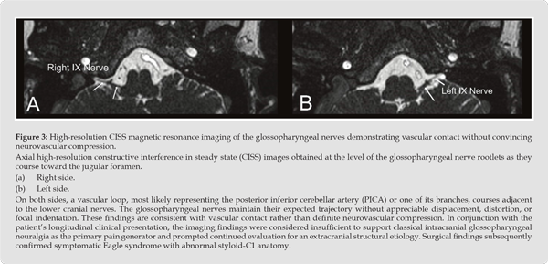
The evolving clinical phenotype suggested a chronic structural pain generator rather than primary neurovascular compression (Chen, et al. [19-21]). High-resolution magnetic resonance imaging of the brain and internal auditory canals, including dedicated evaluation of the lower cranial nerves, demonstrated vascular contact involving the glossopharyngeal nerve without convincing evidence of neurovascular compression or displacement. Representative MRI findings are shown in Figure 3. Although intracranial glossopharyngeal neuralgia remained within the differential diagnosis, the imaging findings were insufficient to justify posterior fossa exploration or microvascular decompression. The absence of definitive radiologic compression encouraged continued investigation of extracranial causes (Gaul, et al. [7,17]).
Figure 3
Rather than terminating the investigation, physician-directed review of the accumulated longitudinal data was supplemented by AI-assisted synthesis of the medical literature. Artificial intelligence organized serial clinical observations, compared competing diagnostic hypotheses, retrieved uncommon publications, and highlighted reports suggesting that symptomatic Eagle syndrome may depend not only upon styloid length but also upon three-dimensional orientation and regional anatomy. All AI-generated observations were independently evaluated and accepted only when consistent with the evolving clinical findings and established medical evidence (Fishman, et al. [2,5,11,16]). Based on this synthesis, dedicated high-resolution computed tomography with multiplanar and three-dimensional reconstruction was obtained.
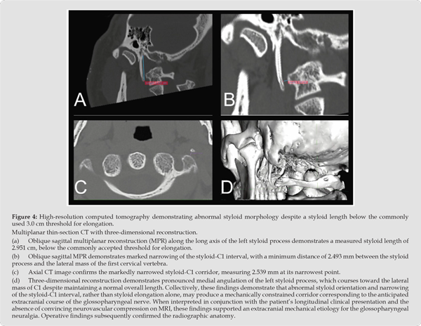
Unlike previous imaging studies, this examination emphasized spatial relationships between the styloid process, the first cervical vertebra, and the anticipated course of the glossopharyngeal nerve. The CT examination demonstrated a left styloid process measuring 2.9 cm, below the commonly used 3.0 cm threshold for elongation, together with pronounced medial angulation and marked narrowing of the styloid-C1 interval on the symptomatic side. Representative CT findings are shown in Figure 4. These anatomical findings closely matched the evolving clinical phenotype documented by the COMPASS framework and established the rationale for definitive surgical treatment. Although current literature recognizes the importance of styloid morphology and three-dimensional anatomy, the present case suggests that abnormal spatial relationships may be clinically significant even in the absence of styloid elongation. This observation should be regarded as a hypothesis generated by the present case rather than an established disease subtype (Badhey, et al. [6,8,9,22]).
Figure 4
The patient underwent minimally invasive transcervical styloidectomy through a short retroauricular incision designed to remain concealed beneath the inferior aspect of the auricle. Continuous facial nerve monitoring was employed throughout the procedure to facilitate safe dissection within the upper cervical and parapharyngeal region. Operative findings closely paralleled the preoperative radiologic assessment. Preoperative CT demonstrated a total anatomical styloid length of 2.9 cm. Intraoperatively, the styloid process demonstrated pronounced medial angulation with marked narrowing of the interval between the styloid process and the lateral aspect of the first cervical vertebra. The operative anatomy was entirely concordant with the preoperative three-dimensional computed tomography findings and supported the proposed mechanical basis for irritation of the glossopharyngeal nerve.
Figure 5
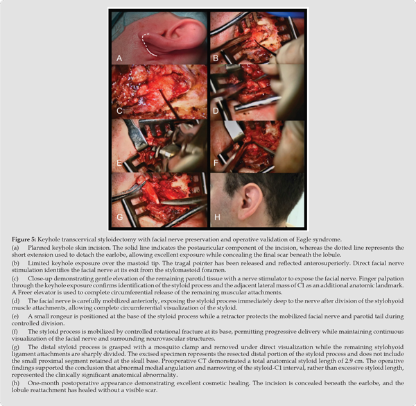
These observations reinforce the concept that styloid orientation and regional spatial relationships may be of greater clinical importance than absolute styloid length alone in selected patients (Badhey, et al. [6,8,9]). Complete Resection of the left styloid process was accomplished without intraoperative complication. Facial nerve function remained intact throughout the procedure, and no vascular or neurologic injury occurred. The patient tolerated the procedure well and was discharged in stable condition. Representative operative findings are shown in Figure 5. Clinical improvement was evident immediately following surgery. The improvement was sustained throughout the early postoperative follow-up period and was accompanied by restoration of normal daily activities without recurrence of the characteristic preoperative deep otalgia.
During the initial postoperative evaluations, the patient reported marked reduction of the deep otalgia that had persisted for approximately six years. At subsequent follow-up, the characteristic deep ear pain had nearly resolved, with only mild discomfort attributable to expected postoperative soft tissue healing. By approximately one month following surgery, the patient described near-complete resolution of his preoperative symptoms and reported that the disabling pain which had dominated his daily life had essentially disappeared. An additional observation emerged during postoperative follow-up. Following resolution of the dominant left-sided symptoms, the patient became aware of milder discomfort on the contralateral side. Review of the preoperative computed tomography demonstrated a similar, although less pronounced, anatomical configuration involving the right styloid process.
Although asymptomatic prior to surgery, the patient recognized that the right-sided symptoms had likely been present but overshadowed by the severity of the left-sided pain. He subsequently expressed interest in staged contralateral styloidectomy following complete recovery from the initial procedure. From a diagnostic standpoint, this outcome demonstrated complete concordance among the longitudinal symptom evolution documented through COMPASS, serial Patient Self-Assessments, physician-directed clinical reasoning, sequential diagnostic interventions, advanced imaging, operative findings, and postoperative clinical outcome. Each stage of the diagnostic pathway progressively refined the differential diagnosis, ultimately converging upon a surgically confirmed anatomical etiology.
The principal contribution of this study is the introduction and preliminary clinical validation of the Comprehensive Patient Assessment and Symptom System (COMPASS), a physician-directed framework for longitudinal diagnostic reasoning. Rather than presenting a new diagnostic algorithm, COMPASS provides a structured methodology for integrating adaptive Patient Self-Assessments, sequential physician assessment, diagnostic interventions, imaging findings, therapeutic response, and AI-assisted evidence synthesis into a continuously evolving clinical model. The surgically confirmed case presented here serves as a proof-of-concept demonstrating the feasibility of this approach in the evaluation of complex craniofacial pain. The convergence of independent clinical observations, targeted imaging, operative anatomy, and successful surgical treatment provides preliminary clinical validation of the COMPASS framework as a physician- directed model for longitudinal diagnostic reasoning.
Importantly, the framework itself was not validated by a single diagnostic test, but rather by the progressive accumulation and integration of multiple independent sources of clinical evidence leading to a correct diagnosis and successful treatment (Committee on Diagnostic Error in Health Care et al. [1,4]) The patient’s six-year diagnostic journey illustrates a recognized limitation of contemporary healthcare delivery. Multiple physicians evaluated the patient, numerous investigations were performed, and several competing diagnostic hypotheses were considered. Although substantial clinical information accumulated during this period, each encounter functioned largely as an isolated event. Information obtained during one consultation was rarely integrated with observations made months or years later.
Consequently, the diagnostic process repeatedly restarted rather than progressively evolved. This fragmentation has been recognized as an important contributor to diagnostic delay, particularly in patients with uncommon disorders and complex symptom patterns (Committee on Diagnostic Error in Health Care et al. [1,3,4]). COMPASS was specifically designed to address this deficiency. Rather than viewing diagnosis as a series of independent clinical encounters, the framework preserves the chronology of symptom evolution and allows each new observation to refine, rather than replace, the working differential diagnosis. The framework therefore functions as a continuously evolving model of physician-directed clinical reasoning in which patient-reported information, physician assessment, diagnostic testing, imaging, therapeutic response, operative findings, and postoperative outcome are interpreted as components of a single longitudinal diagnostic narrative.
One of the distinguishing features of COMPASS is its adaptive use of structured Patient Self-Assessments. Conventional symptom questionnaires generally provide a static representation of a patient’s condition at a single point in time. In contrast, the Patient Self-Assessments employed within COMPASS evolve together with the diagnostic process. Information obtained during one stage of evaluation directly informs the design of subsequent assessments, allowing progressively more focused characterization of symptom localization, provoking factors, temporal evolution, treatment response, and functional impact. Rather than serving solely as instruments for documentation, these assessments become active participants in clinical reasoning. Although formal psychometric validation remains necessary, this adaptive methodology represents one of the principal conceptual innovations of the COMPASS framework (Cella, et al. [14,15]).
The present case also demonstrates how longitudinal assessment may reveal clinically significant information that would otherwise remain unrecognized. Partial suppression of the patient’s dominant otalgia with oxcarbazepine exposed persistent pain localized to the ipsilateral tongue base, an observation that substantially redirected the diagnostic investigation. Importantly, this symptom was not newly acquired. Rather, it had been obscured for years by the severity of the ear pain and became clinically apparent only after the dominant symptom was partially relieved. Recognition of this evolving clinical phenotype redirected subsequent investigation toward glossopharyngeal pathology and ultimately toward an extracranial structural etiology (“Headache Classification Committee of the International Headache Society (IHS) The International Classification of Headache Disorders, 3rd Edition,” [10,18,20,21]).
Artificial intelligence functioned exclusively as a physician-supervised clinical support system throughout the patient’s evaluation. Its greatest contribution was not autonomous diagnosis, but rather assistance with organization of longitudinal clinical information, retrieval of uncommon medical literature, comparison of competing diagnostic hypotheses, and synthesis of evidence accumulated over several years. Every AI-generated observation underwent independent physician review before influencing subsequent diagnostic decisions. Clinical interpretation, selection of investigations, operative planning, and definitive treatment remained entirely under physician supervision (Fishman, et al. [2,5,11,16]). This physician-directed model differs fundamentally from many contemporary discussions of artificial intelligence in healthcare, which frequently emphasize autonomous diagnostic performance.
The experience described in this report suggests that AI may provide greater clinical value when incorporated into an iterative physician- guided workflow than when functioning as an independent diagnostic system. Rather than replacing physician judgment, artificial intelligence functioned as a force multiplier, allowing the treating physician to organize large volumes of longitudinal clinical information, retrieve uncommon literature, recognize subtle diagnostic patterns, and continually refine the differential diagnosis as new evidence emerged. The final diagnosis resulted from physician interpretation of converging clinical evidence rather than from any independent recommendation generated by AI. From an anatomical perspective, this case reinforces the concept that symptomatic Eagle syndrome cannot be defined solely by absolute styloid length. Although the patient’s styloid process measured 2.9 cm on preoperative CT, below the commonly used 3.0 cm threshold for elongation, high-resolution three-dimensional imaging demonstrated pronounced medial angulation and marked narrowing of the styloid-C1 interval.
These anatomical findings correlated closely with the patient’s evolving clinical phenotype, operative findings, and postoperative outcome. Contemporary anatomical and radiologic literature increasingly recognizes that styloid orientation and spatial relationships may be clinically more relevant than length alone in selected patients (Badhey, et al. [6,8,9,23,24]). Nevertheless, caution is warranted when interpreting these findings. Current evidence supports consideration of styloid morphology in addition to absolute length but does not yet establish a distinct syndrome characterized exclusively by a normal-length styloid process associated with a reduced styloid-C1 interval. The present report should therefore be viewed as generating a testable clinical hypothesis rather than establishing a new diagnostic entity. Future prospective studies incorporating standardized imaging measurements and clinical outcome analysis will be required to determine whether this anatomical relationship represents a reproducible subtype of Eagle syndrome.
The present report has several limitations. First, it represents a single proof-of-concept case and therefore cannot establish diagnostic accuracy, reproducibility, or generalizability of the COMPASS framework. Second, the adaptive Patient Self-Assessments described herein remain developmental instruments that have not yet undergone formal psychometric validation. Third, although artificial intelligence contributed substantially to literature synthesis and longitudinal organization of clinical information, its specific incremental contribution relative to expert physician reasoning cannot be quantified within the context of a single case. Finally, the proposed relationship between styloid-C1 spatial anatomy and symptom generation requires validation in larger prospective patient cohorts before broader conclusions can be drawn.
An additional observation arising from this case concerns the potential role of artificial intelligence in healthcare navigation. After years of unsuccessful consultations, the patient independently used publicly available AI platforms to identify a fellowship-trained neurotologist with expertise in complex craniofacial pain. Although this represents a single anecdotal observation and cannot be generalized, it suggests that future AI applications may extend beyond physician decision support to facilitate referral pathways and improve patient access to highly specialized centers for uncommon disorders. This potential application warrants prospective investigation. Future studies of COMPASS should focus on prospective clinical validation, psychometric evaluation of adaptive Patient Self-Assessments, assessment of diagnostic accuracy and efficiency, interobserver reproducibility, integration into routine clinical workflows, and evaluation across a broader spectrum of neurotologic and craniofacial pain disorders.
Despite these limitations, the diagnosis in this case was independently corroborated by concordant longitudinal symptom evolution, serial Patient Self-Assessments, physician-directed diagnostic reasoning, advanced imaging, operative findings, and near-complete postoperative symptom resolution. The convergence of these independent observations provides preliminary support for the feasibility of the COMPASS framework as a physician-directed model for longitudinal diagnostic reasoning. More broadly, this proof-of-concept study suggests that the greatest future contribution of artificial intelligence in medicine may lie not in replacing physician judgment, but in strengthening longitudinal clinical reasoning through structured integration of evolving clinical information.
This report presents the first reported clinical application and preliminary clinical validation of the Comprehensive Patient Assessment and Symptom System (COMPASS), a physician-directed framework designed to support longitudinal diagnostic reasoning in patients with complex neurotologic and craniofacial pain disorders. Rather than functioning as an autonomous diagnostic platform, COMPASS integrates structured Patient Self-Assessments, serial physician evaluation, diagnostic procedures, imaging studies, therapeutic response, operative findings, and artificial intelligence-assisted evidence synthesis into a continuously evolving clinical model. By preserving the chronology of clinical information and facilitating progressive refinement of diagnostic hypotheses, the framework supports physician reasoning throughout extended diagnostic journeys (Committee on Diagnostic Error in Health Care [1,4,5]).
The present proof-of-concept case demonstrates how longitudinal integration of evolving clinical observations led to recognition of an atypical anatomical presentation of Eagle syndrome characterized by a styloid process measuring 2.9 cm on preoperative CT, together with abnormal medial angulation and narrowing of the styloid-C1 interval. Although this anatomical relationship requires validation in larger patient populations, the diagnosis was independently corroborated by targeted imaging, operative findings, and near-complete postoperative symptom resolution (Badhey, et al. [6,8,9,22-24]). This experience also illustrates an appropriate role for artificial intelligence in clinical practice. Rather than replacing physician judgment, AI served as a physician-supervised tool for organization of longitudinal clinical information, literature retrieval, evidence synthesis, and refinement of competing diagnostic hypotheses.
Clinical interpretation, diagnostic decision making, and patient management remained entirely under physician supervision (Fishman, et al. [2,5,11]). Although limited to a single proof-of-concept case, this study establishes the conceptual foundation for prospective evaluation of the COMPASS framework. Future investigations should evaluate diagnostic accuracy, reproducibility, physician workflow integration, psychometric validation of adaptive Patient Self-Assessments, and clinical implementation across a broader spectrum of neurotologic and craniofacial disorders. More broadly, COMPASS provides a framework for investigating how physician-directed artificial intelligence may strengthen longitudinal clinical reasoning without replacing physician judgment.
Ethics Statement
The patient provided written informed consent for publication of de-identified clinical information, radiologic images, operative photographs, and postoperative clinical photographs. All identifying information has been removed in accordance with accepted standards for clinical case reporting. This manuscript describes a single clinical case arising from routine clinical care with retrospective analysis of prospectively collected clinical information. Institutional Review Board or Ethics Committee approval was not required under local institutional policy because this report describes a single de-identified clinical case.
Consent for Publication
Written informed consent for publication was obtained from the patient.
Author Contributions
Andrew J Fishman, MD
Conceptualization; Methodology; Investigation; Clinical care; Formal analysis; Visualization; Writing, original draft; Writing, review and editing; Supervision.
Dušica Ilić, MD
Investigation; Data curation; Literature review; Writing, review and editing.
Andrzej Krzyżaniak, MD
Investigation; Data curation; Literature review; Writing, review and editing.
Funding
No external funding was received for this work.
Conflict of Interest
Andrew J. Fishman is the developer of the Comprehensive Patient Assessment and Symptom System (COMPASS) described in this manuscript. This relationship is disclosed in the interest of transparency. Dr. Dušica Ilić, Andrzej Krzyżaniak declares no competing interests.
Artificial Intelligence Disclosure
Artificial intelligence was incorporated as a physician-supervised clinical support tool within the COMPASS framework. AI assisted with literature retrieval and language editing under direct physician supervision. Artificial intelligence did not independently diagnose the patient, interpret radiologic studies, formulate treatment recommendations, or make clinical decisions. All medical judgment, interpretation of diagnostic findings, operative planning, and patient management remained exclusively the responsibility of the treating physician. All AI-generated outputs were independently reviewed, verified against the medical literature, and accepted or rejected solely at the discretion of the treating physician.
Acknowledgment
The authors thank the patient for his willingness to participate in development of the COMPASS framework and for granting permission to publish his clinical course to advance understanding of complex craniofacial pain disorders.

International Association of Landscape Archaeology, Czech Glass Society, Czech Republic

Department of Chemistry, Semenov Institute of Chemical Physics, USSR Academy of Sciences, Moscow, Russia

Department of Educational Sciences, Research Centre Qualities of Teachers, School of Education – HAN, Nijmegen, Netherlands

Associate Professor at Department of Breast and Thyorid Surgey, Chongqing General Hospital, China

Neurology, LA BioMed Research Institute, USA





 Abstract
 PDF
PDF



























