info@biomedres.us
+1 (502) 904-2126
One Westbrook Corporate Center, Suite 300, Westchester, IL 60154, USA
Site Map
 ISSN: 2574 -1241
ISSN: 2574 -1241
ISSN: 2574 -1241
Received: July 07, 2026; Published: July 15, 2026
*Corresponding author: Sriharsha Kanuri, Resident, St Francis Emory Medical Center, 2200 Manchester Avenue, Columbus, MS 31904, Georgia
DOI: 10.26717/BJSTR.2026.66.010306
Introduction: After the epidemic of COVID-19 infection, there has been uptick in the percentage of population
receiving COVID vaccination. The mortality rate of COVID-19 [Coronavirus 2019] infection has been ascribed to
respiratory failure, multiorgan dysfunction and cardiac complications. Cardiovascular complications including
myocarditis, arrhythmia, fulminant cardiac failure, cardiomyopathy and cardiogenic shock have been reported
in the literature. This case report highlights the clinical significance of COVID-19 induced cardiomyopathy whose
mechanism is currently obscure.
Clinical Case: 73-year-old male presented to ED with progressive weakness, and dyspnea following viral syndrome.
Further workup for elucidating the possible causes of cardiomyopathy did not reveal any possible except
COVID-19 infection. Cardiology was consulted. Appropriate management of broken heart syndrome was initiated
and LVAD [Left ventricular assisting Device] is considered as a last resort to assist with left ventricular pump
function.
Conclusion: COVID-19 induced cardiomyopathy is a rare presentation of post-COVID-19 syndrome. It is a diagnosis
of exclusion after all the possible causes of cardiomyopathy are excluded. A run and hit hypothesis unfold
after prior COVID-19 infection where CD8 [Cluster Differentiation] T-lymphocytes mediated myocardial inflammation
is the likely mechanism for transpiration of cardiomyopathy. High degree of awareness, extensive cardiac
work up including will help to delineate this syndrome after all the possible causes are excluded. Inotropes, and
LVAD] to support cardiac pump function will ensure adequate cardiac output, tissue blood flow and oxygen. Most
of these cases recover within few weeks, with rare cases needing cardiac transplantation.
Clinical Case: 73-year-old male with advanced chronic systolic HF s/p LVAD (~2.5 years ago), asthma, CAD
[coronary artery disease], DM2 [Type 2 Diabetes Mellitus], obesity, admitted 1/5/26 with progressive weakness,
cough, poor oral intake, and dyspnea following a prolonged viral syndrome. His echocardiography showed EF
[Ejection Fraction] of 0%. He recently recovered from COVID019 few weeks ago. Further workup for elucidating
the possible causes of cardiomyopathy did not reveal any possible except COVID infection. Cardiology consultation
was made and he was implanted with LVAD for supporting his cardiac pump function.
Keywords: COVID-19; Cardiomyopathy; Broken Heart Syndrome; Left Ventricular Failure; Cardiogenic Shock
Abbreviations: ARBs: Angiotensin Receptor Blockers; EF: Ejection Fraction; CAD: Coronary Artery Disease; DM2: Type 2 Diabetes Mellitus; CD: Cluster Differentiation; LVAD: Left Ventricular Assisting Device; ACE2: Angiotensin Converting Enzyme 2; CCL2: C-C Motif Chemokine Ligand 2; ATP: Adenosine Tri Phosphate; CRP: C-Reactive Protein; BNP: Brain Natriuretic Peptide; NTproBNP: N-Terminal Pro-B-Type Natriuretic Peptide; ECG: Electrocardiogram
During COVID-19 pandemic, there has been an uptick in the incidence and prevalence of cardiovascular complications [1]. The spectrum of cardiovascular disorders provoked COVID-19 includes myocarditis, pericarditis, arrhythmias, heart block, and coronary artery disease [1]. Takutsubo cardiomyopathy is also reported in few clinical case studies as a complication of COVID-19 infection [2-4]. Takutsubo cardiomyopathy is reversible and transient left ventricular dysfunction [2,5]. The clinical syndrome is first recognized in Japan and its name is derived from octopus like trap used by Japanese fisherman to catch octopuses [5]. It is more prevalent in age groups 50 years and above [6]. Females (71%) are commonly affected as compared to males (63%) [6]. Caucasians (59%) are more prone as compared to African Americans (12%) and Hispanics (19%) [6].
We present a clinical case of COVID-19 induced takutsubo cardiomyopathy where patient presented with shortness of breath and orthopnea. All of the probable causes of systolic heart failure had been excluded except previous COVID-19 infection. Here we briefly describe the risk factors, pathophysiology, symptomatology, diagnosis, treatment and prognosis of COVID-19 induced cardiomyopathy There are five clinical variations in the clinical presentation of Takutsubo cardiomyopathy namely apical (most common), basal, mid-ventricular, biventricular and focal. In the most common type, the apical ballooning and dilation of left ventricular wall leads to weakening of the muscle, thus opening the door for myriads of clinical complications ranging from congestive cardiac failure, cardiogenic shock, mitral regurgitation, atrial fibrillation, and cardiac arrest.
The probable risk factors for materialization of Takutsubo cardiomyopathy described in the literature include emotional & physical stressful event, inflammation, estrogen deficiency, genetics, endothelial dysfunction, decreased vagal tone, increased sympathetic drive and spasm of coronary vessels [7]. Additional risk factors proposed to be instrumental in materialization of Takusubo cardiomyopathy includes hypertension, diabetes, hyperlipidemia, obesity and chronic kidney disease [8] After the recent COVID-19 pandemic, there are few clinical reports published in the literature implicating COVID-19 virus as well as COVID-19 vaccination as predisposing factors for provoking takutsubo cardiomyopathy [9-12]. Although the exact mechanism by which COVID-19 virus engenders Takutsubo cardiomyopathy is currently obscure, there are various hypotheses put forward by clinical researchers, which might be partially responsible for origination of this clinical syndrome.
The various mechanisms by which COVID-19 virus precipitates this syndrome are postulated as follows. COVID-19 acts through ACE2 [Angiotensin Converting Enzyme 2] enzyme on the plasma membrane of the cardiomyocytes to instigate direct myocardial injury via sarcomere destruction and myofiber loss [13-15]. Tandemly, COVID-19 also infects pericytes and macrophages thus provoking coronary endothelial dysfunction and microvascular damage and inciting vasoconstriction, myocardial ischemia and necrosis [16-18]. Furthermore, cytokine storm with upregulation of IL-2 [Interneluekin-2], IL-6 [Interneluekin- 6], IL-7 [Interneluekin-7], IL-16 [Interneluekin-16], IL-17 [Interneluekin-17], IL-22 [Interneluekin-22], IFN-gamma (Interferon gamma) TNF-α (Tumor Necrosis factor alpha), CXCL10 [C-X-C motif cytokine 10] and CCL2 [C-C Motif Chemokine Ligand 2] might stimulate lymphocytes, thence advancing myocardial injury [15,19,20].
Furthermore, increased production of prothrombin, fibrin and D-dimer can facilitate the formation of microthrombi during COVID-19 infection, a factor that usually can contribute to hypoxia and ischemia induced myocardial damage [15,21,22]. Lastly, systemic inflammatory response with increased activation of sympathetic nerves with excess catecholamine surge might accentuate myocardial destruction. A combination of above-mentioned mechanisms might act synergistically to provoke the occurrence of Takusubo cardiomyopathy [23]. Endomyocardial biopsy in Takutsubo cardiomyopathy might reveal multiple foci of necrosis, hemorrhage, inflammatory cellular infiltration (monocytes & neutrophils), interstitial edema, excessive lipid accumulation and reduced apo-B expression [24]. Ultrasonographic analysis had revealed sarcomere disruption, mitochondrial fragmentation, degenerated nuclei, decreased ATP [Adenosine tri phosphate] formation, and lipid deposition [24].
It has been documented that the degree of wall motion abnormalities and contractile arrest in Takusubo cardiomyopathy are directly proportional to the metabolic dysfunction [24]. In a typical clinical scenario, a patient who had recently recovered from a preceding COVID-19 infection present after period of weeks months with shortness of breath, orthopnea, chest pain, palpitations, and fever [25]. These symptoms are emblematic of left ventricular systolic dysfunction, which stems from the underlying deep-rooted pathophysiology of Takutsubo cardiomyopathy. These patients develop regional wall motion abnormalities that encompass the ventricular segments exceeding the limits of single epicardial vascular distribution, thence effecting relatively large portions of ventricular myocardium [26].
In the most common apical form, there is a flabbiness and akinesia of left ventricular myocardium, thus smoothing the path for apical ballooning in association with basal ventricle hypercontraction [26,27]. In reversible Takusubo cardiomyopathy, there occurs basal akinesia in conjunction with apical hypercontraction [26,27]. In a third variant, there is mid-ventricular akinesia with apical/basal hypercontraction [26,27]. Pertinent workup of these cases might reveal elevated WBC count, CRP [C-reactive protein], BNP [Brain natriuretic peptide], NTproBNP [N-terminal pro-B-type natriuretic peptide], and cardiac troponin [25,26]. ECG [Electrocardiogram] might reveal non-specific and reversible ST segment elevation, ST-segment depression, left bundle block, T-wave inversion and QT prolongation [26].
As this a diagnosis of exclusion, it is prudent to exclude plaque rupture, thrombosis, coronary dissection, hypertrophic cardiomyopathy and myocarditis by performing coronary angiography and cardiac MRI [Magnetic Resonance Imaging] [26]. Treatment of Takutsubo cardiomyopathy is primarily focused on management of heart failure, cardiogenic shock and prevention of complications that arise [12,28]. Treatment with colchicine might attenuate IL-11 [Internleukin-11] and IL-17 [Interleukin-17] levels, thus minimizing COVID-19 induced inflammatory response and myocardial complications [29]. Usage of spironolactone, ACE inhibitors, and ARBs [Angiotensin Receptor Blockers] reduces aldosterone mediated myocardial fibrosis, thence protecting against myocardial damage during COVID-19 infections [29-33]. Taking this into account, ACE inhibitors, and ARBs should be medications that should be primarily used to manage cardiogenic shock in Takutsubo cardiomyopathy for better clinical outcomes. According to Schreiber A. et al, tocilizumab and corticosteroids resulted in clinical improvement of COVID-19 induced cardiomyopathy and myocardial stunning [34].
The presence of risk factors like obesity, hypertension, diabetes, and rheumatoid arthritis might be associated with poor clinical outcomes [29]. These systemic risk factors perpetuate systemic inflammation, thereby lowering the threshold for myocardial damage, thus smoothening the path for transpiration of Takutsubo heart syndrome during COVID-19 infections. Other clinical characteristics associated with higher mortality include male, age > 65 years, atrial fibrillation and reduced renal function [35]. Accordingly, these risk factors should be adequately addressed to prevent its occurrence. Furthermore, patients with COVID-19 induced Takutsubo cardiomyopathy are more likely to have higher in-patient mortality as they are more prone to develop complications including acute kidney injury, vasopressor use, mechanical ventilation, worsening heart failure, ventricular arrythmias, cardiac arrest and cardiogenic shock [6,35].
• COVID-19 is one of the new additions to family of risk factors that instrumental in the transpiration of Takutsubo cardiomyopathy.
• It triggers cardiomyopathy due to direct myocardial damage, hypoxia, vasoconstriction, endothelial dysfunction, microvascular thrombosis cytokine storm, and sympathetic storm.
• Most common presentation is apical ballooning of Left ventricle with basal hyperkinesis, thus leading the patient to present with systolic heart failure and cardiogenic shock. Less common types are basal, mid-ventricular and focal.
• It is diagnosis of exclusion which requires ruling of coronary artery disease, myocarditis, and hypertrophic cardiomyopathy.
• Its management mainly is focused on treatment of cardiac failure, cardiogenic shock and prompt attention to associated complications.
• In most cases, it is transient and reversible with good prognosis except in few instances where it follows a protracted clinical course accompanied by complications such as cardiogenic shock, ventricular arrythmias, cardiac arrest and sudden cardiac death.
Figure 1
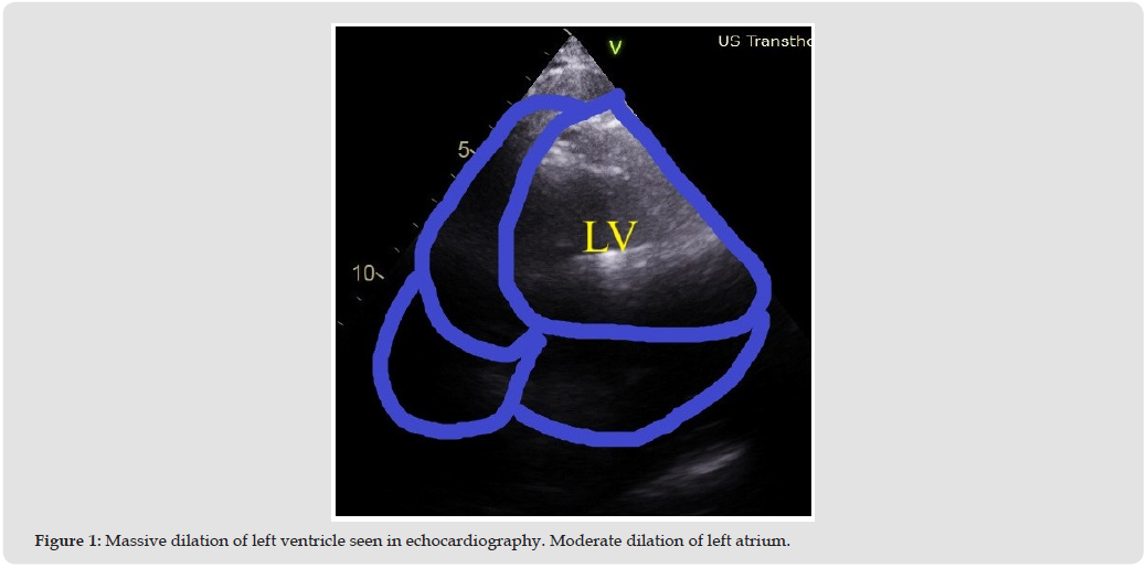
Echocardiography. There is severe global hypokinesis of the left ventricular muscle. Moderately dilated left atrium. Estimated EF = 0-15%.
Ethical Approval and Consent to Participate
Not Applicable.
Consent for Publication
Consent taken.
Availability of Data and Materials
Not Applicable.
Competing Interests
Not Applicable.
Funding
Not Applicable.
Acknowledgement
Not applicable.
Authors’ Contributions
Conceptualization, S.H.K; Methodology, S.H.K; Software, N.G.; Validation, N.A; Formal Analysis, N.A.; Investigation S.H.K& AKC; Resources, N.A.; Data Curation, N.A.; Writing– Original Draft Preparation, S.H.K, AKC, SP; Writing– Review & Editing S.H.K, AKC, & SP; Visualization, S.H.K; Supervision, AR; Project Administration, AR.

International Association of Landscape Archaeology, Czech Glass Society, Czech Republic

Department of Chemistry, Semenov Institute of Chemical Physics, USSR Academy of Sciences, Moscow, Russia

Associate Professor at Department of Breast and Thyorid Surgey, Chongqing General Hospital, China
Professor of Nuclear Medicine, Faculty of Medicine and Surgery, University of Milan, Milan, Italy

Department of Pharmaceutical Sciences, South University School of Pharmacy, USA





 Abstract
 PDF
PDF



























