info@biomedres.us
+1 (502) 904-2126
One Westbrook Corporate Center, Suite 300, Westchester, IL 60154, USA
Site Map
 ISSN: 2574 -1241
ISSN: 2574 -1241
ISSN: 2574 -1241
Received: June 09, 2026; Published: June 25, 2026
*Corresponding author: Anstasiia Mikhailova Plekhanov Russian University оf Economics, 36, Stremyanny per., Moscow,117997, Russia
DOI: 10.26717/BJSTR.2026.66.010276
Using in vitro models, nutraceuticals of different chemical nature (curcumin, berberine, quercetin, and lycopene) were tested for their effectiveness in relation to key processes of metabolic syndrome. For each agent, antioxidant activity (AOA) and the degree of influence on macrophage target genes associated with inflammation were determined. Transcriptomic analysis identified 13 pro-inflammatory and 3 anti-inflammatory genes differentially expressed by each nutraceutical. Multitarget action of nutraceuticals was confirmed; however, using four nutraceuticals as examples, no correlation was found between AOA and inflammation suppression. It is evident that preliminary evaluation of therapeutic potential based solely on AOA is insufficient and should be refined using more complex cellular models.
Keywords: Metabolic Syndrome; Nutraceuticals; In Vitro Models; Inflammation; Antioxidants; Transcriptomic Analysis
Diagnosis, prevention, and treatment of metabolic syndrome (MS) represent a major yet unresolved global issue. Typically, MS includes obesity, hypertriglyceridemia, elevated blood pressure, and hyperglycemia [1]. MS is reasonably considered a driving force of the modern epidemics of diabetes and cardiovascular diseases [2]. To date, the involvement of reactive oxygen species and free radicals in forming oxidative stress in pathological states that constitute MS has been established. In particular, a pronounced imbalance between pro-oxidants and antioxidants is observed, along with accumulation of oxidized proteins and lipids in cells. Increased oxidative activity also leads to alterations in molecular pathways, mitochondrial dysfunction, cell cycle dysregulation, chromosomal aberrations, and destabilization of antioxidant systems [3,4]. Among the main mechanisms underlying MS, chronic inflammation leading to atherosclerosis and diabetes is also highlighted [5]. Macrophages, the largest immune-cell population in adipose tissue, play a key role in inflammation development [6]. MS is characterized by increased expression of many proteins, including pro-inflammatory cytokines such as TNF, IL 1, IL 8, and others that can serve as biomarkers of inflammation [7,8]. Consequently, genes encoding these cytokines may serve as therapeutic targets for conditions associated with MS. Thus, the search for agents possessing both antioxidant and anti-inflammatory properties is relevant for inhibiting MS progression. However, pharmacotherapy of MS, despite its variety, remains insufficiently effective and often associated with side effects [9].
For this reason, researchers have turned their attention to nutraceuticals (Nc), which may become useful tools in combating MS. Unlike monofunctional drugs, nutraceuticals are largely polyfunctional, allowing some authors to consider them promising candidates for therapeutic and preventive strategies targeting MS [10-12].
However, considering either the natural origin or complex composition of nutraceuticals, extensive work is needed to scientifically validate preliminary selection of the most effective agents and refine administration protocols. Evidently, in vitro model experiments provide insights into metabolic pathways and cellular responses to nutraceuticals at the molecular level [13]. Such models are increasingly recognized as predictive for in vivo outcomes. The aim of our study was to develop a methodological model for comprehensive comparative preliminary evaluation of nutraceutical effectiveness, taking into account the information provided in the literature on the molecular and cellular mechanisms of the development of metabolic syndrome.
The experimental samples included nutraceuticals of diverse natural sources and chemical compositions. Commercially available dietary supplements with known active ingredient content were used: the polyphenol curcumin (Cm), the alkaloid berberine (Bb), the flavonoid quercetin (Qc), and the carotenoid lycopene (Lp). The suppliers of these dietary supplements in capsule form were NaturalSupp, Solaray, California Gold Nutrition and GLS Pharmaceuticals, respectively. Capsules were ground and dissolved in dimethyl sulfoxide (DMSO, a standard 10% aqueous solution) to the required concentration. Based on the known supplement composition, all active ingredient concentrations in the extracts were standardized to 100 μg/mL after dissolution in DMSO. Macrophage-like THP 1 cells (MAC) were used as the cellular model. THP-1 cells of monocytic origin, obtained from the collection of vertebrate cell cultures at the Institute of Cytology of the Russian Academy of Sciences (Saint Petersburg), were cultivated in the supplier-recommended growth medium using suspension culture. THP 1 cells were cultivated in suspension in RPMI 1640 medium with 10% FBS and differentiated into MAC with 50 ng/mL PMA (Sigma, USA) for 48 h at 37°C. The exposure time to MAC was the same for all nutraceuticals – incubation after addition to the cell model lasted 24 h.
Antiradical activity was determined spectrophotometrically using 2,2 diphenyl 1 picrylhydrazyl (DPPH, Sigma-Aldrich). Trolox was used as the standard antioxidant [14,15]. For each extract, 0.25 mL was added to 2.85 mL of 5×10⁻⁴ M DPPH solution. after 30 minutes of incubation, optical density values were recorded at a wavelength of 515 nm on a Shimadzu UV-3600 spectrophotometer. Samples were analyzed in triplicate. The control was a working DMSO solution without the test compounds, while a 0.1 M Trolox solution (6-hydroxy- 2,5,7,8-tetramethylchroman-2-carboxylic acid) served as the reference standard (positive control with AOA taken as 100%). AOA level was expressed as a percentage of Trolox.
MAC were pretreated using a standard protocol. The culture medium was replaced with DMEM with 10% lipid-depleted serum. Nutraceutical extracts (100 μg/mL) were added. Control cells (absence of nutraceutical samples in the incubation medium) were obtained under identical conditions. Total RNA was isolated by phenol-chloroform extraction and precipitated with isopropanol. Isolated RNA was treated with Turbo DNase (Life Technologies, USA) to remove trace amounts of genomic DNA, and purified with the RNeasy Mini Kit (Qiagen, Netherlands). RNA samples quality was assessed with the Agilent Bioanalyzer RNA 6000 Nano/Pico (Agilent, USA). To remove ribosomal RNA, the RiboZero Epidemiology kit (Illumina, USA) was used. RNA was fragmented using RNA Fragmentation Reagent (Life Technologies, USA). cDNA libraries were prepared using NEBNext Ultra II Directional RNA Library Prep Kit (NEB, USA) and their quality was analyzed according to the manufacturer’s recommendations. cDNA library sequencing was performed by parallel paired-end sequencing on a HiSeq 2500 sequencer (Illumina, USA). Initial processing of sequencing data was performed using Torrent Suite software (Thermo Fisher, USA). Sequencing quality was assessed using the FASTQC quality assessment program (Babraham Institute, UK). The H. sapiens genome assembly GRCh38 was used as the reference sequence. Reads were mapped using the Tophat2 program [16]. The Htseq-count program was used to count reads [17]. Limma [18] and RankProd [19] methods were used to identify differentially expressed genes. The Limma method allowed identification of genes with statistically significant expression, while RankProd identified genes with increased and decreased expression. To determine the biological significance of the observed changes, differentially expressed transcripts were annotated according to Gene Ontology database categories associated with various biological processes. Statistics. Statistical analysis was performed using GraphPad Prism 8. Student’s t test was used; p < 0.05 was considered significant.
Antioxidant Activity of Nutraceuticals
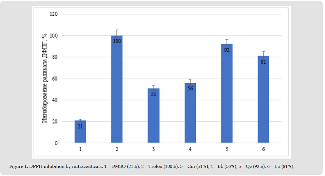
In accordance with the stated objective, at the first stage of the study, antioxidant activity (AOA) of nutraceuticals was determined using a relatively simple and commonly practiced method. All four test objects are known as agents that inhibit oxidative stress; nevertheless, it seemed interesting to compare their AOA under identical conditions at equivalent concentrations of the active ingredient. The Nc solvent served as the negative control, while the positive control was the standard antioxidant Trolox, whose AOA was taken as 100%. The results obtained are presented in Figure 1. Quercetin exhibited the strongest antioxidant activity, followed by lycopene, while berberine and curcumin showed moderate but statistically significant lower AOA.
Figure 1
Effects of Nutraceuticals on Inflammation-Associated Target Genes
The second stage was dedicated to the assessment of the influence of nutraceuticals on inflammatory processes mediated by macrophages, which is a commonly accepted approach in similar studies [20]. It is known that data obtained at the transcriptomic level provide insight into the molecular response of the immune cell system relatively quickly after exposure to the agents under study (24 hours in our experiments). Such information expands and refines understanding of their mechanisms of action. In our experiments, it seemed logical to focus primarily on macrophage target genes encoding both pro-inflammatory and anti-inflammatory cytokines. Transcriptomic analysis results are shown in Table 1.
Table 1: Differentially expressing macrophage genes associated with inflammatory processes, the activity of which significantly decreases (-) or increases (+) under the action of Nc.
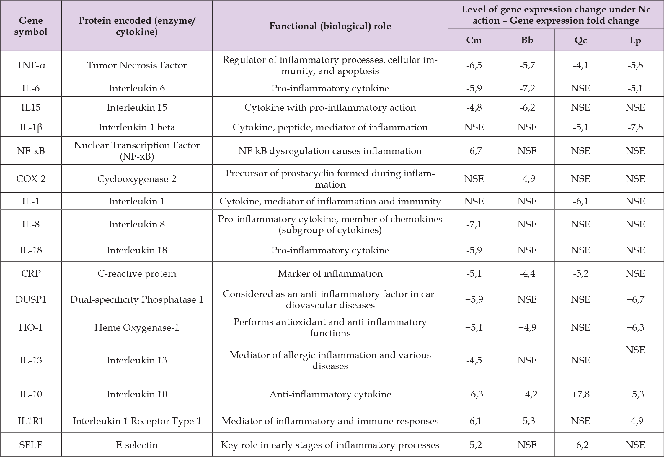
Note: gene expression change levels were calculated using the Limma/Voom method; the control was cells not exposed to Nc; a minus sign (-) indicates a decrease in gene expression of at least 4.0-fold; a plus sign (+) indicates an increase in gene expression greater than 4.0-fold. NSE – no significant effect on gene expression.
Transcriptomic analysis revealed how gene expression in MAC changed under the influence of each Nc. The total number of inflammation- associated genes whose activity significantly increased or decreased under the action of the studied agents was 16. Of these, 3 genes encode anti-inflammatory cytokines and transcription factors, while 13 genes are pro-inflammatory. As expected, all Nc were able to increase the expression level of anti-inflammatory genes and reduce the activity of pro-inflammatory genes, but did so with a fairly high degree of selectivity. Thus, Cm inhibited the largest number of pro-inflammatory genes (10), followed by Bb (6), Qc (5), and Lp (4). For anti-inflammatory genes (increased expression), the following pattern was observed: Cm (3 genes), Lp (3), Bb (2), Qc (1). As can be seen from the table, the sets of target genes significantly affected by Nc action were different; complete overlap in the direction of agent action (ability to reduce expression) was observed only for TNF-α. Overall, Cm possessed the most favorable anti-inflammatory ability to influence target genes (13 genes), while Bb, Lp, and Qc acted on 8, 7, and 6 genes, respectively.
For this study, we selected nutraceuticals of substantially different chemical nature for comparison. The planned experiments were based on the possibility and high probability of combining antioxidant and anti-inflammatory effects in Nc, which was confirmed at the model level. The effects themselves are unlikely to be considered unexpected given the large number of publications on the use of these agents for therapeutic and preventive purposes. At the same time, the literature is dominated by the view that clinical evidence for the use of these Nc has not yet been achieved.
Nevertheless, considering the study objective, attention can be focused on the authors’ attempts to address the following tasks:
• Clarifying mechanisms of nutraceutical action at a molecular- cellular level
• Identifying causal links between antioxidant and anti-inflammatory effects
• Comparing in vitro effectiveness of nutraceuticals
• Evaluating whether simple chemical assays can substitute complex transcriptomic analysis
The nutraceutical with the highest AOA (92%) was quercetin, which suggests a corresponding ability to prevent the development of oxidative stress at the organismal level. This characteristic of Qc fully agrees with data from other authors, e.g., [21]. As it is known, activation of inflammation genes involves numerous proteins, signaling cascades, and transcription factors. Their systemic interaction leads to changes in expression of both pro-inflammatory and anti-inflammatory genes. We found that Qc’s ability to prevent inflammation development is lower than that of Cm and Bb, although Qc is a stronger antioxidant. Overall, several authors [22,23] consider Qc a promising therapeutic agent for MS. Lycopene, as shown by the chemical model, is somewhat inferior (AOA=81%) to Qc but surpasses Bb and Cm in this respect. However, as in the case of Qc, it is inferior to these two Nc in its ability to inhibit inflammation. Nevertheless, Lp attracts attention due to low cytotoxicity, relative affordability, and raw material availability, which stimulates further research on this Nc [24,25]. Bb AOA in our chemical model (56%) was significantly lower than the corresponding value for Qc and Lp, although several authors characterize this Bb capability as fully satisfactory [26]. We demonstrated that Bb inhibits inflammatory processes by reducing expression of six pro-inflammatory genes and increasing expression of two anti-inflammatory genes. These data were fully expected given numerous publications on the immunomodulatory and anti-inflammatory effects of Bb at cellular and organismal levels, including for MS [27-29].
Overall, it seems probable that the mechanisms of Bb action on oxidation and inflammation development are interconnected, affecting both processes within similar timeframes, which precludes determining primacy of either one. Curcumin AOA (51%) is comparable to the corresponding parameter for Bb and substantially lower than the antioxidant activity of Qc and Lp. At the same time, transcriptomic analysis data highlight Cm for the diversity and magnitude of anti-inflammatory effects. Apparently, the direct antioxidant effect of Cm is relatively modest, but its role in the organism as a coordinator of systems opposing MS development is important. Cm possesses the ability to modulate different intracellular signaling pathways, exhibits antioxidant, anti-inflammatory, and immunomodulatory effects, and reduces insulin resistance. Cm produces a hypoglycemic effect by blocking the pancreatic amylase enzyme and stimulating insulin secretion by Langerhans cells [30-31]. As with Bb results evaluation, comparison of importance and primacy (sequence) of Cm action on oxidative stress and inflammation is currently not possible; a close interconnection of these mechanisms and processes is quite probable. Summarizing the obtained data of the study objectives, the following generalized conclusions can be stated. Evaluation of the ability of nutraceuticals to prevent MS development using chemical and cellular models appears undoubtedly feasible and useful.
The obtained results allow not only predicting the magnitude of the expected effect in vivo but also differentiating Nc interaction with different sets of target genes, in this case of the immune system, thereby indicating differences in mechanisms of action of the studied compounds. At the same time, the causal relationship between antioxidant and anti-inflammatory protection effects, and identification of the most critical, initial disruption points, remains unclear, including considering the longer incubation time of Nc with macrophages versus the shorter time with the DPPH reagent. In this regard, the possibility of preliminary evaluation of many Nc using only a fast and inexpensive chemical model remains questionable, and other in vitro testing methods on biological objects remain relevant. Nevertheless, considering the complexity and high cost of in vivo experiments using both experimental animals and clinical trials, the model level of Nc research, including foodomics, remains highly demanded [13,32]. It is evident that within the next few years, the methodological level of Nc effectiveness evaluation will not reach the corresponding level of drug evaluation according to evidence-based medicine criteria. At the same time, it should be taken into account that a large volume of data has already been accumulated on molecular mechanisms of food biologically active substances’ influence on target gene expression [33]. However, interpretation of in vitro transcriptomic data is complicated by the use of cell cultures. Evidently, that cell culture cannot respond to stimuli as occurs in the organism, where cells are exposed to a whole range of factors interacting with each other to trigger some process [34].
The multitarget (polyfunctional) nature of nutraceuticals is, apparently, a necessary condition for their use to ensure therapeutic and preventive effectiveness against metabolic syndrome. The differences discovered in molecular-cellular mechanisms of action of four popular nutraceuticals indicate the possibility of combined application of corresponding substances as functional food ingredients. At the same time, information obtained from preliminary evaluation of nutraceuticals based on a simple chemical model is unlikely to be considered adequate and should be refined by testing on more complex cellular models, which is valid, at least, for the pathology under consideration.
None.
The authors declare no conflicts of interest.

International Association of Landscape Archaeology, Czech Glass Society, Czech Republic

Department of Chemistry, Semenov Institute of Chemical Physics, USSR Academy of Sciences, Moscow, Russia

Department of Educational Sciences, Research Centre Qualities of Teachers, School of Education – HAN, Nijmegen, Netherlands

Associate Professor at Department of Breast and Thyorid Surgey, Chongqing General Hospital, China

Neurology, LA BioMed Research Institute, USA





 Abstract
 PDF
PDF



























