info@biomedres.us
+1 (502) 904-2126
One Westbrook Corporate Center, Suite 300, Westchester, IL 60154, USA
Site Map
 ISSN: 2574 -1241
ISSN: 2574 -1241
ISSN: 2574 -1241
Received: June 09, 2026; Published: June 22, 2026
*Corresponding author: Kwang Nho, SunBio, Inc.129 Eunbong-ro, Namdong-gu, Incheon-si, South Korea
DOI: 10.26717/BJSTR.2026.66.010272
Background: SBX is a novel oxygen-delivery biomaterial with potential neuroprotective effects in ischemic
stroke.
Objective: To evaluate the efficacy of SBX in a rat middle cerebral artery occlusion (MCAO) model, in which SBX
is administered intravenously 30 minutes prior to induction of ischemia.
Methods: Male Sprague-Dawley rats received saline, SBX 5 mL/kg, or SBX 10 mL/kg intravenously 30 minutes
before permanent MCAO. Neurological deficits (mNSS) and infarct volume (TTC staining) were assessed at 24
hours.
Results: High-dose SBX reduced infarct volume versus control (48.17 ± 13.54% vs. 58.91 ± 9.02%, p = 0.044),
while low-dose showed less significant effect. No improvement in neurological function was observed.
Conclusion: SBX provided histological neuroprotection without functional benefit. Further studies are needed
to assess mechanism, safety, and translational potential.
Keywords: Hemoglobin-Based Oxygen Carriers (HBOCs); PEGylated Hemoglobin; PEG-Hb; MCAO; Ischemic Stroke
Abbreviations: SD: Sprague-Dawley; SPF: Specific Pathogen-Free; PEG: Polyethylene Glycol; MCAO: Middle Cerebral Artery Occlusion; CBF: Cerebral Blood Flow; TTC: Triphenyltetrazolium Chloride; PFA: Paraformaldehyde; LSD: Least Significant Difference; mNSS: Modified Neurological Severity Score; MCAO: Middle Cerebral Artery Occlusion; HBOCs: Hemoglobin-Based Oxygen Carriers
Stroke is a leading cause of mortality and long-term disability worldwide, with ischemic stroke accounting for approximately 85% of all cases. It results from an interruption of cerebral blood flow, leading to oxygen and glucose deprivation, rapid neuronal injury, and subsequent neurological deficits. Despite advances in acute stroke care, therapeutic options remain limited. Tissue plasminogen activator (tPA) is currently the only widely approved pharmacological treatment; however, its clinical utility is constrained by a narrow therapeutic window and risk of hemorrhagic complications. Consequently, there remains a critical need for novel therapeutic approaches that can protect brain tissue during ischemia and extend the therapeutic window [1,2].
Preclinical investigation of stroke therapies commonly relies on animal models that replicate the pathophysiology of human ischemic stroke. Among these, the middle cerebral artery occlusion (MCAO) model is the most widely used and well-validated [3,4]. It closely mimics focal cerebral ischemia observed in humans, as the MCA territory accounts for the majority of ischemic strokes, and enables evaluation of both histological and functional outcomes. Both permanent and transient MCAO models are widely used to assess infarct development, reperfusion injury, and neurological deficits [3,5]. A promising therapeutic strategy for ischemic stroke is the use of oxygen carriers to enhance oxygen delivery to hypoxic brain tissue. Hemoglobin- based oxygen carriers (HBOCs) have been extensively investigated as artificial oxygen therapeutics due to their ability to transport and release oxygen independently of red blood cells.
Their smaller size compared to erythrocytes allows them to perfuse the microvasculature and deliver oxygen to ischemic regions, particularly within the penumbra where tissue viability is oxygen-dependent [6]. Several generations of HBOCs have been evaluated in experimental stroke models. Early studies using cross-linked and polymerized hemoglobin formulations demonstrated reductions in infarct volume; however, these effects were often accompanied by vasoconstriction due to nitric oxide scavenging and other safety concerns [7]. Subsequent modifications, including PEGylation, improved pharmacokinetics and reduced vasoactivity. PEGylated hemoglobin derivatives have been shown to improve cerebral perfusion and reduce infarct size in MCAO models, partly by enhancing oxygen delivery and preserving collateral circulation [8]. More recent approaches include hemoglobin-based vesicles and albumin-bound hemoglobin constructs such as HemoAct, which demonstrated neuroprotective effects in transient MCAO models through improved oxygen transport and reduced oxidative injury [9].
Additionally, nanostructured HBOCs, including stroma-free hemoglobin nanoparticles, have shown promising results with reduced infarct volume, decreased oxidative stress, and preservation of blood– brain barrier integrity in preclinical stroke models [10]. The effects of carboxyhemoglobin-based HBOC transfusion were evaluated in a rat transient MCAO model. Treatment reduced infarct volume and helped preserve cerebral blood flow, likely through enhanced oxygen delivery and maintenance of vasodilation. The carboxy state was suggested to attenuate vasoconstrictive effects and improve microvascular perfusion [11]. SBX is a novel oxygen-delivery biomaterial designed to enhance oxygen transport under ischemic conditions. Unlike traditional HBOCs, SBX aims to improve oxygen delivery while minimizing the adverse effects associated with earlier hemoglobin-based systems. The present study aimed to evaluate the preclinical efficacy of SBX in a rat permanent MCAO model of ischemic stroke, focusing on its ability to reduce infarct volume and improve neurological outcomes following pre-treatment administration.
This study was designed to determine whether systemic preloading with an oxygen carrier prior to complete arterial occlusion can enhance oxygen availability during the early phase of ischemia and thereby reduce subsequent brain injury. In contrast to most previous experimental stroke studies involving oxygen carriers, which administer the test material after vascular occlusion or during reperfusion, the present study evaluates the effect of pre-ischemic systemic administration of SBX on outcomes following permanent middle cerebral artery occlusion (MCAO). Accordingly, this approach represents a novel prophylactic or pre-ischemic oxygen-carrier paradigm and should be interpreted as a high-risk prevention or preconditioning model rather than a conventional acute stroke treatment strategy.
Approval of Animal Study Protocol
All animal experiments conducted in this study were approved by the Institutional Animal Care and Use Committee (IACUC) of Hanyang University in Seoul, South Korea.
Experimental Animals
Male Sprague-Dawley (SD) rats were obtained from Koatech in Gyeonggi-do, South Korea. All experiments were performed at the Hanyang University ERICA Animal Facility under specific pathogen-free (SPF) conditions. The animals were acclimated for one week prior to the experiments. After acclimation, rats weighing between 240 and 280 g were selected for the study. The animals were randomly assigned to experimental groups, and all procedures, including drug administration, surgical intervention, behavioral assessment, and histological evaluation, were performed under blinded conditions. Regarding anesthesia, animals were anesthetized with isoflurane (2.5–3.0% for both induction and maintenance) administered via an inhalation chamber followed by a nose cone. Anesthetic depth was verified by the absence of the pedal withdrawal reflex. For euthanasia, following completion of behavioral assessments, rats were deeply anesthetized with 3% isoflurane. After confirming loss of consciousness and absence of pain reflexes, euthanasia was performed by exsanguination. Tissues were immediately harvested postmortem and processed for subsequent histological analysis.
Preparation of SBX
SBX is a polyethylene glycol (PEG)-modified hemoglobin-based oxygen carrier (PEG-Hb solution) developed for intravenous administration. It is derived from bovine hemoglobin that has been covalently conjugated with multiple PEG molecules, resulting in a final average molecular weight of approximately 137 kDa. SBX is formulated at physiological pH (7.2) in a buffered solution containing sodium chloride and sodium phosphate. The final formulation has a total solute concentration of approximately 9.4 g/dL, of which hemoglobin accounts for 4.4 g/dL, with the remainder contributed by PEG. The viscosity of the solution at 37°C is approximately 8–9 centipoise.
Experimental Groups
The animals were divided into three groups: a control group receiving saline (10 mL/kg), a low-dose SBX group (5 mL/kg), and a high-dose SBX group (10 mL/kg). Each group included 15 animals for final evaluation. Statistical analyses were conducted based on these group sizes.
Administration of Test Material Prior to MCAO
To induce ischemic stroke, animals were randomly assigned to groups and administered either saline or SBX via the tail vein at a rate of 1 mL/min. Middle cerebral artery occlusion (MCAO) was performed 30 minutes after administration to induce ischemia. All administrations were carried out in a blinded manner by a researcher who was not involved in the surgical procedures.
Induction of Ischemic Stroke Using Middle Cerebral Artery Occlusion (MCAO)
Ischemic stroke was induced using the well-established middle cerebral artery occlusion (MCAO) model, which is widely used in preclinical studies. A permanent occlusion model, designed to induce irreversible ischemic damage, was employed in this study. Prior to surgery, the body weight of each animal was measured. Anesthesia was induced and maintained using 2.5–3% isoflurane delivered via inhalation. The scalp area for cerebral blood flow measurement and the neck region for surgical incision was shaved and disinfected with 70% ethanol. The animals were then positioned on a surgical table and maintained under continuous inhalation anesthesia throughout the procedure. Surgery was performed on a temperature-controlled heating pad maintained at 37°C, and body temperature was recorded before and after the operation. After exposing the bregma at the top of the skull, cerebral blood flow (CBF) was measured using a laser Doppler flowmeter. Measurements were obtained both before and after MCAO induction for comparison. The animal designated for occlusion was placed in a supine position, and the surgical site was disinfected with 70% ethanol. A careful incision was made to avoid damage to subcutaneous tissues and blood vessels. The left common carotid artery was exposed, and the external and internal carotid arteries were carefully separated from surrounding tissues, including muscle, blood vessels, and fat.
The common carotid artery and external carotid artery were permanently ligated using sterile 6-0 surgical sutures. A small incision was then made in the common carotid artery to allow insertion of the occlusion suture. A silicone-coated occlusion suture (Doccol Co.) was inserted into the common carotid artery and advanced into the internal carotid artery to induce occlusion of the middle cerebral artery (see Figure 5). The insertion length was standardized by marking 20 mm from the tip of the suture, ensuring consistent placement at the internal carotid branch across all animals. Following insertion, the internal carotid artery was permanently ligated using sterile 6-0 sutures, and the surgical site was closed using 3-0 sutures. Cerebral blood flow was measured again after occlusion, and a reduction to less than 30% of baseline was considered successful induction of ischemia.
After completion of the surgery, anesthesia was discontinued, and the animals were monitored until they regained consciousness and were able to move freely. The animals were then returned to their cages and maintained for 24 hours.
Behavioral Assessment
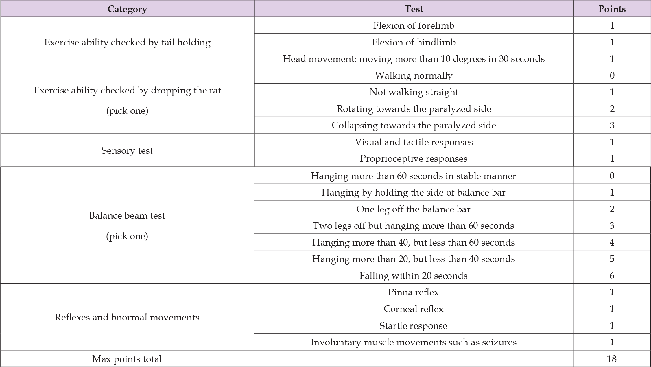
Behavioral evaluation was performed 24 hours after MCAO surgery to assess the degree of functional impairment caused by ischemia. Neurological function was assessed using the modified Neurological Severity Score (mNSS), a widely established and internationally accepted evaluation system. The mNSS has a maximum score of 18 points, with higher scores indicating more severe neurological impairment. All behavioral assessments were conducted in a blinded manner, such that the evaluator was unaware of the treatment group assigned to each animal (Table 1).
Table 1: Behavioral Test Chart.
Brain Tissue Isolation and Histological Analysis
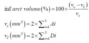
After completion of the behavioral assessment, the rats were anesthetized and euthanized by exsanguination, after which histological evaluation was performed. The brains were carefully removed from the skull and sectioned at 2 mm intervals. The sections were subsequently stained using 2,3,5-triphenyltetrazolium chloride (TTC) solution. TTC staining differentiates viable and infarcted tissue, with viable tissue appearing red due to enzymatic activity, while infarcted (damaged) tissue remains pale or white. Staining was performed for 10 minutes at 37°C under light-protected conditions. Following staining, the brain sections were fixed in 4% paraformaldehyde (PFA) for 24 hours. The fixed sections were then arranged and scanned for analysis. The scanned images were analyzed using ImageJ software to quantify the areas of normal and damaged tissue. The infarct region (white area) was calculated and expressed as infarct volume (%), which served as the primary histological endpoint. All image analyses were conducted under blinded conditions. Infarct volume was calculated using a formula that corrects for edema associated with ischemic stroke, based on area measurements obtained from each brain slice. Each slice had a thickness of 2 mm, and a total of five slices were used to determine the overall infarct volume. The parameters used in the calculation were defined as follows: A (mm2), contralateral hemisphere area; B (mm2), ipsilateral hemisphere area; C (mm2), infarct area; and D (mm2), intact ipsilateral hemisphere area (B − C) (Figure 1).
Figure 1
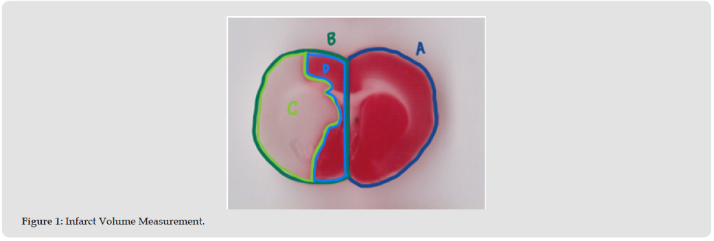
A (mm2): Contralateral Hemisphere
B (mm2): Ipsilateral Hemisphere
C (mm2): Infarct
D (mm2): Intact ipsilateral hemisphere (=B-C)
Statistical Analysis
Data including cerebral blood flow (CBF) changes, histological outcomes (infarct volume), and behavioral scores (mNSS) were collected and used for statistical analysis. All statistical analyses were performed using SPSS software. Comparisons between the control group and the treatment groups (low-dose and high-dose SBX) were conducted using one-way analysis of variance (ANOVA) followed by the least significant difference (LSD) post-hoc test. A p-value of less than 0.05 was considered statistically significant.
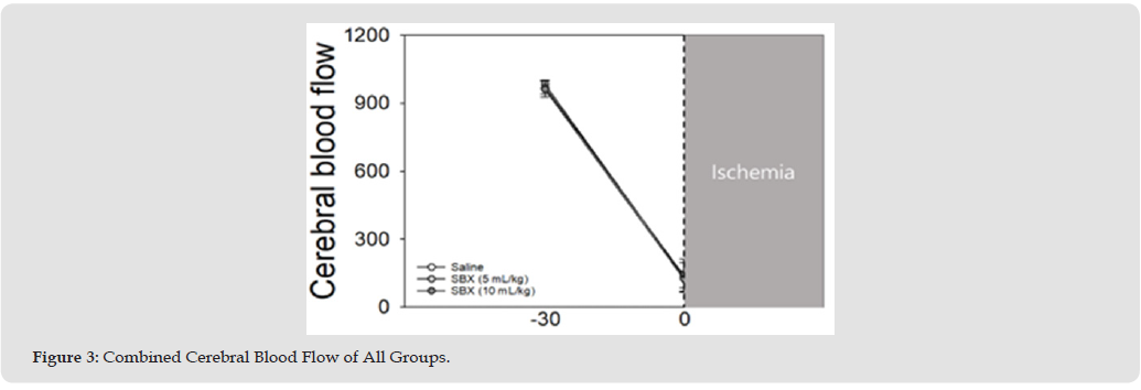
Changes in Cerebral Blood Flow by Group Changes in cerebral blood flow (CBF) were evaluated across all groups, including the control, low-dose SBX (5 mL/kg), and high-dose SBX (10 mL/kg) groups. Prior to MCAO, the mean CBF values were 984.0 ± 17.7 in the control group, 971.5 ± 30.2 in the low-dose group, and 962.0 ± 34.6 in the high-dose group (mean ± standard deviation). Following MCAO, CBF was markedly reduced in all groups due to arterial occlusion. The post-occlusion CBF values were 133.2 ± 63.4 in the control group, 137.6 ± 74.6 in the low-dose group, and 124.3 ± 37.6 in the high-dose group. No significant differences in CBF were observed among the groups either before or after MCAO, indicating that a comparable level of ischemia was successfully induced across all experimental conditions (Figures 2 & 3).
Figure 2
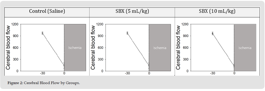
Figure 3
Changes in Histological Damage by Group
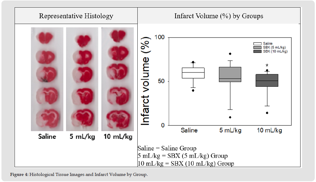
Histological damage was assessed using representative tissue images and a group comparison analysis. Statistical evaluation showed that the low-dose SBX group (5 mL/kg) did not exhibit a significant difference compared to the saline control group (p = 0.217), whereas the high-dose SBX group (10 mL/kg) demonstrated a statistically significant difference (p = 0.044). Following isolation of brain tissues from each animal, the infarcted regions were measured to determine the extent of ischemic brain injury. The infarct volume (%) was calculated as follows: 58.91 ± 9.02 in the control group, 52.42 ± 18.43 in the SBX low-dose group (5 mL/kg), and 48.17 ± 13.54 in the SBX high-dose group (10 mL/kg) (mean ± standard deviation). Based on these data, the SBX low-dose group showed an 11% reduction compared to the control group, while the SBX high-dose group showed an 18% reduction in infarct volume relative to the control group. Statistical analysis using one-way ANOVA confirmed that the reduction in infarct volume was not significant in the low-dose group (p = 0.217), whereas a significant reduction was observed in the high-dose group (p = 0.044). These findings indicate that administration of high-dose SBX (10 mL/kg) resulted in a significant decrease in ischemic brain damage compared to the control group (Figure 4).
Figure 4
Changes in Behavioral Impairment by Group
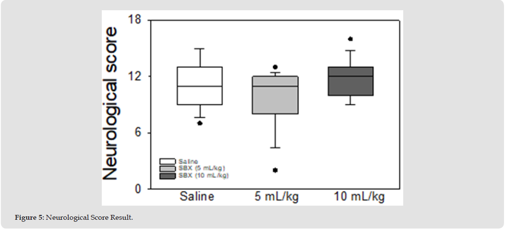
Behavioral impairment following MCAO-induced ischemia was evaluated using the modified Neurological Severity Score (mNSS) across all dose groups. The mean mNSS scores were 11.0 ± 2.42 in the control group, 9.67 ± 3.04 in the low-dose SBX group (5 mL/kg), and 11.67 ± 1.88 in the high-dose SBX group (10 mL/kg) (mean ± standard deviation). These results indicate that SBX treatment did not result in a significant improvement in behavioral outcomes in either the low-dose or high-dose groups (Figure 5).
Figure 5
In the present study, SBX was administered prior to induction of ischemia to evaluate whether systemic preloading with an oxygen carrier could mitigate brain injury following permanent middle cerebral artery occlusion (MCAO). This pre-infusion strategy differs fundamentally from most previous studies of hemoglobin-based oxygen carriers (HBOCs), which have primarily focused on post-ischemic or peri-reperfusion administration paradigms. The rationale for this design was to determine whether the presence of an oxygen carrier in circulation at the onset of vascular occlusion could enhance oxygen availability during the critical early phase of ischemia, when residual collateral circulation may still support partially perfused tissue. Mechanistically, pre-ischemic administration of SBX may facilitate more efficient oxygen delivery through compromised microvasculature and leptomeningeal collateral pathways, thereby delaying the progression of the ischemic penumbra to irreversible infarction. This concept is supported by the observed reduction in infarct volume in the high-dose SBX group, indicating that enhanced oxygen delivery can attenuate tissue damage even in the absence of reperfusion.
However, this histological protection did not translate into significant improvement in neurological function, which may reflect the limitations of the permanent MCAO model. In this model, the absence of reperfusion restricts restoration of bulk blood flow, thereby limiting the potential for functional recovery despite partial preservation of tissue viability. Previous HBOC studies have largely demonstrated beneficial effects when oxygen carriers are administered after ischemic onset, particularly in transient MCAO models where reperfusion occurs. In such settings, HBOCs have been shown to improve cerebral perfusion, reduce infarct size, and, in some cases, enhance functional recovery by augmenting oxygen delivery and maintaining microvascular flow. For example, carboxyhemoglobin-based HBOCs have been reported to preserve cerebral blood flow and reduce infarct volume through improved oxygen transport and reduced vasoconstriction. Compared to these studies, the present work evaluates a distinct therapeutic window, highlighting the importance of treatment timing in determining the extent and nature of neuroprotection. From a clinical perspective, the pre-infusion approach explored in this study represents a novel and less conventional paradigm. While most stroke therapies are administered after symptom onset, pre-ischemic administration of an oxygen carrier may be relevant for high-risk populations, such as patients with severe carotid or intracranial stenosis, recurrent transient ischemic attacks, or those undergoing surgical or interventional procedures associated with increased stroke risk. In these scenarios, prophylactic or pre-emptive oxygen delivery could potentially buffer the effects of sudden vascular occlusion and preserve tissue viability during the early ischemic phase. Thus, SBX may have utility not only as an acute therapeutic agent but also as a preventive or preconditioning strategy. Nevertheless, several limitations should be considered.
The use of a permanent MCAO model may underestimate the potential functional benefits of SBX, as reperfusion is a key determinant of neurological recovery in clinical stroke. Additionally, the lack of behavioral improvement despite reduced infarct volume suggests that further investigation is needed to clarify the relationship between tissue-level protection and functional outcomes. Future studies incorporating transient MCAO or large-vessel occlusion models with reperfusion, as well as detailed evaluation of cerebral oxygenation, microcirculation, and underlying mechanisms, will be important to fully define the therapeutic potential of SBX.
In this study, SBX, developed by SunBio, was administered intravenously 30 minutes prior to stroke induction to evaluate its efficacy in mitigating ischemic brain injury. Histopathological analysis revealed that ischemic damage induced by MCAO was significantly reduced in the high-dose SBX group (10 mL/kg). Specifically, the lowdose group showed an 11% reduction in infarct volume compared to the control group, while the high-dose group exhibited an 18% reduction. However, despite these histological improvements, behavioral impairments resulting from MCAO were not significantly improved in either treatment group. This discrepancy between reduced tissue damage and the lack of functional recovery, along with the underlying mechanisms, warrants further investigation through additional biochemical analyses and evaluation in alternative ischemic stroke models, such as the transient MCAO model. In conclusion, pre-ischemic administration of SBX, an oxygen carrier, can attenuate histological brain injury following permanent cerebral ischemia, supporting its potential as a prophylactic or preconditioning strategy. These findings underscore the importance of treatment timing and suggest that oxygen carriers like SBX may play distinct and complementary roles in both preventive and acute stroke management.
No funding was received.
No potential conflict of interest was reported by the authors.
The authors are research scientists employed by SunBio Inc., a biopharmaceutical company in South Korea.
K. Nho conceived the project and designed the experiments. M.A., B.S., S.H., Y.K., J.L., and C.H. conducted syntheses of the materials and physicochemical measurements. All authors participated in analyses and confirmed data. K. Nho wrote the manuscript.

International Association of Landscape Archaeology, Czech Glass Society, Czech Republic

Department of Chemistry, Semenov Institute of Chemical Physics, USSR Academy of Sciences, Moscow, Russia

Department of Educational Sciences, Research Centre Qualities of Teachers, School of Education – HAN, Nijmegen, Netherlands

Associate Professor at Department of Breast and Thyorid Surgey, Chongqing General Hospital, China

Neurology, LA BioMed Research Institute, USA





 Abstract
 PDF
PDF



























