info@biomedres.us
+1 (502) 904-2126
One Westbrook Corporate Center, Suite 300, Westchester, IL 60154, USA
Site Map
 ISSN: 2574 -1241
ISSN: 2574 -1241
ISSN: 2574 -1241
Received: April 29, 2026; Published: May 07, 2026
*Corresponding author: Stojarov AN, Department of Radiation Medicine and Ecology, Belarusian State Medical University, Minsk, 220116, Minsk, pr. Dzerzhinskogo 83, Belarus
DOI: 10.26717/BJSTR.2026.65.010211
The study analyzed the spectral properties of serum albumin from individuals exposed in utero to radioactive iodine in their thyroid glands following the 1986 Chernobyl disaster. Protein from unirradiated children served as a control. It was shown that, based on its spectral characteristics (fluorescence peak, fluorescence quantum yield, etc.), serum albumin from irradiated individuals has a slightly different tertiary structure organization. This may explain its reduced bilirubin retention capacity, its passage through the blood-brain barrier, and its damage. The authors believe that such changes in albumin transport function may be the result of a mutation in its gene due to exposure to ionizing radiation.
Keywords: Ionizing Radiation; Serum Albumin; Protein Conformation; Spectral Methods of Research; Tryptophan; in Utero Irradiation; Bilirubin; Brain; Chernobyl Disaster
Abbreviations: ICD-10: International Statistical Classification of Diseases and Related Health Problems 10th Revision; PAGE: Polyacrylamide Gel; DS-Na: Sodium Dodecyl Sulfate SDS-PAGE: Sodium Dodecyl Sulfate–Polyacrylamide Gel Electrophoresis; DNA: Deoxyribonucleic Acid
As is well known, the issue of prenatal exposure is a priority in studying the medical consequences of the Chernobyl nuclear power plant disaster. It has been the subject of numerous studies, both experimental and clinical [1]. Intrauterine irradiation manifests itself in various symptoms: mental retardation, delayed physical development, epilepsy, seizures, decreased mental development, etc. Various mechanisms for the development of these phenomena have been proposed; however, in this publication, we propose a different mechanism for the development of this pathology during radiation exposure.
Previously we have shown that children born to women irradiated with radioactive iodine during pregnancy experience characteristic changes in their health. First, this concerned changes in the incidence of nervous system diseases. The cumulative incidence of morbidity in children irradiated in utero during the observation period from 1986 to 2016 significantly exceeded that of children who were not exposed to radiation [2]. Secondly, there were striking differences in the incidence of mental and behavioral disorders (Chapter V, ICD-10). In the group of children irradiated in utero with I-131, a linear increase in the incidence rate was observed in subsequent years, almost three times higher than in the group of unirradiated individuals. Moreover, in the latter group, the incidence rate tended to plateau after 2001 [3].
Analyzing a group of in utero-irradiated children from the Stolin district of the Brest region (Belarus), which was exposed to the radioactive cloud from the Chernobyl nuclear power plant disaster, we observed an increased concentration of indirect, i.e., free, bilirubin in the blood serum of these children, while the level of bound bilirubin remained constant [4]. This may be due to various factors, including a disrupted mechanism for bilirubin transport by serum albumin. Bilirubin is a toxic compound capable of passing through the blood-brain barrier and causing central nervous system dysfunction. Therefore, we hypothesized that prenatal brain damage may be caused by genetic changes in the structure of serum albumin, a decrease in its bilirubin- binding capacity, which led to the release of this component into the brain, altering its functions. Therefore we analyzed the conformational features of this serum protein in irradiated and non-irradiated children.
The main group of individuals was comprised of residents of the Stolin district of the Brest region, born to women who lived there in late April–early May 1986 and were exposed to a radioactive cloud that passed through this region of Belarus. The cloud contained iodine radionuclides, including I-131. The women absorbed it through inhalation, ingestion with food, and through the skin. Once inside the body, it was distributed, accumulated in the thyroid gland, and passed through the transplacental barrier into the fetus, concentrating in its thyroid gland. The main cohort included 123 individuals, including 62 women and 61 men. Birth dates ranged from June 3, 1986, to February 6, 1987. The average absorbed dose to the thyroid gland was 32.2±3.31, median 22 mGy, in males - 35.4±5.4, median 23 mGy, in females - 29.1±3.9, median 22 mGy.
When analyzing the impact of radiation on specific population groups, it is always necessary to include a comparison group consisting of individuals who were not exposed to radiation. In this study, the comparison group also included residents of the Stolin district of the Brest region, but those born later. In other words, their mothers were not exposed to the “iodine shock”. It included 121 individuals from the same district, identical not only in residence but also in social status, including 60 males and 61 females. Their dates of birth ranged from January 3, 1988, to December 31, 1988. The comparison group was selected based on the half-life of I-131, which is approximately 8 days. Over 10 half-lives, i.e., after 80 days, only trace amounts of radioactive iodine remained in the environment, and therefore, the mothers of these children did not receive thyroid radiation during pregnancy.
Serum albumin was isolated and purified from blood serum using a three-step method. In the first step, the serum was desalted by loading it onto a Sephadex G-25 column equilibrated with a solution consisting of 35 mM NaCl and 25 mM Tris-HCl, pH 8.8. The eluate was collected and used for subsequent chromatography on DEAE-Sephadex G-25, equilibrated with the same buffer. Elution was continued until immunoglobulin G was released. Proteins adsorbed to the matrix were washed off with a solution of 0.6 M NaCl and 25 mM Tris- HCl, pH 8.8, and used for the third chromatography step. For this, the collected eluate was loaded onto a blue agarose column equilibrated with the above-mentioned solution. Components unbound to the affinity matrix were washed away with a solution of 0.6 M NaCl, Tris-HCl, pH 8.8, and the albumin fraction was eluted with 3 M NaCl, Tris-HCl, pH 8.8. The protein was concentrated and washed to remove salts by ultrafiltration under pressure using PM-10 filters.
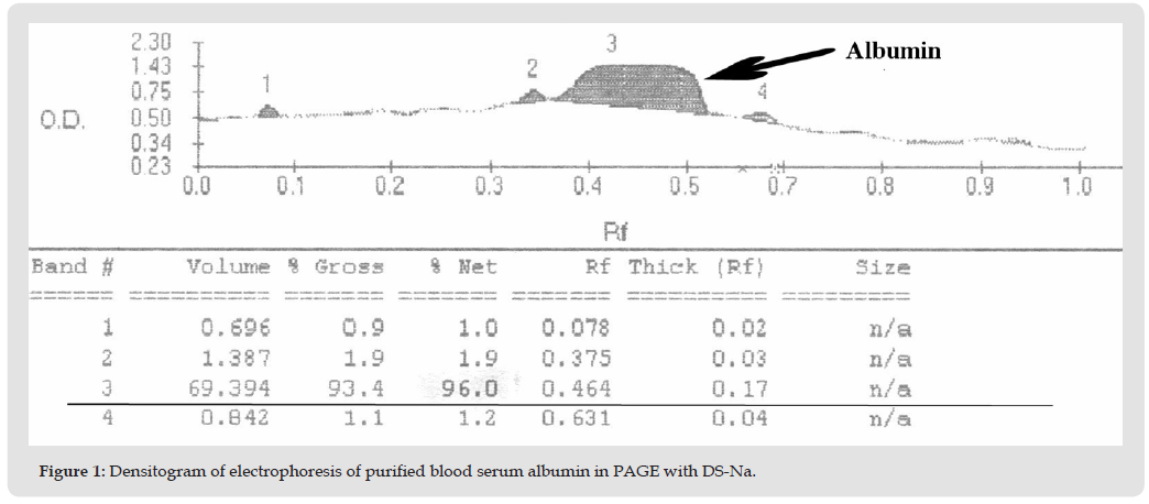
The homogeneity of the resulting protein preparations was monitored using disk electrophoresis in polyacrylamide gel (PAGE) plates with sodium dodecyl sulfate (DS-Na). The homogeneity of the resulting protein preparations was monitored using SDS-PAGE. The purity of the preparation was at least 96% (Figure 1). Luminescence spectra and luminescence excitation spectra of serum albumin were recorded at excitation wavelengths of 280 and 296 nm using an SFL 1211A spectrofluorimeter (SOLAR, Belarus). Files with the (.txt) extension were used for further spectral analysis using the GraphPad Prism and ORIGIN19 software programs. Spectra were processed using nonlinear regression in the wavelength ranges of 300-500 nm (for an excitation wavelength of 280 nm), 315-500 nm (for an excitation wavelength of 296 nm), and 270-305 nm (fluorescence excitation spectra). The correlation coefficient (R), the χ2 criterion, and the absolute sum of squared deviations served as criteria for determining the approximation of the calculated curve to the experimental one. Statistical data processing was performed using the software programs Statistika 10.0 (StatSoft.Inc, USA) and SigmaPlot 12.5 (Systat Software Inc., Germany).
Figure 1
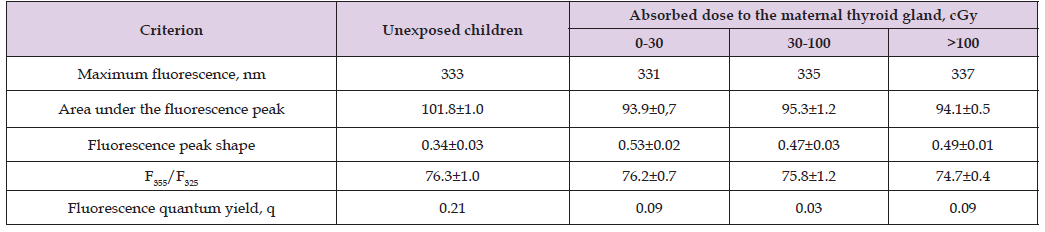
The study showed that intrauterine irradiation of a child altered some spectral parameters of the protein, which consisted primarily of a Stokes shift of the luminescence maximum, a decrease in the area under the spectral curve, and a decrease in the spectral half-width (Figure 2, Table 1). Such changes may be due to the transfer of the tryptophan residue to a more hydrophilic microenvironment. Calculation of the quantum yield (q) in isolated and purified albumins revealed a relatively high value for the control protein (0.21), which is consistent with previously obtained data and lower values for this value in the group of proteins isolated from the blood of irradiated children. Low q values may also indicate the presence of tryptophanyls in an aqueous microenvironment, which confirms previously presented data. We have previously shown that an increase in the value of this parameter may reflect a decrease in the probability of electron transitions with energy values close to the average. This process may depend on various factors: the microenvironment of the chromophore, its fixation in the macromolecular organization of the protein, the possibility of relaxation, etc. Lower values of this indicator in albumins obtained from children irradiated in utero may indicate a slightly different structural organization of the protein.
Figure 2
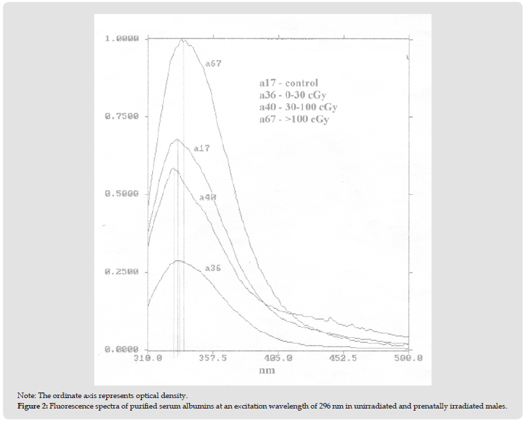
Table 1: Spectral parameters of fluorescence at 296 nm of serum albumin of unexposed and irradiated in utero children with I-131 depending on the absorbed dose to the thyroid gland.
Human serum albumin is known to contain a single tryptophan residue at position 214 of the polypeptide chain, and its spectral characteristics are sensitive to changes in the conformational state of this molecule. Conversely, genetically determined polymorphisms of serum albumin are known, resulting from mutations in the albumin gene and resulting in changes in the primary structure of the protein. To date, more than 55 molecular variants of albumin have been identified, most of which are caused by substitutions of monoaminodicarboxylic acids (Glu, Asp) in three domains of this molecule [5]. Ionizing radiation is well known to induce DNA defects, which can cause subsequent mutations. Calculated data indicate that external irradiation can modulate mutations in five protein variants that were recorded in victims of the Hiroshima and Nagasaki bombings, changes in which do not affect the region of the macromolecule that binds bilirubin [6]. However, the binding site for this pigment may be the arginine residues Arg222 and Arg218, which are located in close proximity to the aforementioned tryptophanyl-214 [6,7]. This may explain the characteristics observed in our spectral studies. In this case, mutations in these amino acids may affect bilirubin binding [8]. If this is indeed the case, then another mechanism for brain damage after intrauterine irradiation can be proposed, associated with a mutation in human chromosome 4, a change in protein conformation, a decrease in the ability to bind bilirubin, and its release into brain structures, leading to their damage. Moreover, in this case, a different type of radiation exposure occurred, different from the Japanese one: the incorporation of iodine with changes in the function of the thyroid gland, the hormones of which are capable of changing the functions of various body systems.
Serum albumin from children irradiated in utero differs from that of non-irradiated individuals in a number of parameters, as determined by spectral analysis. These changes include alterations in its tertiary structure, which may affect the protein’s transport functions, including bilirubin binding. Altered bilirubin binding capacity may affect its passage across the blood-brain barrier, leading to brain damage.

International Association of Landscape Archaeology, Czech Glass Society, Czech Republic

Department of Chemistry, Semenov Institute of Chemical Physics, USSR Academy of Sciences, Moscow, Russia

Department of Educational Sciences, Research Centre Qualities of Teachers, School of Education – HAN, Nijmegen, Netherlands

Associate Professor at Department of Breast and Thyorid Surgey, Chongqing General Hospital, China

Neurology, LA BioMed Research Institute, USA





 Abstract
 PDF
PDF



























