info@biomedres.us
+1 (502) 904-2126
One Westbrook Corporate Center, Suite 300, Westchester, IL 60154, USA
Site Map
 ISSN: 2574 -1241
ISSN: 2574 -1241
ISSN: 2574 -1241
Received: March 29, 2026; Published: May 07, 2026
*Corresponding author: Univ.-Prof. Dr. med. habil. Wolf D Grimm, Periodontology, Department of Dental School, Faculty of Health, University of Witten/Herdecke, Germany
DOI: 10.26717/BJSTR.2026.65.010175
Background
Immediate implant placement in the maxillary premolar region remains clinically challenging due to post-extraction ridge remodelling and soft tissue instability. Autologous platelet concentrates such as platelet-rich fibrin (PRF) and platelet-rich plasma (PRP) have been proposed to enhance early wound healing and support regenerative processes.
Objective
To present a reproducible, biologically driven clinical protocol for immediate implant placement with PRF/ PRP-assisted augmentation.
Methods
A structured clinical workflow is described in a 65-year-old female patient requiring extraction of an endodontically compromised maxillary first premolar. Following atraumatic extraction, a digitally guided immediate implant placement (4 × 11 mm) was performed. Simultaneous augmentation included autogenous bone, bovine- derived xenogeneic graft material (Bio-Oss®), and autologous PRF/PRP prepared using a standardized separation protocol. Primary stability of 45 Ncm was achieved. Soft tissue management and graft stabilization were supported by autologous fibrin matrices.
Results
Healing was uneventful, and prosthetic rehabilitation with a screw-retained crown was completed after 3 months. Clinical and radiographic outcomes demonstrated stable peri-implant bone conditions and favorable soft tissue integration. The combined use of PRF/PRP contributed to enhanced wound healing and graft stabilization without postoperative complications.
Conclusion
Immediate implant placement combined with PRF/PRP-assisted augmentation represents a biologically sound and clinically feasible approach in selected cases. The presented protocol supports predictable hard- and soft-tissue outcomes while potentially reducing the need for additional regenerative procedures. Further controlled clinical studies are required to validate these findings.
Keywords: Dental Implants; Platelet-Rich Fibrin; Immediate Implant Placement; Guided Bone Regeneration; Autologous Blood Concentrates: Digital Implant Planning; Case Report
Digitally guided immediate implant placement combined with autologous platelet-rich fibrin (PRF)-assisted augmentation in the maxillary premolar region resulted in predictable hard- and soft-tissue healing. A systemically healthy patient underwent atraumatic extraction followed by immediate implant placement. Simultaneous regeneration was achieved using autogenous bone, xenogeneic particulate graft material, and liquid PRF stabilized with an autologous fibrin membrane. Digital implant planning enabled prosthetically driven positioning and primary stability. Final restoration demonstrated stable peri-implant tissues, preserved crestal bone levels, and favorable esthetic outcomes without biological or technical complications.
Key Concept
Immediate implantation + PRF-assisted biologic augmentation → accelerated healing, graft stabilization, and predictable clinical outcomes.
• Scientific Rationale: PRF delivers autologous growth factors and fibrin scaffolding that may enhance wound healing and graft integration during immediate implant placement
• Principal Findings: Immediate implantation combined with PRF-assisted augmentation resulted in stable peri-implant tissues and successful osseointegration
• Practical Implications: PRF-supported immediate implant placement may reduce treatment time while promoting predictable regenerative outcomes in appropriately selected patients.
A 65-year-old female patient (born October 25, 1957) presented in July 2022 with mild discomfort in the region of the maxillary left first premolar (tooth 24, European Classification). The patient was systemically healthy, reported no relevant medical conditions, and was not taking regular medication. Family history was negative for hereditary dental or periodontal disorders.
General Findings
The patient was in good general health. She appeared cooperative and demonstrated a positive attitude toward further treatment.
Extraoral Findings
Extraoral examination revealed no abnormalities.
Intraoral Findings
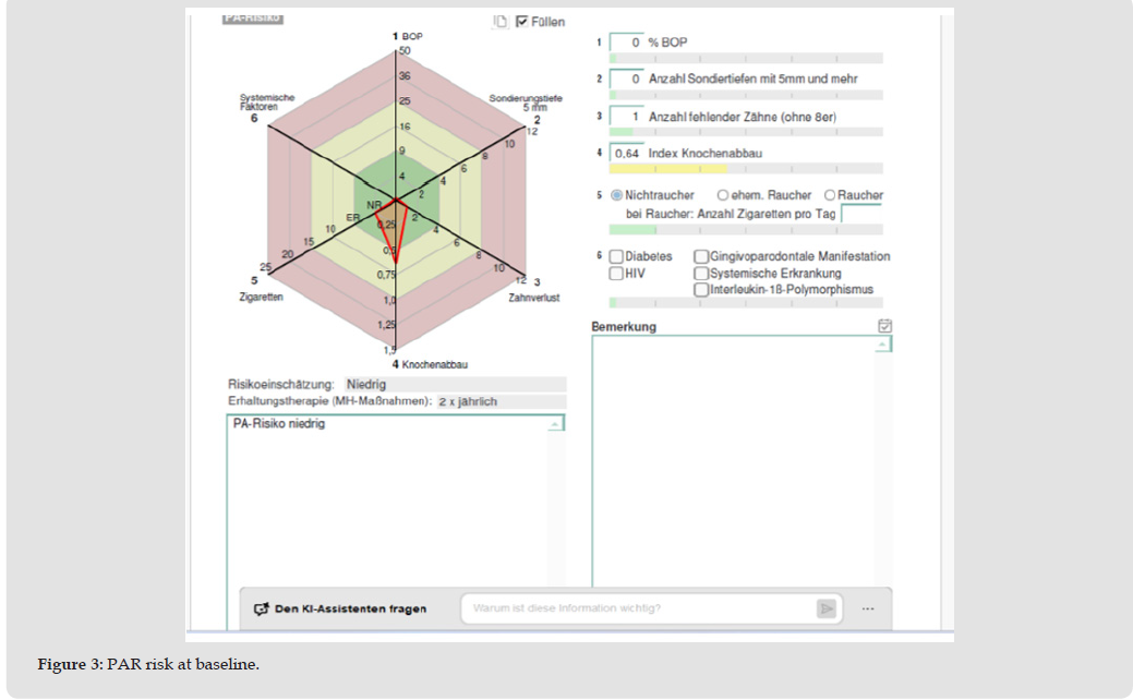
Inspection of the hard and soft palate, floor of the mouth, and oral mucosa revealed no pathological findings. Maximum interincisal opening measured 57 mm. The tongue was freely movable and exhibited normal sensitivity. Labial and lingual frenula were fully developed. The ducts of the major paired salivary glands (parotid, submandibular, and sublingual glands) showed no abnormalities, and salivary flow appeared normal in both quantity and quality. Gingival tissues exhibited reddish-livid discoloration with mild edematous swelling. No hard or soft deposits were detected. Overall oral hygiene was assessed as excellent, see Figures 1-3. Panoramic radiography demonstrated generalized horizontal alveolar bone loss of approximately 38% and an unfavorable periapical condition of tooth 24 following unsuccessful endodontic treatment. Based on these findings, extraction followed by immediate implant placement with adjunctive platelet-rich fibrin (PRF)-assisted augmentation was indicated.
Figure 1
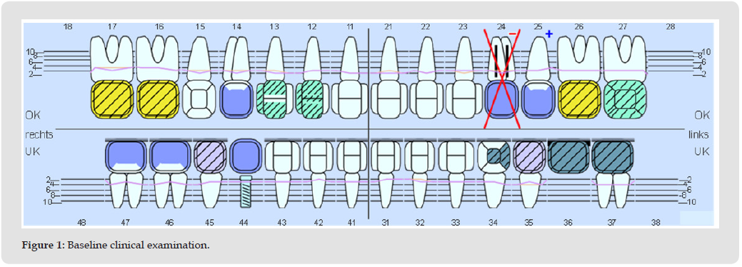
Figure 2
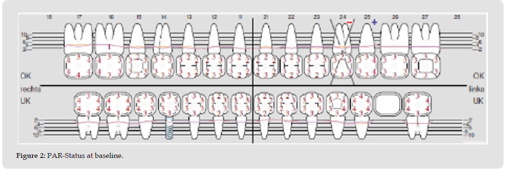
Figure 3
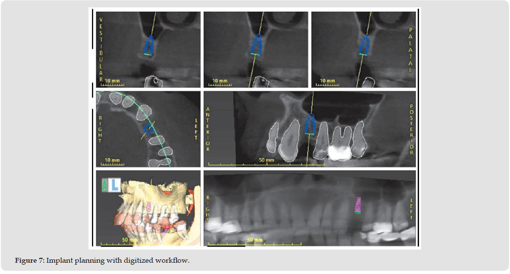
Tooth 24 was atraumatically extracted in January 2023. Post-extraction healing was uneventful. Cone-beam computed tomography performed in April 2023 confirmed sufficient bone volume for implant placement. A digitally assisted workflow was used for prosthetically driven implant planning, see Figure 7.
On May 10, 2023, immediate implant placement with simultaneous augmentation was performed under local anesthesia. Autologous blood was collected intraoperatively and processed to obtain PRF. After elevation of a mucoperiosteal flap, osteotomy preparation was completed, and a 4.0 × 11 mm implant was inserted in region 24 with a final insertion torque of 45 Ncm. Autogenous bone chips combined with xenogeneic particulate graft material were applied to the peri-implant defect and biologically stabilized using liquid PRF and an autologous fibrin membrane prior to primary closure. Postoperative radiographic control confirmed correct implant positioning and adequate augmentation. Sutures were removed after one week, revealing timely and uncomplicated healing, following a three-month osseointegration period, second-stage surgery with laser-assisted exposure was performed. After soft-tissue conditioning, definitive impressions were taken, and a screw-retained single crown was delivered in August 2023.
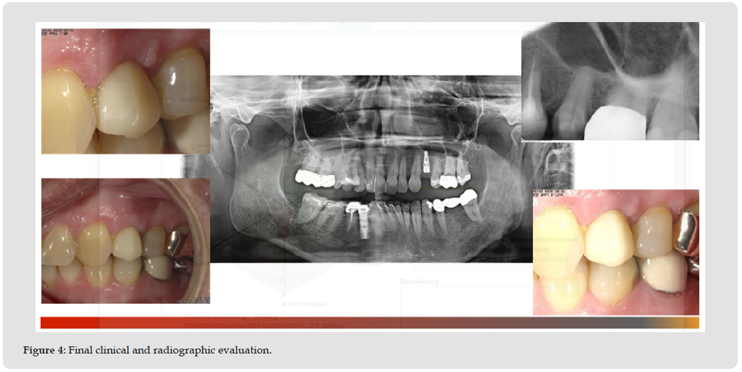
Adjunctive regenerative protocols incorporated standardized PRF preparation and application techniques, while digital implant planning and workflow optimization were performed using coDiagnostiX ™ software. Final clinical and radiographic evaluation demonstrated stable peri-implant soft tissues, preserved crestal bone levels, and satisfactory esthetic integration of the implant-supported restoration (Figure 4). No biological or technical complications were observed during the documented follow-up period.
Figure 4
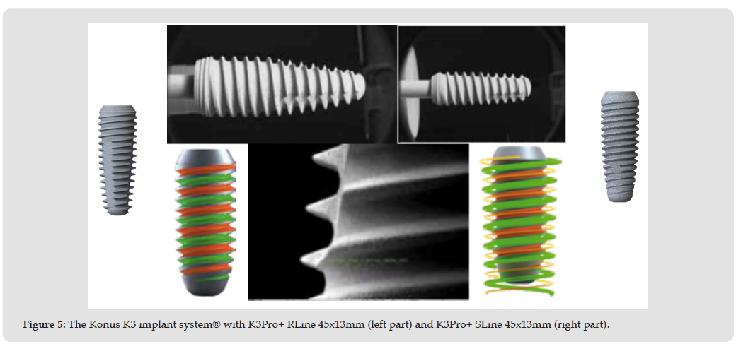
A variety of surface modification strategies for immediate implants have been introduced to improve outcomes in challenging clinical scenarios, including ion-beam-assisted deposition, sputter coating, pulsed laser deposition, electrostatic spray deposition, photofunctionalization, and platelet-rich plasma (PRP). PRP promotes early vascularization of the implant site, particularly during the first 20 days of healing. Studies supporting PRP use emphasize its regenerative potential for both hard and soft tissues. The Konus K3 Pro implant system (K3 Rapid, Argon Dental, Germany) features a bacteria- tight connection at crestal and subcrestal levels with a reduced emergence profile. The Rapid cutting thread design with 0.1% pitch enhances primary stability and incorporates an apical compression thread for bone condensation. Combined with the OsteoActive® surface, this design promotes rapid osseointegration and resistance to vertical and horizontal occlusal forces, see Figure 5.
Figure 5
Dental implant rehabilitation of edentulous areas is well documented and has been shown to be predictable. The literature demonstrates that the alveolar ridge undergoes substantial resorption and volume loss following tooth extraction or tooth loss for other reasons [1-4]. Only a limited number of authors advocate immediate implant placement as a strategy to limit this resorptive process. The literature demonstrates that the alveolar ridge undergoes substantial resorption and volume loss following tooth extraction [1-4]. However, several preclinical studies have reported conflicting results, indicating that immediate implantation does not prevent post-extraction bone resorption [5-18]. PRP promotes early vascularization of the implant site, particularly during the first 20 days of healing. Studies supporting PRP use emphasize its regenerative potential for both hard and soft tissues [19-23].
In Germany, allogeneic cell and tissue transplants are regulated
as medicinal products and therefore require regulatory approval; accordingly,
their manufacture, biological safety, and clinical application
are subject to continuous oversight by German regulatory authorities.
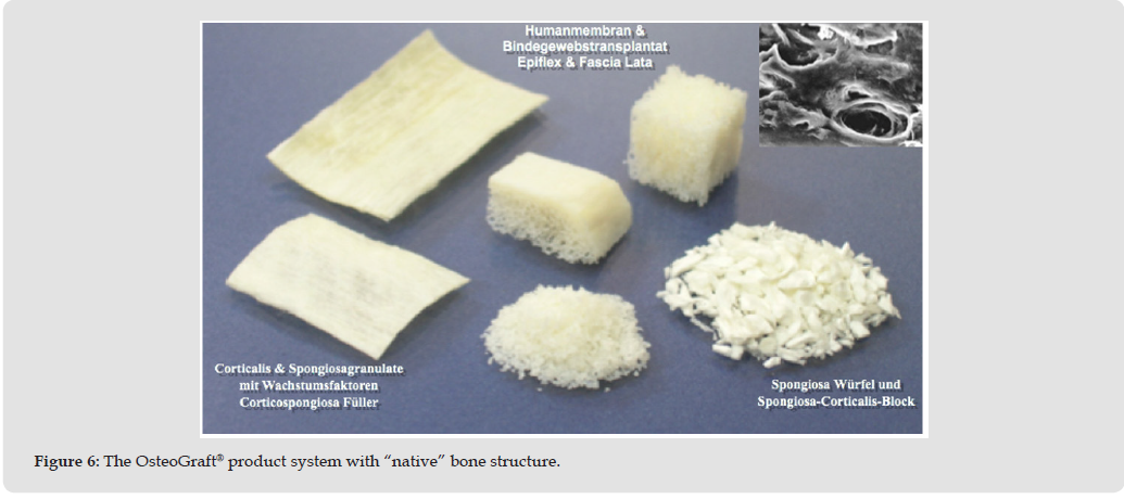
The allogeneic Osteograft® bone substitute material (ABM) is
approved by the German Institute for Cell and Tissue Replacement
(DIZG) as a medicinal product for human tissue transplantation (Figure
6).
The DIZG biological safety system for tissue transplants comprises:
• Comprehensive donor selection criteria,
• Extensive serological donor screening exceeding the requirements
of EU Directive 23/2004, including four viral genome
assays,
• Application of validated procedures for the removal and/or
inactivation of viruses, bacteria, and fungi,
and
• In-process and final quality control of the graft material.
Both DIZG and Argon Dental Vertriebs Gesellschaft mbH & Co. KG
(Bingen am Rhein, Germany) are certified according to DIN EN ISO
13485 and adhere to the Ethical Code and quality standards of the
European Association of Tissue Banks (EATB).
Figure 6
For prosthetically driven implant planning, three-dimensional imaging and planning systems are currently available that allow precise assessment of available bone volume relative to the intended prosthetic restoration and facilitate accurate transfer to the surgical site. In this case presentation, the coDiagnostiX 3D imaging and planning system™ was utilized, enabling preoperative simulation of implant positioning and identification of necessary augmentation procedures, see (Figure 7). The digitally guided workflow allowed prosthetically driven implant positioning and contributed to primary stability, which is a prerequisite for successful immediate implantation. Digital implant planning and guided surgery have been associated with enhanced positional accuracy and improved prosthetically driven outcomes [10-13].
Figure 7
As we began working with autologous blood plasma in minimally invasive regenerative periodontology and implant dentistry approximately five years ago, it quickly became evident that not all PRP formulations are equivalent. Autologous plasma rapidly emerged as a viable option for multiple regenerative indications. A key factor appears to be calcium ion activation within the coagulation cascade. Plasma consists not only of growth factors but also of fibrin and platelets, forming one of the most complex enzymatic systems in human physiology. Our clinical concept aims to utilize the biological potential of coagulation for stabilization of particulate graft materials across multiple indications. This approach is demonstrated in the accompanying surgical video.
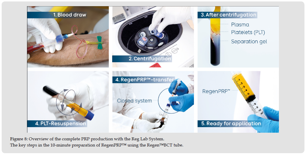
All RegenLab® products employ patented separation gel technology for standardized preparation of cell therapy products with reproducible cellular profiles. During centrifugation, the separation gel forms a stable physical barrier within the buffy coat, isolating platelets and plasma in the upper portion of the tube while binding erythrocytes and most neutrophils below. The resulting RegenPRP™ is a homogeneous plasma PRP characterized by high platelet recovery (>70-80%), low leukocyte concentration, near-complete removal of pro-inflammatory neutrophils, and virtually no erythrocytes. The closed system ensures sterile and safe preparation for both patients and clinicians, see Figure 8.
Figure 8
Immediate implant placement has been extensively investigated over the past decade and is considered a predictable treatment modality in appropriately selected cases, provided that primary stability, intact socket walls, and meticulous surgical protocols are ensured. Recent meta-analyses have demonstrated survival rates comparable to delayed protocols while emphasizing the importance of buccal bone thickness and gap management for long-term crestal bone preservation [6-9]. Simultaneous guided bone regeneration using particulate graft materials can reduce horizontal ridge contraction and improve peri-implant tissue stability following extraction [15-18]. Furthermore, digital implant planning and guided surgery are associated with enhanced positional accuracy and improved prosthetically driven outcomes, particularly in immediate placement scenarios [10-13].
Immediate implant placement in conjunction with biologically active blood concentrates is increasingly supported by clinical evidence. Systematic reviews and consensus reports indicate that adjunctive PRF may enhance early soft-tissue healing, angiogenesis, and graft stabilization when applied during immediate implant protocols [19-23]. Randomized clinical trials and prospective studies further suggest that PRF-supported augmentation may reduce postoperative morbidity and improve peri-implant soft-tissue outcomes [24-27]. Overall, the combined approach of immediate implantation, guided bone augmentation, and autologous PRF application resulted in predictable hard- and soft-tissue healing with favorable functional and esthetic outcomes [28-34].
The authors would like to thank all team members of the Haßlinghausen Practice Team for their help and support in treating the patient. In addition, the authors would like to thank RegenLab GmbH, Muenich, Germany, for the video material, some of which could be used in this publication.
Ethical approval was not required for this case report according to institutional guidelines using Practice Patient Information System “Charly” with pseudonymous patient data. Written informed consent was obtained from the patient for all clinical procedures and for publication of anonymized clinical data and images.
The authors declare that they have no competing interests.
This study received no external funding.
• WDG: Conceptualization, surgical treatment, data acquisition,
manuscript drafting.
• MAF: surgical treatment, data acquisition.
All authors reviewed and approved the final manuscript.

International Association of Landscape Archaeology, Czech Glass Society, Czech Republic

Department of Chemistry, Semenov Institute of Chemical Physics, USSR Academy of Sciences, Moscow, Russia

Department of Educational Sciences, Research Centre Qualities of Teachers, School of Education – HAN, Nijmegen, Netherlands

Associate Professor at Department of Breast and Thyorid Surgey, Chongqing General Hospital, China

Neurology, LA BioMed Research Institute, USA





 Abstract
 PDF
PDF



























