info@biomedres.us
+1 (502) 904-2126
One Westbrook Corporate Center, Suite 300, Westchester, IL 60154, USA
Site Map
 ISSN: 2574 -1241
ISSN: 2574 -1241
ISSN: 2574 -1241
Received: July 25, 2025; Published: August 05, 2025
*Corresponding author:
Yongqiang Yao, Department of Breast and Thyroid Surgery, Ward Three, Zhongshan Hospital Affiliated to Dalian University, Dalian, 116001, China
Zuowei Zhao, Department of Breast Surgery, The Second Hospital of Dalian Medical University, Dalian, 116023, China
Meiling Wang, Department of Breast and Thyroid Surgery, Ward Three, Zhongshan Hospital Affiliated to Dalian University, Dalian, 116001, China
DOI: 10.26717/BJSTR.2025.62.009811
Malignant adenomyoepithelioma(M-AME) is a breast tumor with biphasic differentiation of adenoepithelial and myoepithelial features, which has been reported in fewer clinical reports and is relatively difficult to diagnose and treat. This article reports a 54-year-old woman diagnosed with M-AME of the breast. Presenting with a six-month history of a left breast mass, preoperative core needle biopsy revealed adenomyoepithelial proliferation with marked cytological atypia. The patient underwent left total mastectomy with sentinel lymph node biopsy and chest wall reconstruction using a local advancement flap on December 27, 2024. Postoperative histopathology confirmed M-AME, characterized by biphasic malignant transformation of glandular and myoepithelial components. Adjuvant therapy recommendations included systemic staging via PET-CT, genetic profiling, and AC-T chemotherapy. The patient declined further treatment, opting for active surveillance. This case highlights diagnostic challenges and therapeutic dilemmas in managing this rare malignancy.
Keywords: Breast Cancer; Malignant Adenomyoepithelioma; Transposition Flap; Histopathological Classification; Triple-Negative Breast Cancer; Immunohistochemistry
Malignant adenomyoepithelioma (M-AME) of the breast is a rare neoplasm characterized by malignant proliferation of both glandular epithelial and myoepithelial cells. Predominantly affecting postmenopausal women, with a peak incidence in individuals over 60 years of age, M-AME accounts for <1% of breast malignancies [1,2]. Histologically, it is defined as a biphasic tumor with dual adenoepithelial-myoepithelial differentiation, displaying biphasic differentiation with morphological diversity. The neoplastic components consist of glandular structures admixed with hyperplastic myoepithelial cells, either or both lineages may demonstrate malignant transformation [3]. Given its clinicopathological rarity, M-AME poses diagnostic and therapeutic challenges.
This article reports a case of primary malignant adenomyoepithelioma (M-AME) of the breast in a premenopausal middle-aged female admitted to Unit 3 of the Department of Breast and Thyroid Surgery at Zhongshan Hospital Affiliated with Dalian University in November 2024. The patient underwent primary surgical resection without prior neoadjuvant therapy, including radiotherapy, chemotherapy, or endocrine therapy. Due to the extensive oncological defect intraoperatively, a rotational flap reconstruction was performed, achieving satisfactory postoperative wound healing. Adjuvant AC-T chemotherapy has been recommended postoperatively. Through integrated analysis of clinical data, histologicalmorphology, immunophenotypic and molecular features, and reviews of relevant studies, this study aims to improve diagnostic recognition and optimize therapeutic strategies for this rare entity.
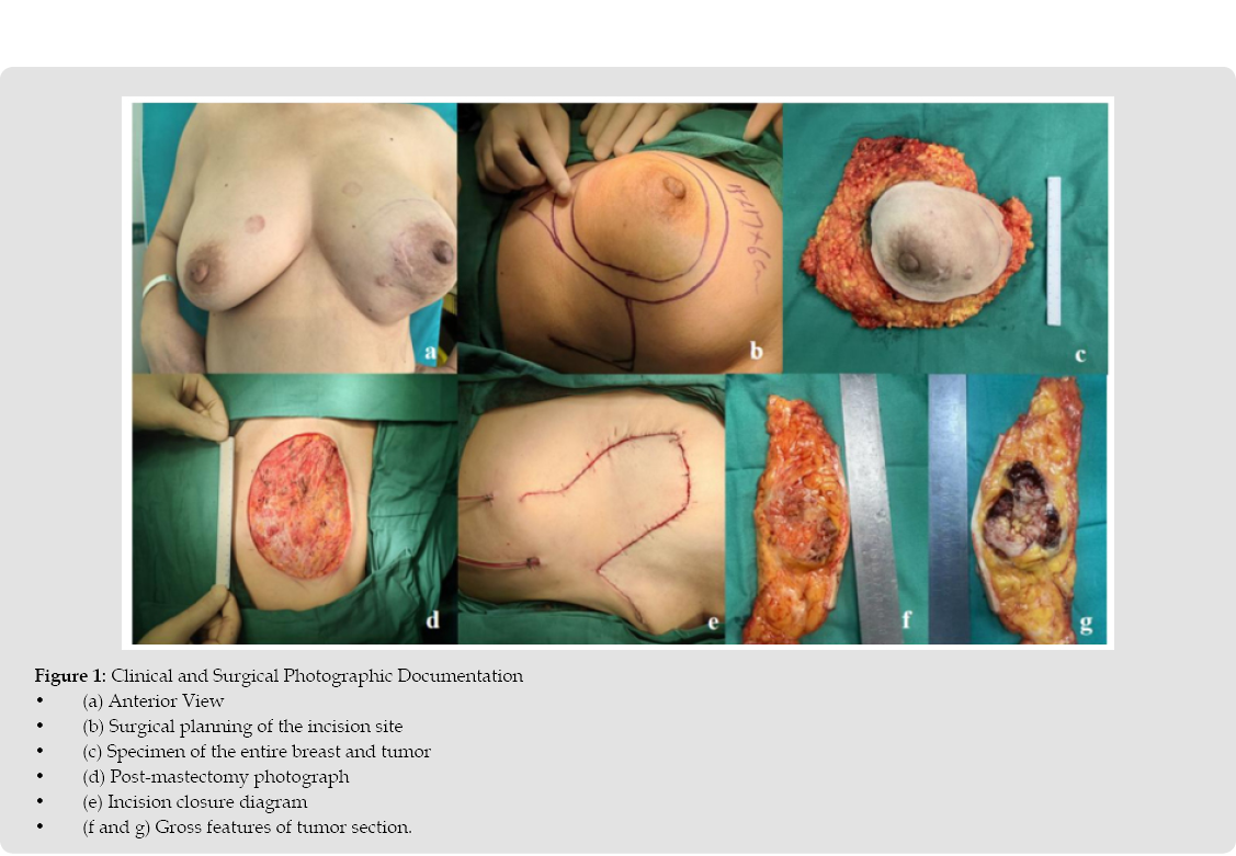
A 54-year-old woman presented to Zhongshan Hospital, Dalian University on November 28, 2024, with a six-month history of a left breast mass. Bilateral breast asymmetry with left breast enlargement and elevation. A raised surgical scar measuring 1.5 cm × 0.5 cm is observed 2.0 cm inferomedial to the nipple in the lower inner quadrant of the left breast, consistent with a post-minimally invasive breast procedure (Figure 1a). The skin demonstrates normal texture without erythema, edema, ulceration, or peau d’orange changes. Both nipples show no retraction or inversion, and the areolae are normal. A dominant mass measuring approximately 14 × 12 cm is palpated in the left breast involving the upper outer quadrant, lower outer quadrant, and subareolar region. The mass is firm with indistinct margins, irregular contour, and mobility without fixation to the chest wall. It adheres to the overlying skin in the subareolar area but shows no signs of ulceration discrete masses are detected in the right breast. Lymph node examination demonstrates no palpable axillary, supraclavicular, or infraclavicular lymphadenopathy. Manual compression of the nipples yields no discharge. Admitted to the hospital, breast ultrasound showed multiple masses in the left breast (BI-RADS category 5), hyperplasia in both breasts, and enlarged lymph nodes in the left axilla. Multiple solid and mixed echogenic masses were detected in the left breast, almost occupying the entire breast, with unclear margins, irregular morphology, and heterogeneous internal echoes (Figure 2a).
Blood flow signals were detected within and around these masses (Figure 2b). A hypoechoic nodule was detected in the left axilla, measuring approximately 2.3×0.9 cm, with clear borders and regular morphology. The corticomedullary structure was poorly differentiated. MRI-enhanced plain scanning of both breasts + vascular image (3.0) shows that both breasts demonstrate a fibro glandular tissue composition. Scattered punctate to patchy hyperintense signals are observed in the glandular tissue on T2-weighted imaging (T2WI) and diffusion-weighted imaging (DWI). Post-contrast images reveal multiple enhancing nodules and small patchy lesions with moderate to marked enhancement. In the left breast: Coalescing patchy areas showing hyperintense to mildly hyperintense signals on T2WI (Figure 2c). Restricted diffusion (DWI hyperintensity with mildly reduced ADC values). Heterogeneous enhancement patterns. Peripheral rim enhancement in focal areas. Non-mass-like heterogeneous marked enhancement (Figure 2d). The right breast shows no suspicious masses. Both nipples, areolae, and skin demonstrate normal architecture. No pathologically enlarged lymph nodes are identified in the axillary regions. Bilateral mammography (MG) reveals heterogeneously dense breast tissue. Both breasts demonstrate patchy, flocculent, and linear hyperdensities with uneven distribution and ill-defined borders, interspersed with irregular hypodense areas.
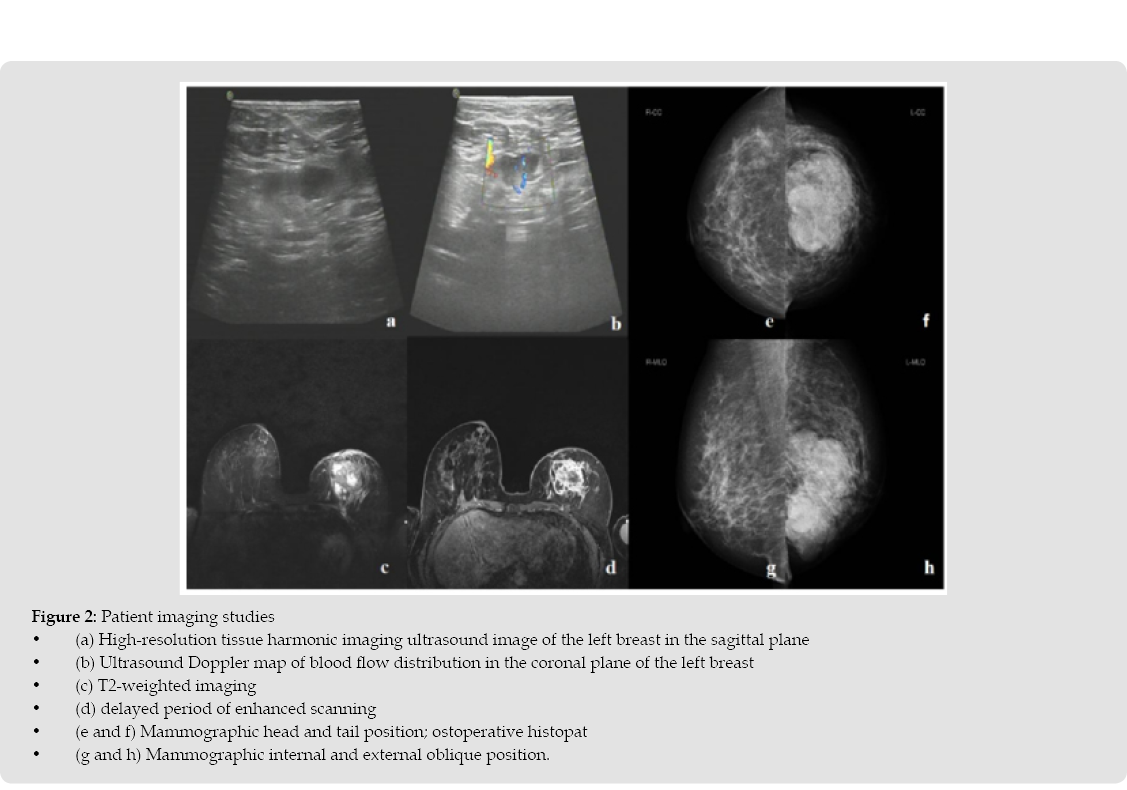
The left breast contains multiple nodular masses with indistinct margins. The largest lesion measures approximately 6.1 cm × 4.8 cm, containing internal calcifications and exhibiting fine spiculated margins (Figures 2f & 2h). The right breast shows no evidence of calcifications or masses (Figures 2e & 2g). Both nipples, overlying skin, and subcutaneous fat appear unremarkable. No significantly enlarged lymph nodes are identified in the imaged axillary regions. On December 10, 2024, the patient underwent core needle biopsy of the left breast mass. Histopathological examination revealed an adenomyoepithelial neoplasm with the following features (Figures 3a & 3b): Proliferative myoepithelial cells arranged in solid nests within a desmoplastic stroma, Marked cytological atypia (nuclear pleomorphism, coarse chromatin), Increased mitotic activity (>3 mitoses/10 HPF), Apoptotic bodies and focal coagulative necrosis, hyalinized stroma with basement membrane-like matrix deposition, Immunohistochemical profile: ER (-), PR (-), Her-2 (score 0), Ki-67 (20%), TOPOII (strong 10%+), E-cadherin (membranous+), P120 (membranous+), CK5/6 (+), EGFR (+), AR (-), p63(+) (Figures 3c-3h). Concurrent left axillary lymph node biopsy showed reactive lymphoid hyperplasia without evidence of malignancy.
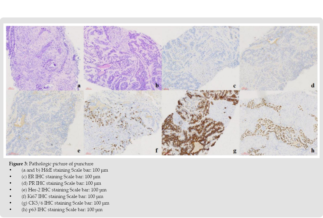
The morphological features combined with immunohistochemical findings are highly suggestive of malignant transformation in adenomyoepithelioma. However, due to sampling limitations inherent to core needle biopsy, complete surgical excision with adequate margins is mandatory for definitive classification. Given the core needle biopsy findings suggestive of malignant adenomyoepithelioma (a rare pathological entity), the following diagnostic opinions were obtained through formal external pathology consultation:
Consultation from Peking University Cancer Hospital: The diagnosis was aggressive fibromatosis with adenomyoepithelial proliferation in the breast ducts.
Consultation from the Department of Pathology, Peking Union Medical College Hospital: The diagnosis favored adenomyoepithelioma with focal consideration of malignant transformation. However, due to the limited amount of tissue and atypical morphology, a definitive diagnosis could not be made. The patient underwent left total mastectomy with sentinel lymph node biopsy and chest wall reconstruction using a local advancement flap on December 27, 2024. Following standard disinfection and draping, a near-circular incision was designed around the primary tumor (14 cm × 15 cm), resulting in a 15 cm × 17 cm skin excision (Figure 1b). The skin and subcutaneous tissues were incised, with superior, inferior, medial, and lateral surgical margins submitted for frozen section analysis. After elevating skin flaps, the breast and retro mammary tissues were dissected mediolaterally (Figure 1c). Critical structures preserved included the axillary vein, long thoracic nerve, thoracodorsal neurovascular bundle, and medial/lateral pectoral neurovascular bundles. The sentinel lymph nodes were harvested, with intraoperative pathology confirming the absence of metastasis. Hemostasis was secured, followed by irrigation with warm distilled water (42°C). Given the substantial tumor size (14 cm × 15 cm) resulting in a 15 cm × 17 cm full-thickness chest wall defect that precluded primary tension-free closure (Figure 3d), intraoperative reconstruction was performed using a medially-based rotational pectoral flap with advancement component (type IB Mathes-Nahai classification) (Figure 1e).
The tumor specimen was submitted to the pathology department for gross sectioning, which revealed papillary architectural features (Figures 1f & 1g). Intraoperative pathology revealed no metastasis in the sentinel lymph nodes. Postoperative paraffin pathology demonstrated the following findings:
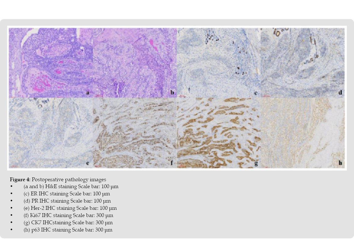
The tumor exhibited diffuse infiltrative growth, with desmoplastic stromal changes characterized by hyalinization and deposition of basement membrane-like material. The proliferative stroma divided the tumor cell nests into irregular sheets and solid patterns. Some nests were located within ducts, presenting as papillary structures. The tumor cell nests showed biphasic features, with central glandular epithelial cells forming luminal structures surrounded by clear, lightly stained myoepithelial cells. The myoepithelial cells were markedly proliferative, forming concentric layers around ducts. These myoepithelial cells exhibited significant atypia and increased mitotic activity (>3/10 HPF), with evidence of apoptosis and extensive necrosis (Figure 4a & 4b). Infiltrative myoepithelial cell nests were observed within the stroma at the tumor margin, which correlated with the immunohistochemical results, consistent with malignant adenomyoepithelioma. Nipple skin and pectoralis major muscle: No tumor involvement was observed. Immunohistochemistry results: ER (-), PR (-), Her-2 (0, with membranous staining), Ki-67 index of 40% (hotspot analysis) E-cadherin (+), P120 (membranous +), EGFR (+), AR (-), CK5/6 (+), P63 (focal +), CD10 (+), CK7 (glandular epithelial +), CD117 (part +) (Figures 4c-4h). Follow-up treatment: The patient was diagnosed with malignant adenomyoepithelioma, with a tumor exceeding 10 cm, an exceedingly rare presentation.
Following multidisciplinary team (MDT) discussion and consultation with specialists at oncology hospitals, recommendations include:
Following comprehensive counseling on all recommended therapeutic strategies, the patient opted solely for active surveillance with regular monitoring, formally declining all other proposed treatment modalities.
Adenomyoepithelial tumor of the breast (AME) is a biphasic tumor composed of glandular epithelium and prominently proliferated myoepithelial cells. While typically benign, this tumor may exhibit malignant potential in rare instances and can progress to breast cancer. AME is clinically uncommon, with only a limited number of cases reported in literature. Itsunique histological features and biological behavior make it associated with high diagnostic error rates in clinical practice. The morphological diversity of AME poses significant challenges for both radiological and pathological diagnosis. As a biphasic neoplasm, it may undergo malignant transformation simultaneously or separately in either component, often leading to confusion with other tumor types. Based on the characteristics of M-AME cases, they can be broadly classified into two categories: one category exhibits malignant changes based on typical adenomyoepithelioma, while the other appears as typical adenomyoepithelioma under low magnification but shows cellular atypia and increased mitotic index under high magnification [4]. Based on the pathological features of malignant adenomyoepithelioma (M-AME), it can be preliminarily distinguished from other breast tumors, such as fibroadenoma, intraductal papilloma, and ductal carcinoma. Notably, due to the tendency of M-AME to invade surrounding tissues, a definitive diagnosis cannot be established based solely on a small biopsy specimen.
In clinical practice, it is necessary to integrate the results of preoperative biopsy, intraoperative frozen sections, and postoperative paraffin-embedded sections to confirm the diagnosis [5], Whether a diagnosis can be made solely based on preoperative or intraoperative biopsy pathology remains to be further discussed. Currently, there is limited research data on the molecular classification of malignant adenomyoepithelial tumors. Relevant reports indicate that malignant adenomyoepithelial tumors may harbor HRAS codon 61 (Q61R/K) and codon 12/13 (G12D/G13R) mutations. Additionally, ER- and PR- negative malignant adenomyoepithelial tumors can simultaneously exhibit the PIK3CA gene H1047R mutation and HRAS gene G12/G13 hotspot mutations [6]. The patient declined postoperative genetic testing, thus the genetic mutation profile of the tumor could not be determined. Only routine immunohistochemical assays, including ER, PR, HER-2, and Ki-67, were performed, and the results were like those reported in the literature for malignant adenomyoepithelioma (M-AME). Currently, there are limited treatment options for breast M-AME, and no authoritative guidelines are available for reference. General treatment strategies for breast cancer include simple surgery, surgery after neoadjuvant t apy, surgery after oral endocrine therapy, or surgery followed by local radiotherapy. The patient underwent left breast simple mastectomy with intraoperative sentinel lymph node biopsy after completing the necessary preoperative evaluations.
The intraoperative lymph node results were negative, and axillary lymph node dissection was not performed. Postoperatively, the patient was advised to schedule radiotherapy and undergo a dose-dense AC-T regimen. Breast M-AME is prone to local recurrence and metastasis. Studies by Bult, et al. [7] and Ahmed, et al. [8] have demonstrated that breast M-AME has a risk of recurrence or distant metastasis. In this case, the patient has not yet undergone postoperative chemotherapy or endocrine therapy. Considering the current treatment plan is not fully established, strict surveillance protocol with 3-monthly clinical exams and annual PET-CT is mandated. The patient was definitively diagnosed with malignant adenomyoepithelioma of the breast. Preoperative core needle biopsy demonstrated an adenomyoepithelial lesion featuring prominent myoepithelial hyperplasia with marked nuclear atypia. Postoperative histopathology with myoepithelial mmunohistochemical markers (p63, CK5/6) confirmed the diagnosis of malignant adenomyoepithelioma. The surgical intervention comprised left total mastectomy with intraoperative sentinel lymph node mapping and local advancement rotational flap reconstruction. Postoperative immunohistochemistry revealed triple negative receptor status. Postoperative immunohistochemistry confirmed triple-negative breast cancer. Follow-up recommendations included completion of PET-CT, genomic profiling, adjuvant chemotherapy, and radiotherapy.
However, the patient has not adhered to the prescribed treatment regimen. Given the tumor’s high-grade biological behavior and metastatic propensity, stringent clinical surveillance was implemented. No evidence of local recurrence or distant metastasis has been observed during follow-up evaluations to date.
The authors report no conflicts of interest in relation to this work.
The study was funded by the basic scientific research project of Liaoning Provincial Department of Education (LJKMZ20221847).

International Association of Landscape Archaeology, Czech Glass Society, Czech Republic

Department of Chemistry, Semenov Institute of Chemical Physics, USSR Academy of Sciences, Moscow, Russia

Neurology, LA BioMed Research Institute, USA

Associate Professor at Department of Breast and Thyorid Surgey, Chongqing General Hospital, China

Professor of Nuclear Medicine, Faculty of Medicine and Surgery, University of Milan, Milan, Italy





 Abstract
  PDF
PDF 



























