info@biomedres.us
+1 (502) 904-2126
One Westbrook Corporate Center, Suite 300, Westchester, IL 60154, USA
Site Map
 ISSN: 2574 -1241
ISSN: 2574 -1241
ISSN: 2574 -1241
Received: May 12, 2025; Published: June 04, 2025
*Corresponding author: Kuan-Hong Lai, Division of Hepatology and Gastroenterology, Department of Internal Medicine, Shin Kong Wu Ho-Su Memorial Hospital, Taipei 111, Taiwan
DOI: 10.26717/BJSTR.2025.62.009705
Cryptococcus neoformans is a life-threatening fungus that primarily affects immunocompromised patients. Herein, we report a case of a man with hepatitis and right upper quadrant pain for 1 week. Abdominal ultrasonography revealed two hypoechoic lesions in the right lobe of the liver. The CT scan was also found two well-defined, hypodense tumors in the liver. An ultrasound-guided liver biopsy confirmed cryptococcal infection. Fluconazole was given orally for 6 months to treat Cryptococcus neoformans infection.
Keywords: Hepatic Cryptococcosis; Ultrasound-Guided Liver Biopsy
Abbreviations: CT: Computed Tomography
Cryptococcus neoformans is an environmental fungus that exists worldwide and has a predilection for immunocompromised hosts, including individuals using corticosteroids or those with HIV infection, malignancy, diabetes, or a history of organ transplantation. Infections in immunocompetent individuals have increased in decades [1]. Individuals without HIV or a history of organ transplantation account for 20%–50% of all cryptococcal infection cases [2]. Antifungal treatment options include amphotericin B deoxycholate, flucytosine, fluconazole, and itraconazole, which are typically administered over several weeks [3]. Herein, we present a case of a racing pigeon breeder with hepatitis who presented with right upper quadrant pain lasting 1 week.
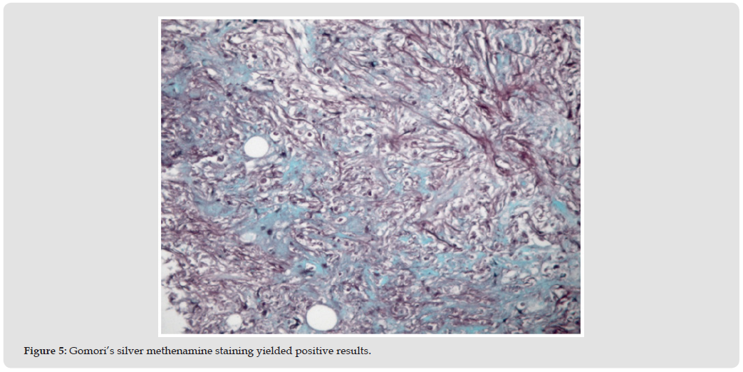
A 45-year-old man with no prior systemic disease presented to our gastroenterology outpatient department with right upper quadrant pain that had persisted for 1 week. He reported no tea-colored urine, fever, weight loss, gastrointestinal bleeding, vomiting, or nausea and maintained a good appetite. Blood tests and abdominal ultrasonography were performed, and the laboratory results are summarized in Table 1. The ultrasonography results (Figures 1 & 2) revealed no gallbladder stones or biliary tree dilation but indicated two hypoechoic lesions in the right lobe of the liver; the lesions measured 1.91 and 1.61 cm in diameter. Abdominal computed tomography (Figures 3 & 4) revealed two closely positioned, hypo vascular, hypodense nodules (1.2 and 1.0 cm) in liver segment 6. An ultrasound-guided liver biopsy (Figure 5) confirmed cryptococcal infection. The patient was subsequently treated with oral fluconazole for 6 months. Follow- up abdominal ultrasonography revealed complete resolution of the liver lesions and normal liver function.
Table 1: Summary of blood and serologic test results.
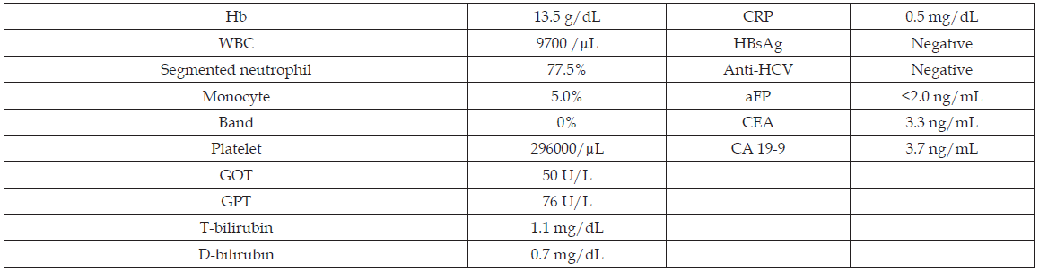
Figure 1
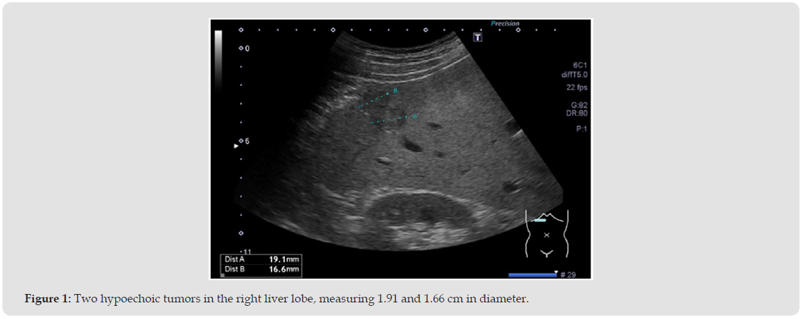
Figure 2
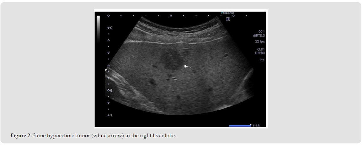
Figure 3
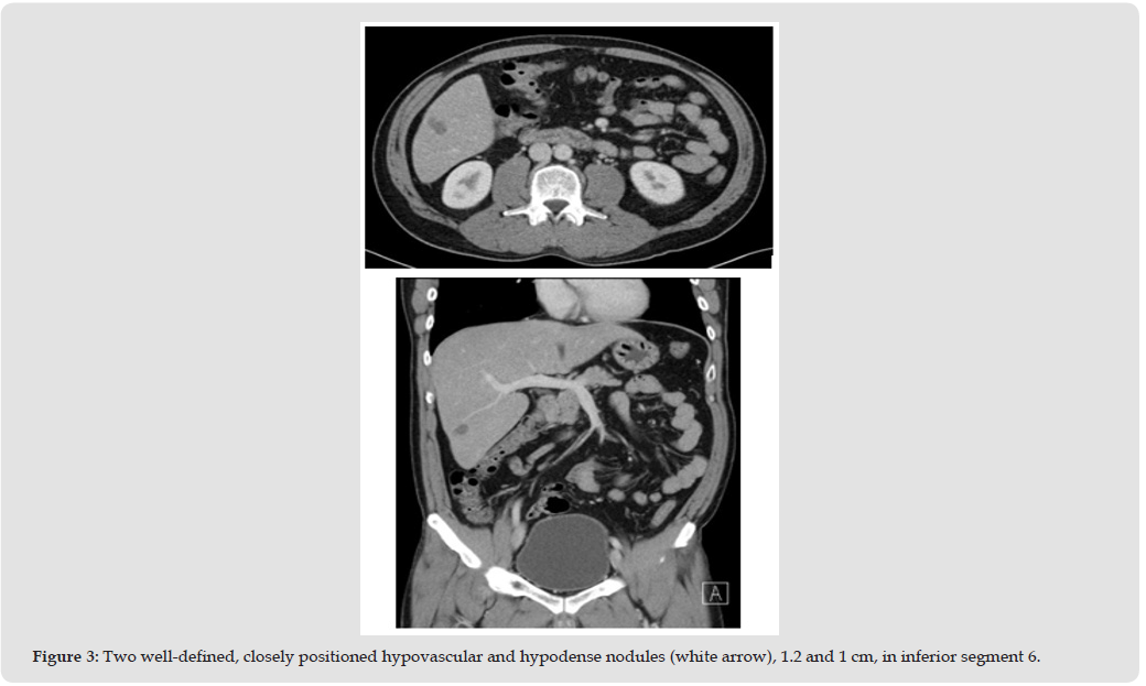
Figure 4
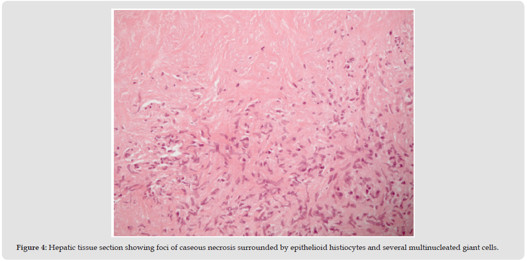
Figure 5
Cryptococcosis primarily affects immunocompromised individuals, but cases in immunocompetent individuals are being increasingly reported. Approximately 95% of all human cryptococcal infection cases are caused by C. neoformans, which is found globally in bird droppings, soil amoebae [4], and certain trees. Infection typically occurs through inhalation of spores into the lungs, but the gastrointestinal tract can also serve as an entry route. The fungus mostly affects the respiratory and central nervous systems [4]. However, the infection can also involve the skin, prostate, eyes, bones, and joints [4]. In patients with severe immunodeficiency, the fungus can spread systemically and involve any organ. In Taiwan, the overall incidence of cryptococcosis has doubled in recent decades among individuals without cryptococcal meningitis, HIV, or a history of organ transplantation [5]. Middle-aged men in the aforementioned cohorts have an elevated risk of cryptococcosis [5]. Studies have reported hepatic nodules in immunocompetent individuals [6,7]. These nodules often exhibit irregular margins and hypodense masses along the hepatoduodenal ligament. Some cases are accompanied by obstructive jaundice and biliary tract dilation [6], mimicking a hepatic neoplasm or primary sclerosing cholangitis. Cryptococcus typically enters through the lungs and spreads to the central nervous system. Treatment aims to eliminate pulmonary infection and prevent or manage central nervous system involvement. For mild to moderate infections in immunocompetent individuals, fluconazole at 200–400 mg daily for 6–12 months is recommended. Severe or progressive infections require amphotericin B at 0.4–0.7 mg/kg daily, achieving a total cumulative dose of 1,000–2,000 mg [8].
In our case, isolated hepatobiliary cryptococcosis might have occurred through retrograde infection via the biliary tract. No other infection sites were identified on the computed tomography scan. The patient’s frequent contact with pigeons is a likely source of exposure. The literature suggests that isolated hepatic cryptococcosis remains a rare clinical entity.


 Abstract
 PDF
PDF






























