info@biomedres.us
+1 (502) 904-2126
One Westbrook Corporate Center, Suite 300, Westchester, IL 60154, USA
Site Map
 ISSN: 2574 -1241
ISSN: 2574 -1241
ISSN: 2574 -1241
Received: May 13, 2025; Published: May 26, 2025
*Corresponding author: Zhong Liu, Department of Neurosurgery, The Second People’s Hospital of Hunan Province (Brain Hospital of
Hunan Province), Changsha; Department of Neurosurgery, Zhongshan Hospital of Xiamen University, School of Medicine, Xiamen University,
China
#These authors contributed equally to this work.
DOI: 10.26717/BJSTR.2025.62.009673
Background: This study examines whether mitochondrial NOX4 (mtNOX4) plays a crucial role in hemorrhagic
transformation (HT) after mechanical thrombectomy (MT) in acute ischemic stroke (AIS).
Methods: The rat HT model and brain microvascular endothelial cells OGD/R model were used to simulate the
process of HT after MT in human AIS. NOX4 siRNA induced NOX4 knockdown in vitro or NOX4 shRNA in vivo.
MtNOX4/ROS was measured in vivo and in vitro. In vitro, tight junction (TJ) proteins and adhering junction (AJ)
proteins were detected in endothelial cells. Infarct volume, HT, BBB damage, and neurological score were determined
24 h after cerebral ischemia in vivo.
Results: The induction of mitochondrial oxidative stress by mtNOX4/ROS disrupted AJ and TJ proteins after OGD/
R. Furthermore, compared to the OGD/R group, NOX4 siRNA decreased the expression of mtNOX4/ROS and
mitigated the downregulation of ZO-1, Occludin, Claudin-5, and VE-Cadherin. In vivo experiments demonstrated
that NOX4 knockdown suppressed mtNOX4/ROS upregulation in the penumbra area compared to the HT group.
Additionally, NOX4 knockdown reduced infarct volume and HT while improving BBB integrity and neurological
outcomes.
Conclusions: Targeting the mtNOX4/ROS pathway may be a potential treatment strategy to improve outcomes
in patients suffering from HT after MT for AIS.
Keywords: NOX4; Mitochondrial Oxidative Stress; Acute Ischemic Stroke; Hemorrhagic Transformation; Mechanical Thrombectomy; Blood-Brain Barrier
Abbreviations: HT: Hemorrhagic Transformation; MT: Mechanical Recanalization; BBB: Blood Brain Barrier; ROS: Reactive Oxygen Species; NADPH: Nicotinamide Adenine Dinucleotide Phosphate; CNS: Central Nervous System; Tmcao: Transient Middle Cerebral Artery Occlusion; mtNOX4: Mitochondrial NOX4; BMECs: Brain Microvascular Endothelial Cells; Gibco: Glucose and FBS; siRNA: Small Interfering RNA; CBF: Cerebral Blood Flow; EB: Evans Blue; TTC: Triphenyltetrazolium Chloride; CCCP: Carbonyl Cyanide M-Chlorophenylhydrazone; DCFHDA: Dichlorodihydrofluorescein Diacetate; TJ: Tight Junction; AJ: Adhering Junction
Acute ischemic stroke (AIS) is an emergency condition characterized by a sudden loss of blood flow to an area of the brain, which contributes to significant mortality and physical disability worldwide [1-3]. As such, a timely reperfusion is critical for patients with AIS.3 Moreover, endovascular therapy with stent retriever devices leads to superior functional outcomes compared to medical care [4,5]. Unfortunately, the MR CLEAN trial showed that 46% of patients experienced hemorrhagic transformation (HT) after mechanical recanalization (MT) [6]. Notably, clinically significant HT occurs at a much lower rate, ranging from 0.6% to 6% [5,7,8]. HT is also associated with worse functional outcomes in patients with AIS due to large vessel occlusion caused by blood brain barrier (BBB) disruption [8-11]. However, the specifics of HT after MT remain unclear [12,13]. Therefore, it is necessary to investigate the potential molecular mechanisms to mitigate HT following MT. Oxidative stress is one of the major mechanisms of AIS. Reactive oxygen species (ROS) are generated during key biological processes, such as mitochondrial dysfunction, disruption of the BBB, and inflammation [14,15]. Mitochondria and nicotinamide adenine dinucleotide phosphate (NADPH) oxidases (NOXs) are the two primary sources of ROS in the central nervous system (CNS), playing critical roles in cerebral ischemia and reperfusion injury [16- 18]. Additionally, mitochondria generate ROS as a by-product of oxidative phosphorylation, whereas the major function of NOXs is the generation of ROS [19-21]. We have successfully established a stable HT model in hyperglycemic rats with intraluminal filament transient middle cerebral artery occlusion (tMCAO).
We found that inhibiting the NOX2 and NOX4 pathways mitigates BBB disruption and reduces hemorrhagic transformation in the HT model after mechanical reperfusion [16,17]. Furthermore, the activation of NOX4 does not require cytosolic regulatory subunits, and its activity is primarily regulated at the expression level, resulting in the generation of H2O2 [20]. Recent reports indicate that NOX4 is localized to mitochondria, contributing to ROS levels [6,22,23]. Moreover, it is likely that mitochondrial-derived ROS enters the cytoplasm and activates intracellular signaling pathways, leading to endothelial dysfunction and inflammation [23]. However, there are no reports on the subcellular localization of NOX4 in the CNS. This study explores the subcellular localization of NOX4 in mitochondria within the CNS and investigates the potential impact of mitochondrial NOX4 (mtNOX4) on BBB integrity. Thus, this study aims to develop protective intervention strategies for HT after mechanical recanalization.
Cell Culture and Oxygen-Glucose Deprivation (OGD)/Reoxygenation (R) in vitro
Rat brain microvascular endothelial cells (BMECs) (C1094; ATCC) were cultured with deoxygenated DMEM without glucose and FBS (Gibco) in a hypoxia incubator chamber (Stemcell, CA), using premixed gas (95% N2 and 5% CO2 ) for 6 h. The cells were then cultured in DMEM containing 10% FBS and placed in a 37℃incubator (95% air and 5% CO2 ) for 24 h. Cells in the control group were cultured with DMEM and 10% FBS for the same incubation times. When the confluence reached 50%, the cells were transfected with NOX4 small interfering RNA (siRNA) according to the manufacturer’s instructions (GenePharma, Shanghai, China) [21,23]. The nucleotide sequences used were as follows:
• Sense 5’-GACCUGGCCAGUAUAUUAUTT-3’,
• antisense 5’-AUAAUAUACUGGCCAGGUCTT-3’.
In vivo HT Model and Analysis of Cerebral Hemorrhage, Evans Blue Staining, Neurological Deficit Scoring, And Measurement of Infarct Volume
All animal studies were executed in accordance with IACUC approved procedure at Xiamen University, China (Animal ethics review number: XMULAC2021). Male Sprague-Dawley rats (200-280 g,8-10weeks) underwent cerebral ischemia/reperfusion injury with MCAO using the intraluminal filament technique as previously described [16,17]. Briefly, hyperglycemia was induced by intraperitoneal injection of 50% dextrose (1.5 mL/kg) 15 min before surgery. Laser Doppler flowmetry monitored regional cerebral blood flow (CBF). After 6 h of ischemia, the filament was removed to allow instant reperfusion. At 18 h after reperfusion, the rats were sacrificed, and HT was confirmed for the subsequent experiments. Coronal brain slices measuring 2 mm in thickness were made and photographed for macroscopic hemorrhage assessment. Penumbra areas were harvested for further measurements. The amount of cerebral hemorrhage was quantified using a spectrophotometric assay. BBB permeability was assessed through Evans blue (EB) extravasation. The rats were blindly examined and scored for neurological deficits, while infarct volume measurements were detected using a 2,3,5-triphenyltetrazolium chloride (TTC) stain. Further details are described previously and provided in Supplemental Materials and Methods [16,17]. One hundred and thirty-one rats were randomly divided into three groups:
1. HT group (n=52): hyperglycemic rats underwent 6 h of MCAO
and received 18 h of reperfusion;
2. HT and NOX4 short hairpin RNA (shRNA) group (n=43);
3. Sham-operated group (n=36).
Intracerebroventricular NOX4 shRNA Injection
The rat NOX4 shRNA-containing retrovirus was designed and synthesized by GenePharma (Shanghai, China). The nucleotide sequence of sh-NOX4 is 5’-GGTATTGTTCCTCATGGTTAC-3’. Rats were anesthetized and fixed using a stereotaxic instrument (David Kopf Instrument, CA, USA). Each rat was injected in the right lateral ventricle 48 h before operation. The injection site was defined relative to the bregma: posterior 1 mm, right lateral 1.5 mm, and depth 3.5 mm. A small hole was placed at this position with a microsyringe at a depth of 2.5 mm below the surface of the skull, and 2 μL of the virus was injected. The incision was closed with sutures, and the rat was allowed to recover.19
Mitochondrial Isolation
Mitochondrial isolation procedures were performed under icecold conditions according to the manufacturer’s instructions (Mitochondria Isolation Kit, KeyGEN). Brain tissues or cells were homogenized in 1 ml lysis buffer for 2 min (for brain tissues) or 1 min (for cells). The homogenate was transferred into a new tube containing 0.2 mL medium buffer and centrifuged at 1,200 rpm at 4 °C for 5 min. The supernatant was then transferred into a new clear tube and centrifuged at 7,000 rpm at 4 °C for 10 min. Subsequently, the pellet was resuspended in 0.3 ml of suspension buffer and centrifuged at 9,500 rpm at 4 °C for 10 min. Finally, the enriched mitochondria were collected and resuspended with store buffer (50 μl/100 mg tissue, 100 μl/5*107 cells) [6].
Intracellular and Mitochondrial ROS (mtROS) Analysis
Measurements of ROS in cultured cells and brain tissues were performed immediately after sample collection. To identify the primary source of ROS in BMECs, we measured H2O2 levels in BMECs pretreated with various inhibitors of ROS-generating systems [23]. Notably, Carbonyl cyanide m-chlorophenylhydrazone (CCCP) (Sigma), an uncoupler of mitochondrial oxidative phosphorylation, and GKT137831 (D&C Chemicals, Shanghai, China), a member of the pyrazolopyridine dione family, are specific inhibitors of both NOX1 and NOX4. As previously described, 2’,7’-Dichlorodihydrofluorescein diacetate (DCFHDA) (Sigma) was used to detect H2O2 levels [17]. For detection of mtROS, mitochondria were isolated from endothelial cells described above. According to the manufacturer’s protocol, H2O2 was assessed with the Amplex Red Hydrogen Peroxide Assay Kit (Invitrogen). In brief, we pipetted 50 μL of the experimental samples into individual microplate wells. Then, we added 50 μ of the Amplex Red reagent/ HRP working solution and incubated it at room temperature for 30 min. Finally, detection at 590 nm using a microplate reader equipped for excitation in the range of 530~560 nm. Background fluorescence from PEG-catalase-treated controls was subtracted to determine final H2O2 levels.6
Quantitative Real-Time (RT)-PCR
The QRT-PCR was performed as described16 and is outlined in Supplemental Materials and Methods. The primer sequences for NOX4 mRNA were as follows: forward primer: 5’ -TGTTTGGCTGTCCCTAAATGTC- 3’, reverse primer: 5’- TGGGTAAAAGGATCAGGCTGC-3’ (RiboBio, Guangzhou, China). The expression levels of NOX4 mRNAs were normalized using GAPDH as an internal control and calculated using the ΔΔCt method. Each sample was tested in triplicates.
Western Blot Analysis
Western blot analysis was performed as described previously [17] and is detailed in Supplemental Materials and Methods. The antibodies anti-NOX4, Anti-COX IV, Anti-beta Actin, anti-ZO1, anti- Occlludin, anti-Claudin-5, and anti-VE-Cadherin were visualized using HRP-conjugated secondary antibody and signals were detected through chemiluminescence (Thermo). The individual protein bands were quantified by densitometry (ChemiDoc XRS; Bio-Rad) using ImageJ software.
Immunohistochemistry and Image Analysis
Immunohistochemistry and image analysis were performed as described17 and are detailed in Supplemental Materials and Methods. The primary antibodies used included anti-NOX4, Mito Tracker, and anti-Claudin-5. Sections or coverslips were probed with the fluorescent secondary antibodies Alexa Fluo 594 or Alexa Fluo 488 for one hour at room temperature. Following mounting, brain sections were examined and photographed using a confocal or fluorescent microscope. Three random images per section at 400× magnification were analyzed using ImageJ software to quantify the positive fluorescence area.
Statistical Analysis
All biochemical experiments were conducted at least three times. Data are expressed as mean ± SD or median±IQR. Continuous data were assessed for normality by the Kolmogorov-Smirnov test; normally distributed continuous data were analyzed using the student’s t-test, and non-normally distributed continuous data were analyzed using the Mann-Whitney U test. Fisher’s exact test compared the neurological scores among groups. SPSS software, version 26.0, was used. Differences were considered significant at P Values < 0.05.
MtNOX4 is Identified as A Major Source of ROS in the in vitro BMECs OGD/R Model
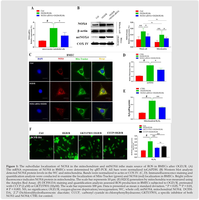
The expression of NOX4 mRNA increased in BMECs after OGD/ R, and the upregulation was inhibited by NOX4 siRNA (Figure 1A). Western blot analysis revealed an increase in NOX4 protein level in the whole cell lysate and mitochondria. Similarly, NOX4 siRNA suppressed the overexpression of NOX4 protein. Interestingly, no differences were observed in NOX4 protein levels between the whole cell lysate and mitochondria (Figure 1B). Furthermore, an upregulation of NOX4 expression in mitochondria was observed by an increase in yellow/orange fluorescence within the mitochondrion due to colocalization of the Mito tracker and NOX4 in BMECs after OGD/R (Figures 1C & 1D). Additionally, mitochondrial H2O2 levels were elevated after OGD/R. However, this elevation was inhibited by NOX4 siRNA compared to the OGD/R group (Figure 1E). Together, these findings confirm the subcellular localization of NOX4 in the mitochondrion and provide evidence for its upregulation in BMECs after OGD/R. To determine whether NOX4 is the primary source of ROS in BMECs after OGD/R, we measured H2O2 levels in cells pretreated with CCCP or GKT137831. Both CCCP and GKT137831 significantly reduced OGD/ R-induced H2O2 levels in BMECs, indicating that both cellular NOX4 and mitochondria are essential sources of ROS in BMECs after OGD/R. Interestingly, no significant difference was observed between the inhibitory effects of CCCP and GKT137831 on H2O2 levels (Figure 1F). Thus, these findings suggest that mtNOX4 may be the primary source of ROS in BMECs after OGD/R.
Figure 1
NOX4 siRNA Alleviates the Disruption of BMEC Junctions After OGD/R
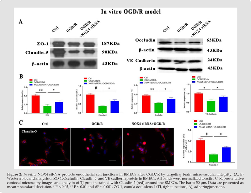
Endothelial cell tight junction (TJ) and adhering junction (AJ) proteins are essential components of the BBB. Western blot analysis showed impaired integrity of TJ and AJ, including down-regulated expression of ZO-1, Occludin, Claudin-5, and VE-Cadherin, compared to the control group in vitro (Figure 2A & 2B). We then investigated the effect of NOX4 siRNA on TJ using immunofluorescence in a BMECs OGD/R model. Claudin-5, a tight junction protein, was upregulated around the BMECs compared to the OGD/R group, suggesting a protective effect of NOX4 siRNA (Figure 2C). Importantly, the addition of NOX4 siRNA partially restored the expressional levels associated with BBB integrity. Therefore, these data highlight claudin-5’s beneficial role in maintaining the alterations caused by OGD/R in BMECs (Figure 2).
Figure 2
Knockdown of NOX4 Suppresses mtNOX4/ROS and Preserves the Integrity of the BBB in vivo
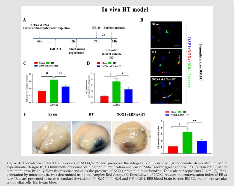
A schematic demonstration of the experimental design is shown in Figure 3A. The immunofluorescence staining of NOX4 and Mito tracker in the penumbra area provided evidence of the subcellular location of NOX4 in mitochondrion in vivo. Furthermore, NOX4 knockdown inhibits the upregulation of NOX4 in the mitochondrion (Figure 3B & 3C). Similarly, NOX4 shRNA inhibited the overexpression of mtROS (Figure 3D). Notably, the BBB is impermeable to EB. Marked extravasation of EB dye is observed in the ischemic penumbra area in the HT group compared with the sham-operated group, suggesting a disruption of the BBB. Furthermore, pretreatment with NOX4 shRNA results in minimal EB extravasation compared to the HT group (1.96 ± 0.24 vs. 3.37 ± 0.48, P <0.01), indicating that knockdown of NOX4 protects the BBB (Figure 3E).
Figure 3
Knockdown of NOX4 Reduces Both HT and Infarct Volume, Leading to Improved Functional Outcomes in vivo
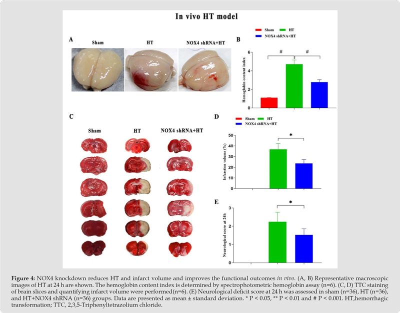
The mechanical reperfusion was performed after 6 h of MCAO-induced HT in all hyperglycemic rats, and most of them had parenchymal hematoma. Knockdown of NOX4 significantly reduced the hemoglobin contents (2.78±0.26 vs. 4.72±0.44, P < 0.001) in survival rats compared with those in the HT group (Figure 4A & 4B). Furthermore, TTC staining analysis demonstrated that NOX4 knockdown significantly reduced the cerebral infarct volume compared to the HT group (23.60± 3.61% vs. 36.79±5.46%, P<0.05) (Figure 4C, D). Furthermore, rats treated with the NOX4 shRNA exhibited better neurological scores (1.52 ± 0.34 vs. 2.24 ± 0.52, Ps<0.05) at 24 h after ischemia compared to those in the HT group (Figure 4E).
Figure 4
This study is the first to demonstrate the colocalization of NOX4 and mitochondria in rat BMECs in vitro and in vivo. MtNOX4 plays an essential role in mechanical reperfusion-induced HT by overproduction of mtNOX4-dependent ROS. Notably, inhibition of mtNOX4 protects the BBB and reduces hemorrhagic transformation after mechanical reperfusion in vivo, targeting brain microvascular integrity. Mitochondria oxidative stress plays an indispensable role in the pathophysiological processes following AIS.14 Several studies have demonstrated the subcellular location of NOX4 within mitochondrion in cardiac myocyte and kidney cortex. Additionally, they reported that mtNOX4/ROS is an essential target for cardiovascular disease and diabetes [22-24]. However, the subcellular colocalization of mtNOX4 in CNS has not been reported. This study first demonstrated the colocation of NOX4 and mitochondria in BMECs. We further confirmed that NOX4-dependent ROS is also upregulated within mitochondria in both the endothelial cell in the OGD/R model in vivo and the HT model in vitro. The hemorrhagic transformation following endovascular mechanical recanalization treatment results from disruption of the BBB. Moreover, endothelial cells’ TJ, AJ, and basement membrane are important components of the BBB [9,17,25]. NADPH oxidase is primarily responsible for the oxidative damage to the BBB in AIS [16,17,26]. Importantly, we found that mitochondrial H2O2 was increased in BMECs. Additionally, the knockdown of NOX4 inhibited the upregulation of mitochondrial H2O2 levels in both in vitro BMECs OGD/R and in vivo HT models.
Furthermore, inhibition of mtNOX4 reduced the disruption of Occludin, Claudin-5, ZO-1, and VE-Cadherin in vitro. The knockdown of NOX4 also reduced the infarct volume and HT, improved BBB integrity, and enhanced neurological outcomes in vivo. Consequently, free radicals can activate ProMMP, which further aggravates BBB damage. [25-27] Thus, inhibiting the overexpression of mtNOX4/ROS may be a potential strategy for protecting the BBB after mechanical thrombectomy in patients with AIS. This study has some noted limitations. Although we confirmed the subcellular localization of NOX4 in mitochondria in rat BMECs in vitro and in vivo, we did not detect the expression of mtNOX4 in neurons and astrocytes. Astrocytes are critical components of the BBB because they are essential for maintaining neural integrity. Additionally, we did not detect AQP-4 and PDGFR- β proteins, which are crucial molecules for BBB integrity. Therefore, further investigation is required to understand the protective effects of the mtNOX4/ROS pathway on HT after mechanical recanalization.
Mitochondrial NOX4/ROS may play a critical role in the occurrence and development of HT by targeting brain microvascular integrity after mechanical reperfusion in AIS. These results suggest that inhibiting the mtNOX4/ROS pathway could be a potential treatment target to improve outcomes in patients suffering from hemorrhagic transformation after mechanical thrombectomy for AIS.
The authors thank AiMi Academic Services (www.aimieditor. com) for English language editing and review services.
Credit
Zhong Liu, Conceptualization, Data curation, Formal analysis, Funding acquisition, Investigation, Methodology, Project administration, Resources, Software, Supervision, Validation, Visualization, Writing–original draft, Writing–review and editing. Xu-xiang Yu, Conceptualization, Data curation, Formal analysis, Investigation, Methodology, Software, Supervision, Validation, Writing–original draft, Writing–review and editing. Xiao-qiong Su, Conceptualization, Data curation, Formal analysis, Investigation, Methodology, Software, Supervision, Validation, Writing–original draft, Writing–review and editing. Feng-lin Zhang, Supervision, Validation, Writing–review and editing. Gui-jiang Dong, Conceptualization, Data curation, Supervision, Writing–review and editing. Jun-jiang Tong, Conceptualization, Data curation, Supervision, Writing–review and editing.
This study was funded by the the National Natural Science Foundation Youth Project of China (82101403), Natural Science Foundation of Fujian Province (2020J05288), and the Xiamen Municipal Bureau of Science and Technology (3502Z20214ZD1050).

International Association of Landscape Archaeology, Czech Glass Society, Czech Republic

Department of Chemistry, Semenov Institute of Chemical Physics, USSR Academy of Sciences, Moscow, Russia

Department of Educational Sciences, Research Centre Qualities of Teachers, School of Education – HAN, Nijmegen, Netherlands

Associate Professor at Department of Breast and Thyorid Surgey, Chongqing General Hospital, China

Neurology, LA BioMed Research Institute, USA





 Abstract
 PDF
PDF



























