info@biomedres.us
+1 (502) 904-2126
One Westbrook Corporate Center, Suite 300, Westchester, IL 60154, USA
Site Map
 ISSN: 2574 -1241
ISSN: 2574 -1241
ISSN: 2574 -1241
Received: March 17, 2025; Published: March 25, 2025
*Corresponding author: M Cholakova, Acibadem City Clinic UMHAT Tokuda Hospital, Bulgaria
DOI: 10.26717/BJSTR.2025.61.009552
Epilepsy is a chronic disease of the brain, characterized by repeated attacks because of hyperexcitation of a certain population of neurons and accompanied by various transient clinical (motor, sensory, autonomic, mental, behavioral) and paraclinical manifestations. An ambulatory surface electrode EEG is the most common diagnostic test used to evaluate seizures. Despite its widespread use, routine EEG has some limitations. Long term video-EEG monitoring has superior diagnostic value but is very expensive. According to our data short-term video-EEG monitoring is highly effective in diagnosis of epilepsy, especially psychogenic seizures.
Keywords: Epilepsy; Video-EEG; Routine-EEG; Antiepileptic Drugs
Epilepsy is a chronic disease of the brain, characterized by repeated attacks because of hyperexcitation of a certain population of neurons and accompanied by various transient clinical (motor, sensory, autonomic, mental, behavioral) and paraclinical manifestations [1]. Epilepsy is prevalent in all races and affects both sexes almost equally. Its frequency varies from 4 to 10 per 1000 population, and the incidence from 20 to 70 per 100,000 population. About 50% of epilepsies begin before the age of 10, and 75% before the age of 20. The average duration of disease is 12-13 years, and the mortality is 0.4-4%, mainly related to status epilepticus [2]. About 30% of patients with drug resistant epilepsy are misdiagnosed. The most common reasons are: the patient may have psychogenic attacks, use of the wrong medication or patients have low compliance to treatment. This necessitates the introduction of the concept of pseudo-resistance. In pseudoresistance, non-response is due to diagnostic errors[3]. Routine EEG studies are often normal between seizures. According to different authors, between 30 and 70% of patients, depending on the type of epilepsy, have abnormal electroencephalograms during routine recordings [4,5]. The diagnostic value of the method is increased by using techniques that stimulate the appearance of electrical responses [6]. Recording of ictal or interictal EEG changes in some patients suspected of epilepsy is impossible, even with repeated routine recordings. Ambulatory EEG monitoring for an extended period increases the likelihood of obtaining an abnormal EEG or recording during an attack. In addition, it provides reliable data for the evaluation of patients with suspected syncope, transient ischemic attacks, psychogenic seizures, and unclear epileptic seizures [7].
Epilepsy is a clinical diagnosis that can sometimes bebased on history, status, and EEG data. Standard EEG has advantages such as an increased number of channels and a variety of montages. In some cases, the diagnosis of epilepsy cannot be confirmed or rejected only based on the routine EEG, due to the differential diagnostic difficulties with physiological and psychogenic seizures [8,9]. An ambulatory surface electrode EEG is the most common diagnostic test used to evaluate seizures [10]. Despite its widespread use, routine EEG has some limitations. In the awake state and during sleep, the routine EEG is of short duration. Therefore, interictal EEG changes are mainly observed, which can be influenced by antiepileptic drugs [11]. The interictal EEG pattern can be an uncertain indicator in the classification of seizures, which also leads to incorrect treatment [12]. Routine EEG can be of very low sensitivity, i.e., no epileptiform changes are registered, only non-specific ones. Paroxysmal changes (potentially epileptiform discharges and nonspecific changes) can be interpreted in patients with nonepileptic seizures [13]. Patients with epileptic seizures, even with treatment-resistant epilepsy, may have a “normal” interictal finding [14,15]. Although interictal epileptiform EEG activity in many cases allows the classification of seizures [16], the sensitivity and specificity of the ictal EEG finding is significantly greater [17-20]. Carrying out standard provocation procedures - photostimulation, hyperventilation, sleep, sleep deprivation increases the diagnostic value of the routine EEG examination [21,22]. Additional dissphenoidal and other scalp electrodes provide additional information on the location of the epileptiform focus [23].Short-term video-EEG monitoring can be performed in an outpatient setting and lasts 1–12 hours. This study is appropriate for patients with frequent and provoked seizures [24]. The disadvantages of this method are the limited duration of monitoring, which does not allow the recording of epileptiform activity, and the need for staff to monitor the patient in the laboratory. Reducing antiepileptic drugs before the study can be dangerous and ineffective and is not appropriate for short-term video-EEG monitoring.
Video-EEG recording with duration 1-2 hours, routine EEG (EEG-R), activation procedures-photostimulation, hyperventilation, sleep, sleep deprivation, saline ingection.
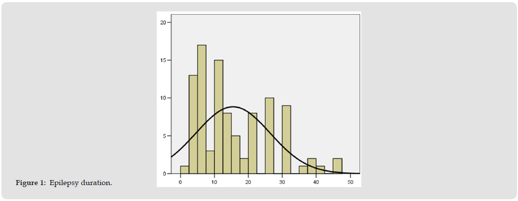
A total of 97 patients were studied, of whom 50 were women, 47 were men with a mean age of 36.9 years (SD±14.268).The patients had epilepsy duration (Figure 1) ranging from 3 to 46 years, with an average of 15.47 years (SD=10.947).According to the type of epilepsy (based on medical documentation), patients were divided into two groups:
1. With generalized epilepsy - 17 patients,
2. With focal epilepsy - 80 patients.
Figure 1
All the selected patients were on polytherapy. According to the type of AED used they were divided into the following groups: oxcarbazepine and others - 46 patients; Carbamazepine and others - 66 patients; Valproic acid and others - 76 patients; Levitracetam and others - 30 patients; Lamotrigine and others - 29 patients.Depending on the type of seizures, depending on the documentation of the patients before hospitalization and the conduct of EEG-P and video-EEG monitoring, they were distributed into two groups.
1. Simple and complex partial seizures with/without secondary generalization - 75 patients.
2. Primary generalized seizures - 22 patients.
The group of patients with primary generalized seizures also includes 2 patients with Juvenile Myoclonic Epilepsy and 1 patient with Juvenile Absence Epilepsy.Video-EEG was performed on a Cadwell 32 CH EEG Amplifier, with Easy II EEG software, with the same filter characteristics. Video-EEG was performed on inpatients, under medical supervision for 2 hours. The studies were performed with the activation procedures described for routine EEG. The study was performed in a lying or sitting position in the awake state. The EEG recordings were visually assessed according to the following criteria:
1. Organization and type of the main activity for the recordings in the awake state.
2. Presence of focal paroxysmal epileptiform activity, expressed in discharges of spikes, sharp waves and/or complexes of sharp-slow or spike-slow wave or slow theta or delta waves, in one or several adjacent leads, which may also appear contralaterally and/or independently asynchronously in other areas, as well as generalize during the recording.
3. Presence of generalized paroxysmal epileptiform activity, expressed in sudden simultaneous appearance of synchronous abnormal graph elements - single or multiple spikes and/or sharp waves, as well as their combinations (complexes) with high slow waves over both hemispheres and over extensive areas of both hemispheres.
4. When receiving an attack - Description of ictal electrophysiological phenomena depending on the clinical manifestations.
5. EEG-R and Video-EEG, on which seizures or interictal paroxysmal activity were recorded, are designated as positive, and EEG-R and Video-EEG with diffuse changes or normal as negative. Study of the Sensitivity of EEG-R and Video-EEG Dependence on the Type of Epilepsy
According to the type of epilepsy, patients were divided into 2 groups:
1. With generalized - 17 patients,
2. With focal - 80 patients.
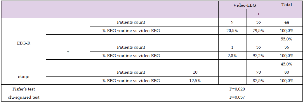
The results show (Table 1) an increase in the number of positive recordings in patients with generalized epilepsy during Video-EEG monitoring (47.1%) versus 11.8% for EEG-R, but without reaching statistical significance (p=0.206).bA total of 80 patients were included in the focal epilepsy group, and the results of both protocols are shown in Table 2. The results show an increase in the number of positive recordings in Video-EEG monitoring (87.5%) versus 45.0% for EEG-R, with Fisher’s exact test showing statistical significance (p=0.020).
Table 1:
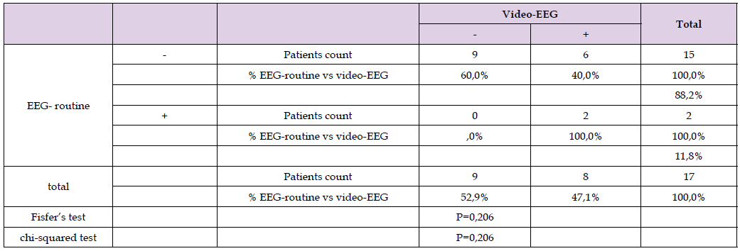
Table 2:
Study of the Sensitivity of EEG-R and Video-EEG Dependence on the Type of Epileptic Seizures
According to the type of seizures, patients were divided into 6 groups:
1. Simple and complex partial seizures with/without secondary generalization - 70 patients;
2. Primary generalized seizures - 18 patients;
3. Non-epileptic seizures - 9 patients Table 2).
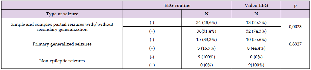
In all types of seizures, video-EEG monitoring shows a higher percentage of positive results compared to routine EEG, and for patients with simple and complex partial seizures with/without secondary generalization the differences reach statistical significance. Positive EEG-P are observed in 34.6%, and positive video-EEG in 80.8% of patients with simple and complex partial seizures with/without secondary generalization (p=0.0023). An increase in positive results of video-EEG (44.4%) compared to EEG-P (16.7%) is also observed in patients with primary generalized seizures. For the diagnosis of patients with non-epileptic paroxysmal states, the benefit of video-EEG monitoring is undeniable, as in 100% of patients it is positive and in none of the patients with EEG-P.In a total of 11 patients, the initial diagnosis was changed after conducting video-EEG monitoring. In 5 patients with an initial diagnosis of simple and complex partial seizures with/without secondary generalization, a non-epileptic etiology of the seizures was proven - in 4 patients, psychogenic seizures were registered, and in 1, action dystonia. In four patients with an initial diagnosis of primary generalized seizures, a seizure occurred during video-EEG and psychogenic pseudoepileptic seizures were proven. 1 patient with an initial diagnosis of complex partial seizures experienced 2 myoclonic seizures during video-EEG monitoring and the diagnosis was changed to juvenile myoclonic epilepsy. 1 patient diagnosed as idiopathic epilepsy experienced a complex partial seizure during video-EEG and was transferred to the group of patients with simple and complex partial seizures with secondary generalization (Table 3).
Table 3:
According to our data short-term video-EEG monitoring with activation procedures has good clinical value and usefulness for drug resistant epilepsies. No matter of better results in generalized epilepsy patient’s group, we consider short term video-EEG is better in diagnosis of focal epilepsies and non-epileptic seizures.

International Association of Landscape Archaeology, Czech Glass Society, Czech Republic

Department of Chemistry, Semenov Institute of Chemical Physics, USSR Academy of Sciences, Moscow, Russia

Department of Educational Sciences, Research Centre Qualities of Teachers, School of Education – HAN, Nijmegen, Netherlands

Associate Professor at Department of Breast and Thyorid Surgey, Chongqing General Hospital, China

Neurology, LA BioMed Research Institute, USA





 Abstract
 PDF
PDF



























