info@biomedres.us
+1 (502) 904-2126
One Westbrook Corporate Center, Suite 300, Westchester, IL 60154, USA
Site Map
 ISSN: 2574 -1241
ISSN: 2574 -1241
ISSN: 2574 -1241
Received: March 17, 2025; Published: March 25, 2025
*Corresponding author: Nikolay Mladenov, Clinic of Neurology, “Acibadem City Clinic Tokuda University Hospital”, Sofia, Bulgaria
DOI: 10.26717/BJSTR.2025.61.009550
Ischemic stroke is a vascular disease with high morbidity and mortality. Not only the acquired risk factors are significant in vascular incidents, but also the genetic. The predisposition to thrombosis could be a result of genetic factors, acquired changes in the clotting mechanism or interaction between them. Impaired fibrinolytic function caused by elevated plasminogen activator inhibitor-1 (PAI-1) levels has been implicated in ischemic stroke. Studies of ischemic stroke show that histological, biochemical and structural composition of the thrombus influence the success rate of the different types of treatment. Erythrocyte-rich thrombus is associated with a favourable outcome and higher recanalization rate, shorter intervention time, and increased r tPA sensitivity. Thrombi that are rich of fibrine could be associated with high plasma PAI-1 levels and low recanalization rates at the first hour and 6 hours after rt-PA infusion. Screening for PAI mutation could be a predictive factor to evaluated risk of recurrent thrombosis especially in absences of other risk factors among young adults with ischemic stroke.
Keywords: Thrombophilia 1; PAI Mutation 2; Ischemic Stroke 3Thrombophilia 1; PAI Mutation 2; Ischemic Stroke 3
Abbreviations: PAI-1: Plasminogen Activator Inhibitor-1; tPA: Tissue-Type Plasminogen Activator; u-PA: Urokinase Plasminogen Activator; rt-PA: Recombinant Tissue Plasminogen Activator; IS: Ischemic Stroke; CT: Computer Tomography; MCA: Middle Cerebral Artery
Thrombophilia is defined as a predisposition to irregular clot formation. It is a polygenic disorder with changeable expressivity. A predisposition to thrombosis may result from genetical factors, acquired changes in the clotting mechanism, or an interaction between them. Homozygous carrier or the combination of two or more heterozygous abnormal factors for thrombophilia can lead to thrombotic predisposition in youngers [1,2]. Homozygous carring of the PAI mutation, 4G/4G is clinically significant for thrombotic events. Increased plasma activity of PAI-1 leads to reduced fibrinolytic activity and increased risk of arterial and venous thrombosis. Plasminogen Activator Inhibitor-1 (PAI-1) is the primary Tissue-Type plasminogen Activator (tPA). Decreased fibrinolytic function due to increased plasma levels of PAI-1 plays an important role in the pathogenesis of thrombotic events [1,3]. Individuals who are homozygous for the 4G allele have increased plasma PAI-1 concentrations compared to those with the 5G allele [4]. In some studies, the prevalence of the 4G allele has been found to be higher in coronary artery disease, meningococcal septic shock, osteonecrosis, severe preeclampsia, Pulmonary Thromboembolism (PTE) [5,6]. The SERPINE1 gene is responsible for the production of PAI-1. PAI-1 is involved in hemostasis by inhibiting the action of plasminogen activators.
These proteins, including Urokinase Plasminogen Activator (u- PA) and tissue-type plasminogen activator (t-PA), convert plasminogen to its active form, plasmin. Plasmin is involved in fibrinolysis. By inhibiting the conversion of plasminogen to plasmin, PAI-1 prevents fibrinolysis. The 4G allele is associated with higher plasma PAI-1 activity. Elevation of plasma PAI-1 activity leads to decreased fibrinolytic activity and increased risk for arterial and venous thrombosis [7]. In different populations, plasma levels of the PAI-1 antigen are associated with the 4/5 guanosine (4G/5G) polymorphism in the promoter region of the PAI-1 gene [8]. Homozygous carriers of the 4G allele have the highest levels of PAI-1 [9,10]. PAI-1 activity levels fall transiently after thrombolysis, while PAI-1 antigen levels remained unchanged. PAI-1 4G/5G polymorphism had no effect on PAI-1 levels and did not influence stroke severity. Thrombolysis by Recombinant Tissue Plasminogen Activator (rt-PA) is the main pharmacological therapy in acute ischemic Stroke (IS). Ischemic Stroke (IS) is a widespread vascular disease also among youngers [11]. Although mechanical thrombectomy has revolutionized stroke care in the past decade the most commonly used pharmacological therapy of IS is still intravenous (i.v.) thrombolysis with recombinant tissue plasminogen activator (rt-PA) [12-14]. Although the relative benefit of i.v. t-PA is undisputed for selected patients with acute IS, in a large proportion of patient’s neurological improvement fails and about 6–8% develop hemorrhagic transformation after thrombolysis as a side effect [15].
In order to improve safety and outcome of i.v. rt-PA therapy, there is a growing interest in finding new biomarkers as potential predictors of post-lysis intracranial bleeding, recurrent thrombosis and treatment failure. As the most important inhibitor of t-PA, the relative balance between PAI-1 and t-PA plays an crucial regulatory role in fibrinolysis [16]. Elevated PAI-1 levels have been recognized as a risk factor for cardiovascular disease and stroke [17]. There are little studies that evaluate changes in PAI-1 levels during thrombolysis in stroke patients and its association with outcomes. PAI-1 levels could be chanced by a number of factors, for example inflammation, obesity, triglyceride levels, and genetic factors [18,19]. Several polymorphisms within the PAI-1 gene have been described to influence PAI-1 levels. It has been shown that both alleles of 4G/5G polymorphism contain a place for tying for a transcription activator, while the 5G allele also contains a binding site for a transcription repressor that partially overlaps with the activator-binding site [20]. The individuals with 4G/4G genotype have the highest, while those with 5G/5G genotype have the lowest circulating PAI-1 levels [21]. An association between the 4G/5G polymorphism and the risk of cardiovascular or cerebrovascular disease has been examined in observational studies, and known meta-analysis has indicated that PAI-1 4G/5G polymorphism may serve as a genetic biomarker for atherosclerotic diseases [4,22].
Testing for thrombophilia, laboratory tests, Computer Tomography (CT), conventional angiography, neurological exam. We presented clinical cases of patients with ischemic stroke, rt-PA and endovascular treatment due to large vessel occlusion.
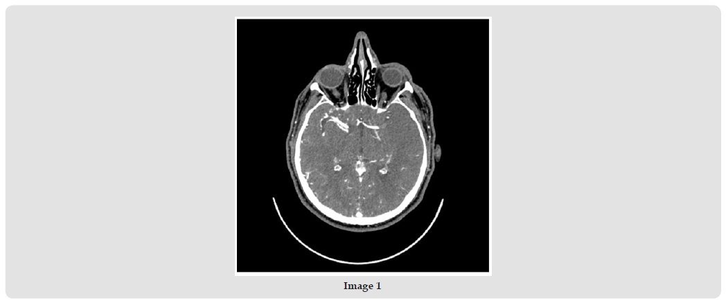
Reduced fibrinolytic capacity due to increased plasma levels of PAI-1 plays an important role in the pathogenesis of thrombotic event [23]. Although intravenous thrombolysis increases the probability of a good functional outcome in carefully selected patients with acute ischemic stroke, a substantial proportion of patients who receive thrombolysis do not have a good outcome. A number of recent trials of mechanical thrombectomy appear to indicate that this treatment may be superior to thrombolysis. Compared with patients treated medically, patients who received mechanical thrombectomy were more likely to be functionally independent as measured by a modified Rankin score. Mortality and symptomatic intracerebral hemorrhage were not significantly different between the two groups. Mechanical thrombectomy significantly improves functional independence in appropriately selected patients with acute ischemic stroke. We present two clinical cases of patients admitted with ischemic stroke with thrombosis of middle cerebral artery. The first patient is 51-year-old male who was hospitalized with acute right hemiplegia and total motor aphasia (NIHSS- 16score). Occlusion of M1segment of left Middle Cerebral Artery (MCA) was found after performed CT angiography (Image 1). The patient was treated by performing intravenous thrombolysis. After that, thrombus aspiration was performed, and full recanalization of MCA was achieved- TICI 3 score.
Figure 1
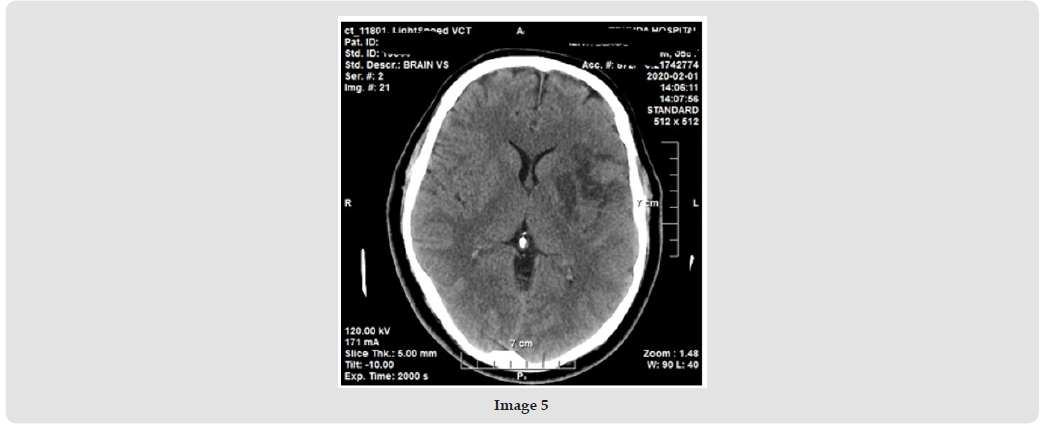
The patient was with mild residual deficit and control CT showed ischemic zone in the area of the left temporal, insular and in the head of the caudate nucleus. (NIHSS- 3, mRS- 1) (Images 2 & 3). The patient was evaluated for risk factors for thrombosis. He was diagnosed with thrombophilia (homozygous carrying of PAI mutation 4G/G, heterozygous for prothrombin mutation G20210A) and hemodynamically insignificant PFO. The following catheters and guide wires were used: 6F Neuron Max, Synchro 0.018, 300 mm, microcatheter- Marksman, aspiration catheter- ACE 64, aspiration pump- Penumbra. The time from the patient’s entry to the hospital to the start of IVT is 40 min, and to the start of the interventional procedure - 75 min. Complete SMA recanalization – TICI 3 was achieved at 3.30. from the onset of symptoms [24]. The second patient was 50-year-old male. He was admitted with acute severe right hemiparesis, somnolence and aphasia (NIHSS- 18, GCS-11). The patient was diagnosed with ischemic stroke caused by thrombosis of M1 segment of left MCA after CT angiography. Bridging therapy was done with r tPA and mechanical thrombectomy with fully recanalization- TICI 3 score (Images 4 & 5). The following catheters and guide wires were used: Synchro 2- 0.014”, microcatheter- Marksman, stent retriever- Solitaire 2. Recanalization is achieved after 2 passes. The patient was with ischemic changes from control CT in the region of lentiform nuclei and insula. He was diagnosed with homozygous carrying for PAI mutation 4G/4G and arterial hypertension [24].
Figure 2
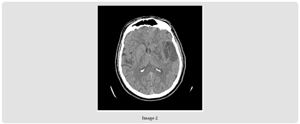
Figure 3
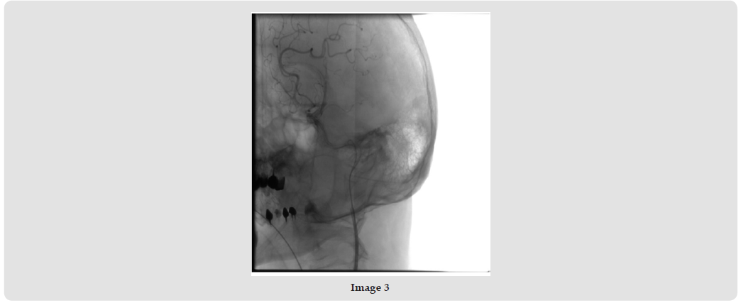
Figure 4
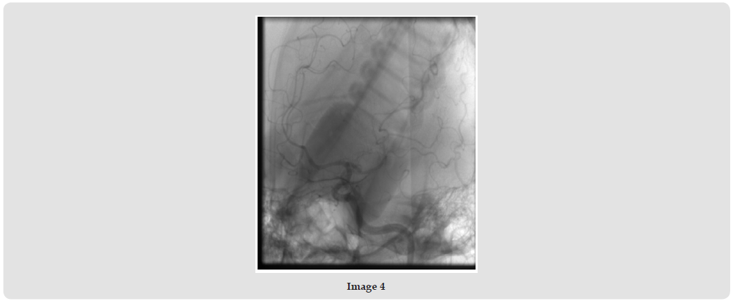
Figure 5
Mechanical thrombectomy is a safe, effective and safe method for the treatment of IS with middle cerebral artery occlusion, both alone and after intravenous thrombolysis. Screening for PAI mutation could be a predictive factor to evaluated risk of recurrent thrombosis especially in absences of other risk factors. An assessment of risk factors is necessary for patients with ischemic stroke. Genetic factors play a leading role in most cases of thrombotic event. The correct determination of risk factors for cerebrovascular accidents, including the presence of thrombophilia, plays a role in the effective prevention and treatment. Further studies are needed to evaluate the role of PAI mutation as a risk factor for complications after treatment for acute ischemic stroke.

International Association of Landscape Archaeology, Czech Glass Society, Czech Republic

Department of Chemistry, Semenov Institute of Chemical Physics, USSR Academy of Sciences, Moscow, Russia

Department of Educational Sciences, Research Centre Qualities of Teachers, School of Education – HAN, Nijmegen, Netherlands

Associate Professor at Department of Breast and Thyorid Surgey, Chongqing General Hospital, China

Neurology, LA BioMed Research Institute, USA





 Abstract
 PDF
PDF



























