info@biomedres.us
+1 (502) 904-2126
One Westbrook Corporate Center, Suite 300, Westchester, IL 60154, USA
Site Map
 ISSN: 2574 -1241
ISSN: 2574 -1241
ISSN: 2574 -1241
Received: March 12, 2025; Published: March 19, 2025
*Corresponding author: Steven M Czop, Director Medical Affairs, OssDsign USA, Columbia MD 21044
DOI: 10.26717/BJSTR.2025.61.009539
Anterior Cervical Corpectomy and Fusion (ACCF) is typically reserved for severe degenerative, neoplastic, and/ or traumatic conditions. Posterior cervical fusion may be required when more than 2 vertebrae are involved to reduce construct failure. Time to fusion is the most important factor to avoid instrumentation failure. There are multiple factors which play a role in time to fusion which include surgical technique, instrumentation selection, bone graft selection, smoking status, and patient selection. A variety of bone grafting options are available such as autograft, allograft, and synthetic bone grafts. In the category of synthetic bone graft there are multiple options. A silicate enriched (5.8wt% silicon) calcium phosphate bone graft substitute (OssDsign Catalyst®) was designed such that the nanoarchitecture and site-specific silicate substitution mimic more faithfully the structure of natural bone mineral and therefore follow the healing pathways more commonly associated with autograft or biologics than with synthetics, including endochondral ossification, which is of particular interest in challenging fusion environments. The following cervical trauma case highlights the careful selection of surgical approach, construct, instrumentation, and selection of bone graft to achieve a positive clinical outcome for a complicated and compromised cervical trauma patient presentation.
Keywords: ACDF: Anterior Cervical Discectomy and Fusion; ACCF: Anterior Cervical Corpectomy and Fusion; Cervical Fusion; Cervical Trauma; Catalyst; Synthetic Bone Graft; Silicate; Calcium Phosphate; Interbody Fusion
Abbreviations: ACCF: Anterior Cervical Corpectomy and Fusion; BMI: Body Mass Index; CT: Computed Tomography Scan; ACP: Anterior Cervical Plate; MRI: Magnetic Resonance Imaging; OTC: Over the Counter; TMC: Titanium Mesh Cage; SA: Segmental Angle; PLL: Posterior Longitudinal Ligament; PT: Physical Therapy
Cervical corpectomy is much less common and is typically reserved for severe degenerative, neoplastic, and/or traumatic conditions [1]. Cervical corpectomy is performed via an anterior cervical approach [2]. However, corpectomy involving more than one level can lead to a higher incidence of pseudarthrosis [3]. Patients with a solely anterior approach have experienced high rates of instability and clinical deterioration which resulted in the recommendation of additional posterior fusion [4]. An anterior cervical locking plate combined with a titanium cage has shown a high degree of anatomical reduction, direct decompression, and stability [5]. The use of Titanium Mesh Cages (TMC), allowing for the addition of bone graft, provides sufficient biomechanical stability, and avoids donor site morbidity. Structural characteristics of TMCs, however, can frequently cause subsidence, and the surface interface of these devices should be optimized to reduce potential subsidence [6]. Expandable titanium cages have been developed and shown to improve segmental angle (SA). This results in improved cervical lordosis but may in certain constructs restrict the amount of bone graft which can be inserted and/or limit the surface area for fusion to occur [7]. Given the advantages of an expandable cage construct, the device chosen should have a variety of components and options designed to fit the anatomical needs of the patient and restore the integrity of the spinal column.
In the following trauma case, the surgeon utilized an expandable titanium cage (Normandy VBR, Zavation Medical, Jackson, Mississippi). Due to the possible limitations of adequate bone graft volume and optimal bone graft placement within the device, the choice of a suitable bone graft substitute should also be carefully considered when faced with more challenging cases. Since the early 2000’s the continued use of synthetic bone graft substitutes has successfully reduced the need and subsequent morbidity of autograft harvest and have the added advantage of having knowledge of the exact composition of materials as opposed to allograft and autograft [8]. Synthetic bone grafts are typically calcium-based substitutes containing Hydroxyapatite (HA) and Tricalcium Phosphate (TCP), which accounts for nearly 70% of the mineral content of naturally occurring bone. Synthetic bone grafts are osteoconductive with some exhibiting osteointegrative properties [9] Recently, technical advances in synthetic bone grafts include nanoscale architecture and/or surface technology, similar in size and crystallinity to human bone mineral, in contrast to the more common sintered ceramic architecture which results in feature sizes many orders of magnitude greater in size.
A silicate enriched (5.8wt% silicon) calcium phosphate bone graft substitute (OssDsign Catalyst®) was designed such that the nanoarchitecture and site-specific silicate substitution mimic more faithfully the structure of natural bone mineral and therefore follow the healing pathways more commonly associated with autograft or biologics than with synthetics, including endochondral ossification, which is of particular interest in challenging fusion environments. This site-specific silicate substitution (5.8wt% silicon) calcium phosphate bone graft substitute (OssDsign Catalyst®) has shown encouraging results in both animal models and early clinical use [10-13]. The following cervical trauma case illustrates the need for a surgeon’s technical skill and knowledge in choosing the most appropriate surgical techniques and adjuncts to achieve the best possible outcome for challenging patients.
The patient was a 48-year-old male (BMI 29.3, overweight) with a history of cardiothoracic disease which included congestive heart failure requiring arterial aneurysm repair and cardiac catheterization in 2019. He was a former smoker (5 pack-year history (i.e., number of packs per day x number of years a smoker)) who had quit three years prior and was imbibing alcohol in the evening and fell down a flight of stairs (12 steps). He was taken to the hospital by ambulance, immobilized and evaluated. He was experiencing neck pain, bilateral upper and lower extremity weakness (left>right) and decreased sensation with paresthesia. Further examination, imaging, and MRI revealed vertebral artery dissection at C3-C4, comminuted fracture of the C5 vertebral body and right facet, lamina fracture at C3, and superior endplate compression fracture at C6. A vascular surgeon evaluated the vertebral artery dissection and recommended treatment with an antiplatelet agent (Aspirin 81mg when able). The consulting neurosurgeon recommended C5-C6 anterior corpectomy and C4-C7 anterior cervical fusion, followed by C2-T2 posterior spinal fusion with posterior screw and rod fixation. The surgery was performed in two stages, the anterior corpectomy was performed initially, followed two days later by posterior cervical fusion.
Anterior Procedure
The patient was administered general endotracheal anesthesia. After careful planning and preparation, an incision was made on the left side and carried down to the spine with blunt dissection. Standard Smith-Robinson technique was used to expose the anterior cervical spine from C4 to C7 [14,15]. The corpectomy was then performed by drilling into the C5 and C6 vertebrae using a diamond bur. Using a microscope the drilling of the C5 and C6 continued, then with microdissection a plane was developed between the dura and the disrupted Posterior Longitudinal Ligament (PLL). The PLL was then removed with a Kerrison rongeur. There was an epidural hematoma present which was then evacuated. Intact dura was observed from the C6- C7 level all the way to the C4-C5 level. At this point, the spinal cord was completely decompressed. A corpectomy cage of appropriate size (Normandy Corpectomy 12x14x30, 10-degree cage) was packed with 5cc of OssDsign Catalyst® synthetic bone graft. The corpectomy cage had been expanded so that it spanned the C4 to the C7 endplates, and under fluoroscopic guidance the cage was tapped into place. A cervical plate (Alphatec Spine, Carlsbad, CA) was used to secure the corpectomy cage. Fluoroscopy was used to assess the construct. Hemostasis was then achieved, retractors were removed, and a drain was placed through a separate stab incision before wound closure. No complications were noted intraoperatively. The posterior cervical fusion was performed 2 days after the anterior cervical corpectomy and fusion.
The patient was brought back to the operating room, given general endotracheal anesthesia, and motor and somatosensory evoked potentials were obtained. The patient was placed in a Mayfield head holder, then carefully turned over onto a Jackson table with bolsters to achieve a neutral position for the head and neck. An incision in the midline was made from C2 to T2 and carried down to the spinous process, lamina, and lateral masses at these levels. An O-arm was brought into the field, and an intraoperative CT scan was obtained, then transferred to the Stealth station (Stealth Station S8 Surgical Navigation System-Medtronic), for intraoperative stereotactic navigation. Placement of instrumentation was done with pedicle screws placed at C2, T1, and T2 in the following manner. A matchstick bur was utilized to create a pilot hole. A navigated tap was used to create a trajectory through the pedicle with the assistance of Stealth navigation. The trajectory was palpated for any breaches, then a screw of appropriate size was then placed into the C2, T1, and T2 pedicles bilaterally. Lateral mass screws were inserted using a matchstick bur to create a pilot hole. A high-speed drill was utilized to create a trajectory through the lateral mass. A ballpoint feeler was used to ensure that there was no breach, then screws of appropriate size were placed on the right at C4, C5, and C6; on the left at C3, C4, and C6.
Rods of appropriate size were placed and connected the tulip heads from C2 down to T2, the setscrews were used to tighten down the rod and were torqued according to manufacturing specifications. The lateral masses at C2, C3, C4, C5, C6, and C7 were decorticated. The transverse process at T1 and T2 were decorticated. 10cc of Catalyst Bone Graft Bone Substitute was placed over all the decorticated surfaces. Hemostasis was achieved and a drain was placed via separate stab incision before wound closure. No complications were noted intraoperatively. Follow-up visits for the patient occurred at 3 weeks, 3 months, 6 months, and 12 months post-operatively with a CT scan of the construct taken at 14 months. Initially, the patient had difficulty swallowing but could eat solid food, and this was resolved in subsequent follow-ups. Both anterior and posterior incisions were healed without erythema or edema. Pain was controlled initially by narcotic medication but at subsequent follow-ups, this was tapered until non-narcotic medications (Lyrica) and OTC medications (Acetaminophen) were sufficient to manage pain. Lower extremity weakness (left leg) was at full strength by 3 months and left upper extremity weakness persisted but continually improved over time as he underwent a regimen of Physical Therapy (PT). Anterior-Posterior and Lateral X-rays showed intact and well-positioned instrumentation at each time point.
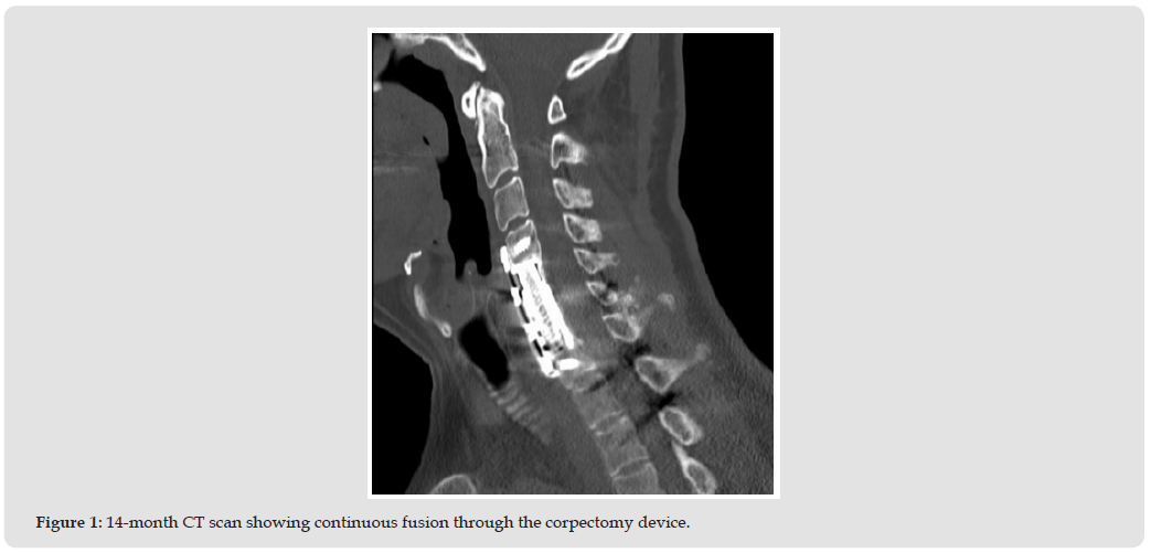
By 12 months the patient had mild residual weakness in the left upper extremities and left leg but was able to do most of the activities of daily life independently. The CT scan at 14 months showed that the Catalyst bone graft had facilitated fusion (i.e., bridging bone) through the corpectomy device from C4 to C7 as well as posterior fusion bilaterally from C2-T2.
The key steps in this patient’s surgical intervention were safe anterior access of the cervical spine, removal of any compression of the spinal cord and nerve roots, restoration of stability via fusion of the spine. Restoring lordosis was also a consideration to reduce incidence of adjacent level disease and improve overall patient outcome. The bone graft used for this case (Catalyst) was selected based on its handling and performance characteristics. The amount of bone graft that could be packed into the corpectomy device was determined, or limited, by its design. One of the most important performance features of Catalyst Bone Graft was shown in a rabbit posterolateral fusion study conducted prior to commercial availability (referred to as Osteo3 ZP Putty). Histomorphometric observations of tissue samples of the progressively healing bone graft at selected time points showed regions of chondroblastic tissue between Catalyst granules. This finding was indicative of new bone forming through endochondral ossification (the formation of cartilage-like precursor tissue) [10]. Endochondral ossification, by which cartilage serves as a template for new bone, is characterized by the initial formation of collagen matrix, followed by hypertrophic chondrocyte-mediated bone formation. This performance characteristic is advantageous when extensive rapid bone formation, mechanical strength, and vascularization are needed to stabilize or reinforce the surgical construct and is not typically observed as a predominant mechanism of bone formation in synthetic grafts [13].
The combination of surgeon skills and experience in selecting the most appropriate and effective surgical procedures and adjuncts, including the choice of bone graft, resulted in a positive clinical outcome in this complicated and compromised patient presentation (Figure 1).
Figure 1

International Association of Landscape Archaeology, Czech Glass Society, Czech Republic

Department of Chemistry, Semenov Institute of Chemical Physics, USSR Academy of Sciences, Moscow, Russia

Department of Educational Sciences, Research Centre Qualities of Teachers, School of Education – HAN, Nijmegen, Netherlands

Associate Professor at Department of Breast and Thyorid Surgey, Chongqing General Hospital, China

Neurology, LA BioMed Research Institute, USA





 Abstract
 PDF
PDF



























