info@biomedres.us
+1 (502) 904-2126
One Westbrook Corporate Center, Suite 300, Westchester, IL 60154, USA
Site Map
 ISSN: 2574 -1241
ISSN: 2574 -1241
ISSN: 2574 -1241
Received: February 20, 2025; Published: February 25, 2025
*Corresponding author: Bolaños Guedes J, Campos E. Emergency Department, Hospital del Mar, Barcelona, Spain
DOI: 10.26717/BJSTR.2025.60.009497
Aortic pseudoaneurysm is a rare entity that can present with nonspecific symptoms, making early diagnosis challenging. We present the case of a patient with hemoptysis and hematemesis as the initial manifestations of a contained rupture of an aortic pseudoaneurysm. Additionally, we review the literature to highlight the importance of differential diagnosis in such cases.
Keywords: Aortic Pseudoaneurysm; Hemoptysis; Hematemesis; CT Angiography; Contained Aortic Rupture; Endovascular Repair; Open Surgery; Differential Diagnosis
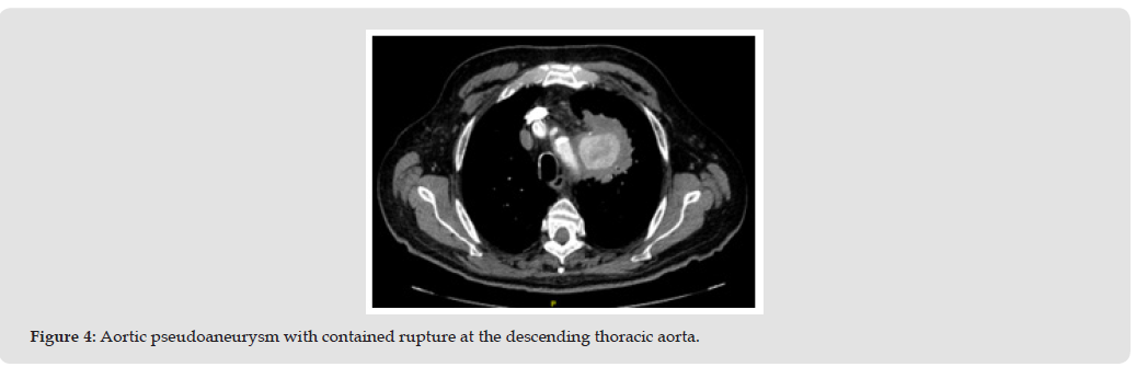
A 63-year-old male, former smoker, with a history of hypertension and chronic obstructive pulmonary disease (COPD), presented with intermittent hemoptysis for one month and dysphonia for two months, without prior evaluation. The patient experienced an episode of frank hemoptysis and hematemesis without dyspnea or fever. Physical examination revealed tachycardia (HR: 123 bpm), low-grade fever (37.3 °C), and pallor. Laboratory findings showed anemia (Hb 9.4 g/dL), leukocytosis with neutrophilia (16.72 x10³/μL, 83.3% neutrophils), and elevated C-reactive protein (15.92 mg/dL). Chest X-ray revealed a mediastinal mass in the left upper lobe (Figure 1). A contrast- enhanced CT angiography confirmed the presence of an aortic pseudoaneurysm with contained rupture at the descending thoracic aorta (Figures 2-4). The patient was transferred to a specialized center for surgical management.
Figure 1
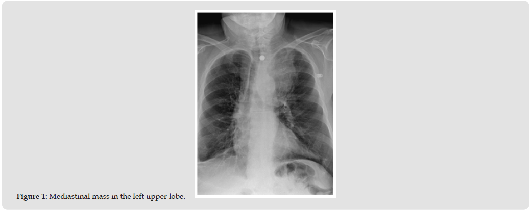
Figure 2
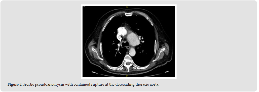
Figure 3

Figure 4
Given the patient’s presentation with hemoptysis, hematemesis, and a mediastinal mass, several differential diagnoses were considered before reaching the final diagnosis:
Pulmonary Neoplasm
Malignancy, particularly lung cancer, is a leading cause of hemoptysis in smokers. The mediastinal mass seen on imaging raised suspicion of a primary lung tumor or metastatic disease.
Tuberculosis or Infectious Etiologies
Chronic cough, hemoptysis, and constitutional symptoms could be indicative of pulmonary tuberculosis or other infectious diseases such as fungal infections (e.g., aspergillosis).
Pulmonary Embolism (PE)
Given the elevated D-dimer and history of smoking, PE was considered. However, CT angiography ruled out acute thromboembolic disease.
Bronchiectasis
Chronic inflammatory airway diseases can lead to recurrent hemoptysis due to dilated and fragile bronchial vessels.
Aortic Pathology (Aneurysm or Dissection)
Although rare, aortic conditions can present with hemoptysis due to vascular erosion into adjacent structures. CT angiography was essential in identifying the pseudoaneurysm.
Goodpasture Syndrome or Vasculitis
Autoimmune conditions leading to pulmonary hemorrhage were considered but were less likely given the clinical and laboratory findings.
Hemoptysis in the context of an aortic lesion is an uncommon but potentially fatal finding. Similar cases in the literature describe hemoptysis associated with aneurysmal fistulization into the airway [1,2]. The combination of respiratory and gastrointestinal symptoms can delay diagnosis, as observed in our case. Computed tomography is the key tool for detection, providing an accurate assessment of lesion size and location [3]. The management of aortic pseudoaneurysms depends on the patient’s hemodynamic stability and the extent of the lesion. According to recent studies [4,5], endovascular repair has proven to be a safe and effective alternative to open surgery in selected cases. However, in the presence of a contained rupture, surgery remains the preferred treatment to prevent fatal outcomes.
It is crucial to consider aortic pseudoaneurysm in the differential diagnosis of persistent hemoptysis with an atypical mediastinal mass. Early diagnosis and timely intervention can significantly impact patient outcomes. Literature supports the importance of CT angiography as a diagnostic tool and highlights the need for a multidisciplinary approach to optimize patient management.


 Abstract
 PDF
PDF






























