info@biomedres.us
+1 (502) 904-2126
One Westbrook Corporate Center, Suite 300, Westchester, IL 60154, USA
Site Map
 ISSN: 2574 -1241
ISSN: 2574 -1241
ISSN: 2574 -1241
Received: September 17, 2024; Published: October 01,2024
*Corresponding author: Patralika Nath, Lecturer, Department of physiotherapy, National institute for Locomotor Disabilities (Divyangjan), Kolkata, India
DOI: 10.26717/BJSTR.2024.58.009217
Squamous cell carcinoma, a common form of skin cancer possibly results from continued exposure to sun and ultraviolet radiations and has a rarer occurrence than basal cell cancer. SCC has a higher tendency to metastasize aggresively, progressing through regional lymph nodes. This case study discusses a male diagnosed with poorly differentiated SCC of axilla who underwent multiple surgical interventions, radiation therapy and chemotherapy in order to contain the repeated relapses of the SCC. A 10 day treatment program was formulated, with the patient receiving 5 Manual lymph drainage sessions followed by kinesiotaping every alternate day and other exercises. The results showed that although inetrventions reduced the swelling immediately following each session, it failed to maintain the reductions achieved by use of kinesiotaping.
Keywords: Squamous Cell Carcinoma; Secondary Lymphedema; K-Tape; Manual Lymph Drainage; Combined Decongestive Therapy
Non melanoma skin cancer(NMSC) is a most frequently encountered human malignancy of the new world, with an expectation to further rise in numbers. Squamous cell carcinoma, a common form of skin cancer possibly results from continued exposure to sun and ultraviolet radiations and has a rarer occurrence(20%) than basal cell cancer(80%).Contrary to the rates of incidence, SCC has a higher tendency to metastasize aggresively, progressing through regional lymph nodes. Certain high risk factors of recurrence and progression for SCC have been outlined based on diameter of tumor, location, depth of invasion, differentiation, perineural involvement and vascular thrombi, presence of immunosuppression, etc. The general treatment choices oscillate between surgical options (excision, mohs surgery, curettage and electrodessication), immunotherapy and radiation therapy based on the severity of the malignancy. Among post surgical complications, the subsequent development of secondary lymphedema is common in case of lymph node resections. While most cases may recover with conservative management, some may progress to develop continued lymphedema which responds poorly to conservative management and hence difficult to relieve. Such occurences are common where multiple nodal resections have been performed or the SCC had a history of recurrence.
This case study discusses a male diagnosed with poorly differentiated SCC of axilla who underwent multiple surgical interventions, radiation therapy and chemotherapy in order to contain the repeated relapses of the SCC. Although the SCC could be contained following multiple excisions and chemotherapy, the patient developed secondary lymphedema of the right upper extremity that was nonresponsive to conventional management protocols and exercises and hence needed a newer and more aggressive approach.
A 45 year old male presented to the outpatient department with complaints of generalized swelling of the right upper extremity since the last 2 years. The patient demonstrated a history of squamous cell carcinoma of right axilla. He first noticed a swelling in his right axilla and with no complaints of trauma, localised tenderness or fever, and consulted with a local physician who prescribed surgical excision of the mass and biopsy. Biopsy reports revealed moderately differentiated Squamous cell carcinoma of axilla. The patient went on to develop repeated relapses of SCC affecting multiple lymph nodes of the same axilla. These were concurrently treated with recurrent excision surgeries and chemoradiotherapy with cisplatin as per CTRT regime for 4 cycles. Following remission, the patient developed lymphedema of right upper extremity and was referred to physiotherapy. Physiotherapeutic interventions included pneumatic compression therapy and active range of motion exercises. However, the relief was short lived with the lymphedema recurring soon. The patient presented to us with generalized lymphedema of right upper extremity without any complaints of pain. The scars at the axilla were healed, non adherent. The appearance of the arm was shiny without any redness. The temperature of the whole extremity was higher compared to the sound limb. Stemmers sign for right dorsum of hand was positive, staging for lymphedema determined as stage 2 (1).
An intervention strategy was tailor-made to address the needs of the patient while accounting for patient availability. A 10 day treatment program was formulated, with the patient receiving 5 Manual lymph drainage sessions followed by kinesiotaping every alternate day. An informed consent was obtained from the patient post explanation of proposed intervention model.
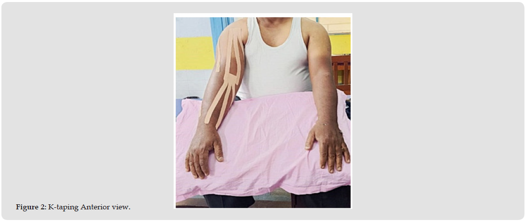
The manual lymph drainage sessions lasted for about 45 mins and involved 3 sets of manual lymph drainage with effleurage technique, distal to proximal, with the limb in elevation(45 degrees) interspersed with active pumping exercises to aid in drainage. Self pumping exercises involved repeated fist making, elbow flexion-extension and shoulder flexion with limb in elevation. Following this, kinesiotaping was applied over the whole right upper extremity. Ktape pieces were cut as per measurement from shoulder to elbow, and from elbow to wrist.2 strips were used for the upper arm and two for the forearm, with each strip being split longitudinally into 3 1/4th inch strips. The terminal 1 inch of strip was keep intact and provided anchorage. The kinesio fan strips were applied with no tension at the distal intact end and 5-15% stretch at the fan tails. The achoring ends were placed near axillary lymph node and cubital node. Measurements of the arm girth were taken before and after the MLD sessions with the help of a tape measure. The measurements were taken at 3 sites- at axilla, 8 cm above the olecranon, 8cm above wrist joint (Figures 1 & 2). For the home exercise program on alternate days, patient was asked to perform self pumping exercises of extremity in elevation, three sets daily lasting around 15 minutes. Further, instructions were given to keep limb in elevation as far as possible and to continue ADL with involved limb, but to refrain from carrying heavy weights. Use of compression garments was discouraged to avoid ambiguity. Skin care practices were encouraged with adequate moisturization to prevent skin breaks.
Figure 1
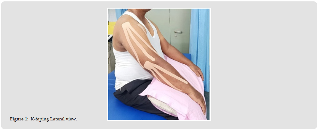
Figure 2
The pre and post intervention data of same day showed reduction in limb girth measurements (Tables 1 & 2), indicating the effectiveness of MLD and pumping exercises in reducing lymphedema. However, the pre-session data recorded on the next session showed increased values of limb girth, indicating the inability to maintain the reduction obtained from previous session (Table 3). The same trends continued for the entire set of data, i.e for the total 10 days of treatment and rest days. The results of MANOVA conducted revealed significant changes across the treatment duration in measures at axilla( p=0.02) and wrist(p=0.03), although Pillai’s trace values recorded were high for all levels. The level of significance at olecranon revealed p=0.1 which suggests no significant difference across the repeated measurements (Table 4).
Table 1: Baseline measures.
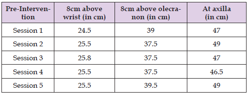
Table 2: Post Intervention measures.
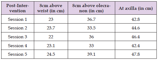
Table 3: Follow up measures.
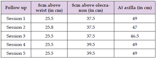
Table 4: Comparison of efficacy of intervention by MANOVA.

Note: *Level of significance p≤0.05
Various literatures have explored the potential efficacy of Kinesiotaping in reduction of lymphedema. Most of these studies focused on secondary lymphedema of upper arm post mastectomy. In women who underwent treatment for breast cancer and had stage 2 and 3 lymphedema, Taradaj et al. examined the impact of KT on the extent of lymphedema and manual dexterity of the upper extremities [1]. The authors came to the conclusion that KT is ineffective in reducing stage 2 and 3 lymphedema in women who had breast cancer treatment after a 4-week therapy regimen. When lymphedema reaches that level, KT cannot take the place of traditional, multi-layered bandaging [2]. Kasawara et al however conducted a meta-analysis of clinical trials that showed efficacy of kinesiotaping in reducing post mastectomy lymphedema, although the superiority of this approach over other conventional ones was not reported [3]. The possible mechanism for the reduction has been proposed to be due to mechanical and sensory changes, enlargement of interfascial space and consequent better lymphatic drainage from the site of application [4]. As per our data, kinesiotaping could not provide the long term maintenance of reduction in volume achieved by MLD. Such deviation from above cited results could be possibly due to a number of factors, including the fact that in this case report, the lymphedema was chronic recurring over a period of greater than one year. The chronic, repetitive nature could have resulted in greater fibroadipose deposition, thereby reducing the effectiveness of KTaping. Whether gender and age could also be a factor in the results differing is a potential area to be explored. The patient was also put on a home exercise program regime and strict adherence to the same by the patient could not be confirmed. We also abstained from the use of pressure garments in order to identify the singular effect of kinesiotape to maintain lymphatic drainage. In retrospection, the use of pressure garments combined with daily sessions of MLD and taping could be beneficial and remains open to research.
Kinesiotaping could not be affirmatively said to aid in lymphedema reduction nor in maintenance of reduction over time, although statistically significant changes were obtained at some levels. The superiority, if any, of kinesiotaping over pressure garments remains open to discussion and research needs to be conducted on a greater sample size to reach an effective conclusion.

International Association of Landscape Archaeology, Czech Glass Society, Czech Republic

Department of Chemistry, Semenov Institute of Chemical Physics, USSR Academy of Sciences, Moscow, Russia

Neurology, LA BioMed Research Institute, USA

Associate Professor at Department of Breast and Thyorid Surgey, Chongqing General Hospital, China

Professor of Nuclear Medicine, Faculty of Medicine and Surgery, University of Milan, Milan, Italy





 Abstract
 PDF
PDF



























