info@biomedres.us
+1 (502) 904-2126
One Westbrook Corporate Center, Suite 300, Westchester, IL 60154, USA
Site Map
 ISSN: 2574 -1241
ISSN: 2574 -1241
ISSN: 2574 -1241
Received: April 14, 2024; Published: June 06, 2024
*Corresponding author: Anubha Bajaj, Consultant Histopathology, AB diagnostics, A-1, Ring Road Rajouri Garden New Delhi, India
DOI: 10.26717/BJSTR.2024.56.008923
Abbreviations: FEA: Flat Epithelial Atypia, ADH: Atypical Ductal Hyperplasia, ALH: Atypical Lobular Hyperplasia, LCIS: Lobular Carcinoma In Situ, PASH: Pseudo-Angiomatous Stromal Hyperplasia
Columnar cell hyperplasia breast is constituted of enlarged terminal duct lobular units [TDLUs] delineating irregular distension of acini. Terminal duct lobular units composed of mammary gland acini are layered by columnar epithelial cells and commonly expound intraluminal secretions and calcification. Generally, lesion is devoid of low grade cytological atypia as observed with flat epithelial atypia or architectural atypia encountered within atypical ductal hyperplasia or ductal carcinoma in situ. As per World Health Organization [WHO] categorization, lesion may be additionally designated as columnar cell change or columnar cell hyperplasia. Notwithstanding, nomenclature as blunt duct adenosis, columnar alteration of lobules, columnar metaplasia, hyperplastic unfolded lobules, hyperplastic enlarged lobular units or enlarged lobular units with columnar alteration are not recommended. Besides, lesions exemplifying cytological atypia are denominated as flat epithelial atypia [FEA]. Commonly implicating breast terminal duct lobular unit, columnar cell lesions as columnar cell changes or columnar cell hyperplasia and flat epithelial atypia may be encountered in ~42% of surgical specimens evaluated for presence of calcification [1,2].
Of obscure etiology, tumefaction may represent as an antecedent non-obligate precursor within low grade breast neoplasia pathway and manifests as an indicator of potential cellular and nuclear atypia within concurrent breast lesions [1,2]. Columnar cell hyperplasia expresses loss of chromosome 16q. Generally, columnar cell hyperplasia enunciates minimal prevalence of molecular alterations. However, progressive accumulation of allelic denaturation through a morphological continuum of columnar cell hyperplasia, atypia and invasive carcinoma breast may emerge [2,3]. Columnar cell hyperplasia is devoid of specific clinical features. Commonly, tumefaction is discerned upon screening mammography, especially lesions associated with micro-calcification [2,3]. Cytological examination expounds variable cellular and nuclear atypia wherein distinction from diverse papillary neoplasms or well differentiated adenocarcinoma may be challenging. Neoplasm is composed of flattened cellular sheets.
Tumor cells configure three dimensional cellular clusters composed of polygonal epithelial cells delineating a distinct cellular perimeter. Tumor cells are permeated with finely granular cytoplasm and enlarged nuclei. Few myoepithelial cells appear commingled with neoplastic cells wherein palisading columnar epithelial cells are disseminated peripherally [3,4]. Grossly, columnar cell hyperplasia is devoid of specific macroscopic features. Upon microscopy, columnar cell change implicates terminal duct lobular units with the occurrence of irregular, variably distended acini. Breast acini are layered with singular or dual epithelial cell layers wherein layering epithelial cells expound uniform, ovoid to elongated nuclei which are oriented perpendicular to basement membrane. Tumor cells frequently depict apical snouts. Intra-luminal secretions and calcification may be observed [3,4]. Columnar cell hyperplasia exemplifies irregular, variably distended acini of terminal duct lobular unit. Breast acini are layered with stratified epithelial cells constituted of ≥ 2 cell layers.
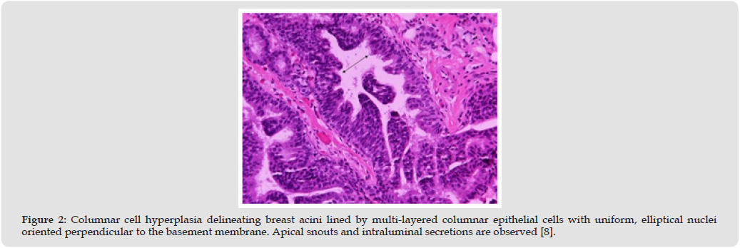
Epithelial cells may configure tufts or mounds. Layering epithelial cells are impregnated with uniform, ovoid to elongated nuclei which appear to articulate nuclear crowding and overlapping. Apical snouts are frequently discerned. Intra-luminal secretions and calcification may be discerned [3,4]. Columnar cells change or columnar cell hyperplasia demonstrating cytological atypia is constituted of tumor cell impregnated with spherical nuclei delineating irregular nuclear chromatin, variably prominent nucleoli, and enhanced nucleocytoplasmic ratio. Aforesaid lesions may be categorized as flat epithelial atypia. Tumor cells expound loss of orientation and appear perpendicular w.r.t basement membrane. Notwithstanding, morphological variations as rigid bars, arcades and true micro-papillary articulations are encountered within atypical ductal hyperplasia or low-grade ductal carcinoma in situ [3,4] (Figures 1 & 2).
Figure 1
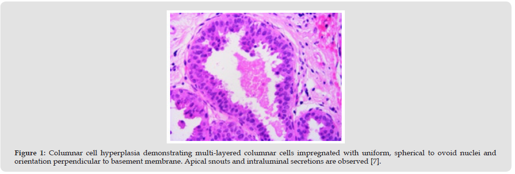
Figure 2
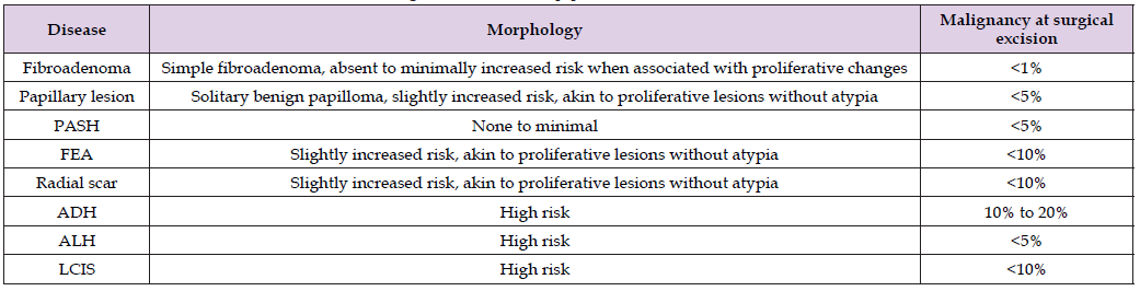
Columnar cell hyperplasia expounds diffuse, intense immune reactivity to estrogen receptors [ER] and progesterone receptors [PR], low molecular weight keratins as CK8, CK18, CK19, BCL2 or E-cadherin. Tumor cells appear immune nonreactive to high molecular keratin as CK5, CK5/6 or 34β E12 [5,6] (Table 1). Columnar cell hyperplasia breast requires segregation from neoplasms as mammary gland cyst, apocrine metaplasia, cystic hyper-secretory hyperplasia, flat epithelial atypia, atypical ductal hyperplasia, low grade ductal carcinoma in situ or ductal carcinoma in situ, clinging subtype [5,6]. Neoplasm may be appropriately ascertained by histological assessment of image guided core needle biopsy or surgical excision specimens. Upon mammographic imaging, heterogeneous, dense breast tissue appears admixed with clusters of fine or amorphous, pleomorphic calcification. However, imaging features as branching of micro-calcification are infrequently observed [5,6]. Surgical excision appears superfluous in instances where columnar cell change or columnar cell hyperplasia devoid of atypia emerges as a significant morphological feature upon evaluation of core needle tissue biopsy specimens [5,6].
Table 1: Lifetime Risk of Carcinoma Breast in Benign Breast Disease [4].
Additionally, adjuvant therapeutic intervention of preponderant columnar cell lesion discerned upon histological assessment of surgical [7,8] excision specimens appears unnecessary. Proportionate emergence of malignant metamorphosis and invasive carcinoma breast is minimally enhanced with a relative risk of ~1.5. However, possible malignant transition of columnar cell changes or columnar cell hyperplasia appears concordant with concurrent proliferative breast lesions [5,6]. Columnar cell hyperplasia may be concordant with diverse breast lesions pre-eminently associated with low grade breast neoplasia pathway as flat epithelial atypia, atypical ductal hyperplasia, low grade ductal carcinoma in situ or lobular neoplasia [5,6].

International Association of Landscape Archaeology, Czech Glass Society, Czech Republic

Department of Chemistry, Semenov Institute of Chemical Physics, USSR Academy of Sciences, Moscow, Russia

Associate Professor at Department of Breast and Thyorid Surgey, Chongqing General Hospital, China

Professor of Nuclear Medicine, Faculty of Medicine and Surgery, University of Milan, Milan, Italy

Department of Pharmaceutical Sciences, South University School of Pharmacy, USA





 Abstract
 PDF
PDF



























