info@biomedres.us
+1 (502) 904-2126
One Westbrook Corporate Center, Suite 300, Westchester, IL 60154, USA
Site Map
 ISSN: 2574 -1241
ISSN: 2574 -1241
ISSN: 2574 -1241
Received:January 26, 2023; Published:February 13, 2023
*Corresponding author: Mayumi Inaba, Department of Pathology, Yodogawa Christian’s Hospital, Japan
DOI: 10.26717/BJSTR.2023.48.007677
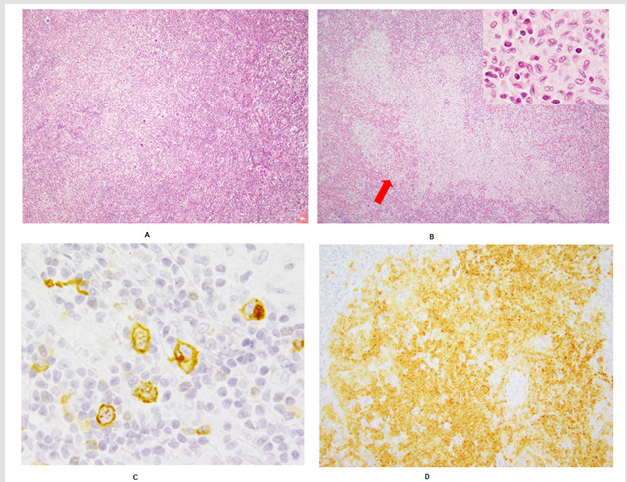
A 79-year-old man was presented with bilateral axillary lymphadenopathy. The biopsied nodes were obliterated by vague nodules, composed of non-cohesive proliferation of giant cells, including Reed-Sternberg cells, and various inflammatory cells (panel A; x10 objective, inset; x100 objective). In addition, a single well demarcated Langerhans cell (LC) cluster was identified as a “tumor in tumor” pattern (panel B; red arrows, x10 objective, inset; x100 objective). Immunohistochemistry and ISH revealed the giant cells to be positive for CD15 (panel C; x40 objective), CD30, PAX5, and EBER. The LCs were positive for CD207 (Langerin) (panel D; x20 objective), and CD1a. BRAF V600E-expressing cells were absent. A diagnosis of classic Hodgkin lymphoma (mixed cellularity type) with possible LC histiocytosis was given (Figure 1A). The patient is free of disease for 27 months after chemotherapy
The interpretation that LCH is a neoplastic disorder derived from LCs has been accepted as in WHO classification, revised 4th ed, (WHO- 2017) [1], for example. For diagnosing LCH, CD207 / Langerin expression should be confirmed immunohistochemically, in addition to histologic findings (Figure 1B). Although clonality of LCH was previously suggested only in a part of the female cases, a recurrent BRAF V600E mutation was recently found in more than a half of patients with LCH [2], and the possibility of its neoplastic nature has been increasing. However, there are still some arguments about the possibility of being an inflammatory process [3]. One study has shown that some of the BRAF V600E-negative LCH cases had BRAF V600E mutation (Figure 1C). It implies that BRAF V600E should be assessed by direct sequencing [4]. Also, the frequency of MAPK2K1 mutation appears to be high in BRAFV 600E mutation-negative LCH cases [5].
The development of LCH with lymphoid and myeloid neoplasms is known as one of a feature of LCH [6]. Egeler, et al. reported 91 patients with LCH having other neoplasia (Figure 1D). In their series, 39 patients had lymphoma consisting of 25 patients with “Hodgkin’s disease” at that time [7]. Reactive or neoplastic nature of LC proliferation and clonal relation between it and CHL in our case are unclear, because BRAF V600E was immunohistochemically negative in both processes [8-14]. Very focal LC proliferation hampered further study. Accumulation of such cases is required to characterize significance of “tumor in tumor” of this combination.
Figure 1.

International Association of Landscape Archaeology, Czech Glass Society, Czech Republic

Department of Chemistry, Semenov Institute of Chemical Physics, USSR Academy of Sciences, Moscow, Russia

Neurology, LA BioMed Research Institute, USA

Associate Professor at Department of Breast and Thyorid Surgey, Chongqing General Hospital, China

Clinical Radiologist (MD) - Department of RADIOLOGY, Cosenza Hospital, Cosenza, Italy





 Abstract
 PDF
PDF



























