Impact Factor : 0.548
- NLM ID: 101723284
- OCoLC: 999826537
- LCCN: 2017202541
Bi Bi Zainab Mazhari1*, Dayanand Agsar2, Mohammed H Saiemaldahr1, Waleed Abu AlSoud1, Ahmad Adam1, Amina Qaeet Alrowaily1 and Intisar Elshek1
Received: September 16, 2018; Published: September 20, 2018;
*Corresponding author: Bi Bi Zainab Mazhari, Department of Microbiology, Jouf University, Al Jouf, Saudi Arabia
DOI: 10.26717/BJSTR.2018.09.001764
In the present investigation, an attempt was made to explore the anticancer activity of extracellular gold nanoparticles from Streptomyces sp. The synthesized gold nanoparticles were characterized by UV-vis spectrophotometry and scanning electron microscopy. The presence of gold nanoparticles after the synthesis was examined by recording the UV-vis spectra of the solution and also by visual biomonitoring of the solution. The peaks recorded in the UV-vis spectra of the solutions was in between the wavelength of 500 to 600nm which confirms the presence of gold nanoparticles. Scanning electron microscopy reveals the synthesis of gold nanoparticles with an average size 27 to 56nm in the solution. Different concentrations of gold nanoparticles (200 - 10^l) were subjected to illustrate the anticancer activity using cell lines namely adeno carcinoma (HT 29), kidney cancer (K 293) and breast cancer (MDA 231). The percent inhibition for these cell lines was recorded and observed to be very high at 200^1. The investigation revealing the anticancer activity of gold nanoparticles has been reported for the first time. Gold nanoparticles could be used as cost effective and efficient means of future devices in medical sciences.
In modern scenario nanotechnology is used to provide more accurate and timely medical information for diagnosing and treating human cancers. Cancers is a complex disease occurring as a result of a progressive accumulation of genetics and epigenetics changes that unable escape from normal cellular and environmental control [1-3]. Cancer is the leading cause worldwide [4]. Traditionally the most common cancer treatment was limited to chemotherapy radiation and surgery. Limitations in cancer treatment are result of current challenges which leads to the development of cancer therapies including lack of early disease detection, nonspecific systemic distribution, in adequate drug concentration reaching the tumor and ability to monitor therapeutic responses [5]. Many attempts have been made to use silver nanoparticles as an anticancer agent and they have all turned up positive [6].
The role of gold nanoparticles as an anticancer agent should open new doors in the field of medicine [7]. In view of this an attempt was made to focus on the role of microbially synthesized gold nanoparticles in cancer diagnosis and therapy. The present study was carried out to prove the anticancer action of gold nanoparticles synthesized using Streptomyces tuirus DBZ39 on Adenocarcinoma (HT 29), Kidney cancer (K 293) and breast cancer (MDA 231) evaluating the number of viable cells after incubation with the microbially synthesized extracellular gold nanoparticles at different concentrations and duration of exposure.
An isolate of Streptomyces tuirus DBZ39 obtained from limestone quarry soil earlier was employed for the synthesis of extracellular gold nanoparticles, as per the standard protocol [68]. A loopful of three days old culture of Streptomyces tuirus DBZ39 was inoculated into starch casein broth containing Starch-1g, casein-0.003g, KH2PO4- 2.0g, KNO3-2.0g, NaCl-2.0g, MgSO4-0.002g, FeSO4-0.001g, CaCo3-0.001g and incubated at 400C for 5 days on rotatory shaker (200rpm). After incubation, the broth culture was centrifuged at 8000 rpm. The biomass obtained was suspended in Aurium Chloride (AuCl4) solution and kept for incubation at 370C on shaker (200rpm) for three days. The gold nanoparticles synthesized in the solution were visually confirmed by the development of deep purple colour and UV-visible absorption spectrum in the range of 500-550nm. The gold nanoparticles produced were characterized by Scanning electron microscopy for their size, shape and dispersion as per the standard protocols [9,10].
Anticancer activity of gold nanoparticles was carried out by following the standard procedure prescribed by Marques-Gallego et al., [11]
HT-29 (human adeno carcinoma), K-293 (human kidney), MDA- 231 (human breast cancer) cell lines were grown as adherent in DMEM media, whereas MCF-7 (Breast cancer lines) were grown in MEM media supplemented with 10% fetal bovine serum, 100μg/ml penicillin, 200μg/ml Streptomycin, 2mm L-glutamine and culture was maintained in a humidified atmosphere with 5% CO2.
Stock solution of 10mg/ml stock solution in DMSO, from the above stock was made with sterile water to get required concentration.
Toxicity of test compound in cells was determined by MTT assay based on mitochondrial reduction of yellow MTT tetrazdium dye to highly coloured blue formazen product. 1x10⁴ cells (counted by Trypan exclusion dye method) in 96 well plates were incubated with compounds with series of concentrations tested for 48 hrs at 370C in DMEM/MEM with 10% FBS medium. Then the above media was replaced with 90μl of fresh serum free media and 10μl of MTT reagent (5mg/ml) and plates were incubated at 370C for 4hrs, thereafter the above media was replaced with 200μl of DMSO and incubated at 370C for 10min. The absorbance at 570nm was measured on a spectrophotometer (spectra mass, molecular devices) IC-50 values were determined from plot: % inhibition (from control) Vs Concentration [11-13].
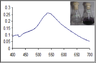
Figure 1: Visual observation and UV-vis absorbance spectra of gold nanoparticles synthesized by Streptomyces tuirus DBZ39.

Synthesis of gold nanoparticles was carried out by Streptomyces tuirus DBZ39 as explained earlier under materials and methods. The presence of gold nanoparticles after the synthesis was examined by recording the UV-vis spectra of the solution and also by visual biomonitoring of the solution. The peaks recorded in the UV-vis spectra of the solutions was in between the wavelength of 500 to 600nm confirms the presence of gold nanoparticles (Figure 1). The development of deep red colour uniformly throughout the solution was recorded as extracellular synthesis (Figure 1). Nanoparticles composed of gold offer, in addition to their enhanced absorption and scattering, good biocompatibility, facile synthesis and conjugation to a variety of biomolecular ligands, antibodies and other targeting moieties, making them suitable for use of biochemical sensing and detection, medical diagnostic and therapeutic applications [14-21].
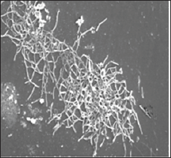
There have been several demonstrations of bioaffinity sensors based on the plasmon absorption and scattering of nanoparticles and their assemblies [23]. UV, XRD, EDAX and electron microscopic (SEM and TEM) studies are important and normally employed for the detection and characterization of nanoparticles. In the present study, Scanning Electron Microscopy is employed to detect and characterize the extracellular gold nanoparticles synthesized by Streptomyces tuirus DBZ39. Scanning electron microscopy of gold nanoparticles produced by Streptomyces tuirus DBZ39 is presented in Figure 2. Production of spherical nanoparticles with an average size of 27-56 nm of were observed.
Figure 2: Scanning electron Microscopy of gold nanoparticles synthesized by Streptomyces tuirus DBZ39.

The development of nanobiotechnology has progressed by leaps and bounds. Engineered and desired nanoparticles have been mass produced microbially and are being widely applied as commercial and emerging biomedical agents. Despite the rapid progress and earlier acceptance of microbial nanotechnology, the potential for adverse health effects due to short and long term exposure has not yet being established. However, gold nanoparticles acting on the living cells at nanolevel resulting not only is biologically desirable, but also generates undesirable affects. Though, many effects of gold nanoparticles have been aimed at exploiting beneficial properties for various purposes, there are limited attempts to evaluate the probable ill effects of these materials. Therefore, it is necessary that, the safety of newly synthesized gold nanoparticles that influence their associated hazards are understood.
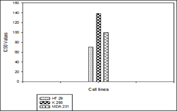
An important area governing regulatory health risk assessment is carcinogenicity of gold nanoparticles. In view of this, gold nanoparticles with a size of 27 to 56nm, at different concentrations were used to know the anticancer activity against different cell lines. Different cell lines to study the anticancer activity of gold nanoparticles, namely adeno carcinoma (HT 29), kidney cancer (K 293) and breast cancer (MDA 231) were subjected at different concentrations (200 - 10μl) of gold nanoparticle solution. The percent inhibition of all the three cell lines was recorded and observed to be very high at 200μl concentrations (Table 1). The anticancer activity of kidney cancer cell lines (K - 293) recorded to be significantly very high with high IC50 value 139 (Figure 3). Nano medicine concerns the use of precision - engineered nanoparticles to develop novel anticancer therapeutic agents for human use. The present report demonstrates the efficacy of gold nanoparticles synthesized by Streptomyces as an anticancer agent.
Table 1: Anticancer activity of gold nanoparticles synthesized by Streptomyces tuirus DBZ39 against different cell lines.

Figure 3: Anticancer activity of gold nanoparticles by Streptomyces tuirus DBZ39 at different IC50 values.

The novel isolate Streptomyces tuirus DBZ39 prove to be potential for the synthesis of extracellular gold nanoparticles was used to evaluate the anticancer activity at different concentrations. Cell lines namely adeno carcinoma (HT 29), kidney cancer (K 293) and breast cancer (MDA 231) were subjected to test the anticancer activity of gold nanoparticles at different concentrations (200 - 10pl). The percent inhibition of these cell lines was recorded to be very high at 200pl concentrations for kidney cancer cell lines (K - 293) with high IC50 value. This investigation reveals notably high anticancer activity of Streptomyces mediated gold nanoparticles at high concentration, which can be considered as good anticancer attribute and promising natural agent against tumor cells which is of important contribution in future for medical therapies.


