Impact Factor : 0.548
- NLM ID: 101723284
- OCoLC: 999826537
- LCCN: 2017202541
Masahiro Kitami*
Received: May 27, 2018; Published: June 05, 2018
*Corresponding author: Masahiro Kitami, Department of Diagnostic Radiology, Tohoku University Graduate School of Medicine, 2-1 Seiryo-cho, Sendai, 980-8574 Japan
DOI: 10.26717/BJSTR.2018.05.001159
Unilateral genital hypertrophy is rare phenomenon, occurring in both boys and girls. due to their rarity and laterality, these can be misdiagnosed as malignant tumors, resulting in unnecessary biopsy or operation. Therefore, correct recognition of these tumor mimickers is important. We presented representative cases of childhood asymmetrical labium majus enlargement (CALME) and unilateral enlargement of the testis. Typical radiological feature of CALME is the unilateral swelling of labia majora with heterogeneous signal intensity with low-intensity strands on magnetic resonance imaging. On the other hand, homogenous enlargement is the key feature for unilateral enlargement of the testis.
Keywords: Unilateral genital hypertrophy, Physiologic finding, Hypertrophy, Labia, Vulva, Testis, Swelling, Enlargement, Overgrowth, Hyperplasia
Abbrevations: CALME: childhood Asymmetrical Labium Majus Enlargement, MRI: Magnetic Resonance Imaging
Unilateral vulval or testicular hypertrophy is peculiar phenomenon in puberty. These are physiologic hormonal response and do not require any surgical procedures. However, due to their rarity and laterality, these can be misdiagnosed as malignant tumors, possibly resulting in unnecessary biopsy or operation. Therefore, recognition of these phenomena is important. We present here the representative cases, and discus the key points for correct diagnosis.
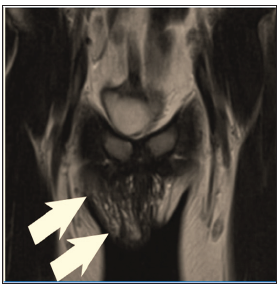
10 year-old girl was referred to our hospital, with unilateral vulval “mass”. 3 years ago, she had noticed right inguinal-vulval bulge, and visited the hospital. At the time of medical examination, the bulge had decreased in size, and an inguinal hernia or hydrocele was suspected. Although conclusive diagnosis was not obtained even after sonography, observation was adopted. She re-visited the hospital because the bulge recently increased. At the time of examination, the bulge had decreased in size once again. Magnetic resonance imaging (MRI) was performed for further examination with suspicion of inguinal hernia or hydrocele. MRI Figure 1 showed the swelling of labia majora with heterogeneous intensity with low-intensity strands on T1-weighted image (WI) and T2WI. Despite of vulval swelling, the margin was obscure and there was no discrete mass. Diffusion weighted image showed no restriction. Sonography also showed heterogeneous echogenicity without discrete mass (not shown). There was no laboratory data suggesting malignancy such as tumor marker elevations. Based on the clinical and radiological findings, the diagnosis of childhood asymmetrical labium majus enlargement (CALME) was obtained.
Figure 1: 10 year-old girl with childhood asymmetrical labium majus enlargement (CALME) Coronal MRI (T2 weighted image) shows right labium majus swelling without discrete mass formation. The lesion shows heterogeneous intensity with internal low intensity strands. Together with clinical course of fluctuating size, and absence of laboratory findings suggesting malignancy, diagnosis of childhood symmetrical labium majus enlargement was obtained.

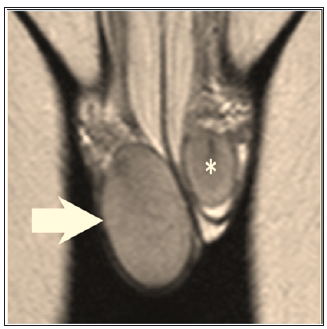
11-year-old boy presented with the complaint of right testicular unpainful swelling from two months ago. Sonography, performed with suspicion of testicular tumors, showed right testicular homogenous swelling without mass formation. MRI also showed right testicular homogenous swelling without mass (Figure 2). There was no laboratory data suggesting malignancy such as tumor marker elevations. Together with radiological and laboratory findings, diagnosis of unilateral enlargement of the testis was obtained.
Figure 2: 11 year-old boy with unilateral enlargement of the testis.Coronal MRI (T2 weighted image) shows right testicular homogenous swelling without mass formation. Together with sonographic homogeneous finding and absence of laboratory findings suggesting malignancy, diagnosis of unilateral enlargement of the testis was obtained.

Unilateral vulval hypertrophy, and testicular hypertrophy are peculiar phenomena in pediatrics. Both entities could be misdiagnosed as malignant tumors, possibly leading to unnecessary surgical procedures. Although these phenomena were not widely recognized by clinicians and radiologists, these recognitions are crucial to avoid unnecessary invasive procedures. Childhood asymmetrical labium majus enlargement or CALME is also known as prepubertal unilateral fibrous hyperplasia (PUFH), vulvar hamartoma, prepubertal vulval fibroma, fibrous hyperplasia of labium majus [1]. This is the characteristic phenomenon showing prepubertal or pubertal unilateral hypertrophy of labia majora. Etiology is not fully understood, but some author suggested that this could be a physiologic enlargement in response to hormonal surges of pre- and early puberty [2]. Peculiar clinical course with enlargement and reduction, which might mislead clinicians to the misdiagnosis as an inguinal hernia as in our case, could be explained by the physiologic hormonal fluctuation.
Pathology shows non-specific findings with spindled fibroblasts and prominent elastic fibers and myxoid extracellular matrix, and without abnormal cells. Therefore, pathological diagnosis is difficult [2,3]. Besides, surgical resection is also difficult because of unclear margin, resulting in 50% recurrence rate [2]. Therefore, biopsy and surgical resection is not recommended and conservative approach has been proposed [4]. Sonography and MRI shows unilateral ill-defined labial enlargement without a definable mass. MRI also shows hypointensity strands on T1WI and T2WI [1]. Unilateral enlargement also occurs in Testis [5-7]. But this phenomenon seems much less recognized and does not have specific nomenclature. This is described simply as unilateral enlargement of the testis, transitory unilateral testis enlargement of puberty, unilateral testicular end-organ hypersensitivity, unilateral asymptomatic testis enlargement and so on.
Although testicular enlargement suggests the possibility of malignant tumors, the completely normal finding on radiological assessment is the strong evidence of the diagnosis, and surgical exploration is not required [7]. Differential diagnoses of unilateral testicular enlargement include schistosomiasis, mastocytoma, Mc- Cune Albright syndrome, and Beckwith-Wiedemann syndromes. Therefore, clinical assessment as well as radiological evaluation is also necessary. Interestingly, this unilateral testicular enlargement could be recognized as the male counterpart of CALME, because this is ascribed to the hypersensitivity to hormone [7], and both phenomena are mimicking malignant tumors.
We discussed two entities of unilateral genital hypertrophies in boys (unilateral enlargement of the testis) and girls (CALME). These are tumor mimickers in pediatrics, and appropriate recognition is crucial to avoid unnecessary surgical intervention.


