info@biomedres.us
+1 (502) 904-2126
One Westbrook Corporate Center, Suite 300, Westchester, IL 60154, USA
Site Map
 ISSN: 2574 -1241
ISSN: 2574 -1241
ISSN: 2574 -1241
Received: August 28, 2023; Published: October 26, 2023
*Corresponding author: Iveta Tasheva, PhD, Hospital SofiaMed, Sofia University, Bulgaria
DOI: 10.26717/BJSTR.2023.53.008411
Aim: The aim of this study is to report our experience in endovascular treatment of superior and inferior vena cava syndrome.
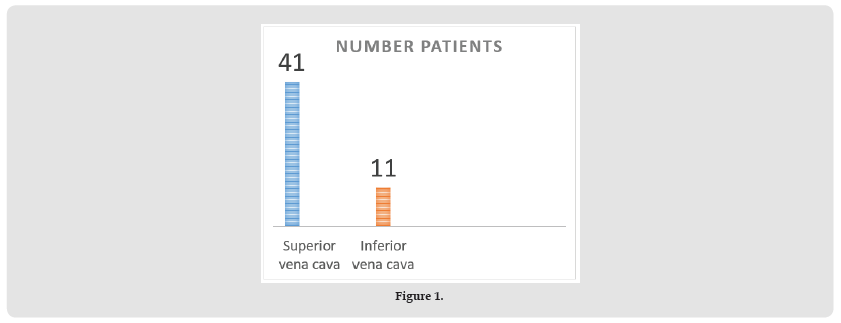
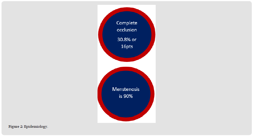
Methods: Between June 2013 and August 2019, 52 patients (mean age 54.75 years; minimum 20yo, maximum 81yo) were endovascularly treated with self-expandable stents of stenosed superior (78.8% - 41 pts) or inferior (21.2% - 11 pts) vena cava. All patients presented with vena cava obstruction syndrome. Complete occlusion was found in 30.8% - 16pts and mean percent of stenosis was 89.92%. The two main causes of the caval obstructions were carcinoma (82.7%, 43pts) and thrombophilia (7.7%, 4 pts) (Figure 1).
Figure 1
• Treatment and complications Successful procedures were 97.8%.
• Balloon predilataion.
• Stent placement – self expandable stents (Sinus XL, Protégé, Abre).
• Balloon postdilation.
• Two major complications (3.8%).
• Thrombus migration causing pulmonary embolism (Figure 2).
Figure 2
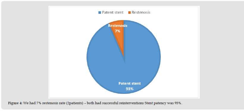
Both of them treated endovascularly with thrombus fragmentation, local fibrinolysis and aspiration. In nearly all patients stents were placed (97.8%) and there was clinical improvement of the caval syndrome- considerable reduction of edema and symptom relief in the treated patients. Of all 52 procedures we had two major intra-procedural complications (3.8%) – both migration of thrombus material causing pulmonary embolism, which was treated with intrapulmonary thrombus fragmentation and thrombolysis. All patients were clinically followed-up; 59.6% were followed-up by CT scan or flow Doppler ultrasound up to 65 months after procedure, with a median follow up period of 1 month (30-1950 days). Stent patency was 93.5% (29 pts); significant stent restenosis or occlusion were found in 2 cases (6.5%) and were successfully treated endovascularly (Figure 3).
• All patients had relief of vena cava syndrome.
• All patients were clinically followed up – up to 65 months.
• Median follow up period was 1 month (30-1950 days).
• CT - scan or Doppler ultrasound follow up was done in 59.6% of patients (Figure 4) [1-4].
Figure 3
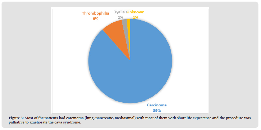
Figure 4
Endovascular treatment with stent implantation for superior and inferior vena cava syndrome is a minimal invasive and safe procedure with favorable clinical effect and satisfactory mid-term result.

International Association of Landscape Archaeology, Czech Glass Society, Czech Republic

Department of Chemistry, Semenov Institute of Chemical Physics, USSR Academy of Sciences, Moscow, Russia

Department of Educational Sciences, Research Centre Qualities of Teachers, School of Education – HAN, Nijmegen, Netherlands

Associate Professor at Department of Breast and Thyorid Surgey, Chongqing General Hospital, China

Neurology, LA BioMed Research Institute, USA





 Abstract
 PDF
PDF



























