Impact Factor : 0.548
- NLM ID: 101723284
- OCoLC: 999826537
- LCCN: 2017202541
Karin Lorinson1*, Sibylle Kneissl2, Alexander Tichy3 and Dragan Lorinson1
Received: April 05, 2023; Published: April 12, 2023
*Corresponding author: Karin Lorinson, Chirurgisches Zentrum für Kleintiere Dr. Lorinson, Marktstrasse 19, 2331 Vösendorf, Austria
DOI: 10.26717/BJSTR.2023.49.007842
Objective: Proximodistal positional changes of the patella can result in patellar luxation, pain or gait abnormalities which is commonly known in humans and suspected in dogs. To evaluate patellar height in radiographs, this study focused on measuring the TPPA (tibia plateau-patella angle) applied for the first time in cats.
Study Design: One hundred and nine pairs of mediolateral stifle radiographs in adult cats without prior stifle surgery and malalignment were reviewed by two independent observers. TPPA and stifle angle were assessed. Joints were classified into three goniometric groups: less than 80 degrees (n = 1), between 80 and less than 130 degrees (n = 51),130 degrees and higher (n = 57).
Results: TPPA ranged from 23 to 67.3 degrees, goniometry varied from 54 to 162 degrees both with a statistically significant inter-observer correlation (p < 0.0001). TPPA and goniometry negatively correlated in both observers (p < 0.01).
Conclusion: Anatomical landmark determination, especially the caudal aspect of the tibial plateau and the distal ending of the articular surface of the patella might be tricky to assess. In contrast to humans, TPPA significantly decreased the more the stifle joint was extended. Therefore, consecutive measurements should only be compared on radiographs with similar joint angulation.
Keywords: Patellar Height; Tibia Plateau-Patellar Angle; Radiography; Cat
The patella is a motorical important part of the feline stifle joint. By gliding proximally and distally inside the trochlear groove, its position continually changes during each step of mobility. Stifle diseases like patellar luxation, cruciate ligament rupture, patella or patellar tendon disease as well as consecutive surgical interventions may cause patellar dislocation. Although mediolateral patellar dislocation is well documented in veterinary medicine, proximodistal positional changes of the patella in feline patients have not been focused on yet. In dogs, radiographic evaluation of the patellar height was performed using several indices well-known in human medicine, eg the (modified) Insall-Salvati index, Blackburne-Peel index or de Carvalho index [1-7]. The tibia plateau-patellar angle (TPPA) is a simple diagnostic tool from Portner and Pakzad (2011) used in humans and dogs [7,9-16]. To eliminate index calculations, this method only requires a single angle measurement without additional calculation. By performing this angular measurement, there is no interference of radiographic magnification or patient size [8]. The purpose of this study was to investigate the feasibility of TPPA measurements in feline mediolateral radiographs, to assess reproducible radiographic landmarks and to evaluate the influence of stifle flexion on angle size. The hypothesis was that TPPA measurements would be reproducible in cats and TPPA would decrease with extension and increase with flexion of the stifle joint.
Radiographs
One hundred and nine mediolateral radiographs of feline stifle joints were retrospectively evaluated. Craniocaudal radiographs of these patients were used to rule out hindlimb malformations or malalignments. Also, radiographs showing immature bone formation with open epiphyses as well as postoperative radiographs were excluded from further evaluation.
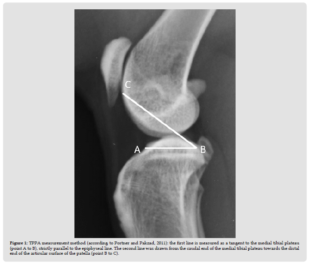
Figure 1 TPPA measurement method (according to Portner and Pakzad, 2011): the first line is measured as a tangent to the medial tibial plateau (point A to B), strictly parallel to the epiphyseal line. The second line was drawn from the caudal end of the medial tibial plateau towards the distal end of the articular surface of the patella (point B to C).

Radiographic Analysis
Using JiveX Dicom Viewer (Visus Health IT GmbH, Bochum, Germany), radiographs were orientated with the patella showing to the left. With the open angle measurement tool, the mid-shaft femoral and tibial longitudinal axes were marked. The resulting stifle angle was calculated and documented as the goniometric measurement. For TPPA measurements, the closed angle measurement tool was used. According to Portner and Pakzad (2011), the tibial plateau was measured as a tangent to the medial tibial plateau comparable to the TPLO measurements (point A to B), strictly parallel to the epiphyseal line. The second line was drawn from the caudal end of the medial tibial plateau towards the distal end of the articular surface of the patella (point B to C, Figure 1). The resulting TPPA was automatically calculated and documented as TPPA measurement. In a pilot study, consensus of anatomical measurement details was established in about 30 cases. Goniometric and TPPA measurements were performed by two different observers. Goniometric measurements were divided into three groups. Group 1 contains mediolateral stifle radiographs with a femoral-tibial angle of less than 80 degrees. Group 2 were goniometric results between 80 and less than 130 degrees and group 3 those with 130 degrees and higher. Group 1 contained only one cat (0.9 %) and was excluded from further statistical analysis. Group 2 contained 51 cats (46.8 %) and group 3 57 cats (52.3 %).
Statistical Analysis
All statistical analyses were performed using IBM SPSS v 27. Correlations between all measurements were calculated using Pearson’s correlation coefficient. The agreement between the two observers in goniometry and TPPA measures was analyzed using intraclass correlation coefficient (ICC). In addition, cases were divided into three groups according to the measured goniometric degrees (< 80; 80 - 129; ≥ 130). The < 80-group was excluded from further analysis due to the low number of cases. The difference in the mean measured goniometry and TPPA between the two groups (80 – 129; ≥ 130) was analyzed using T-test for independent samples. The assumption of normal distribution for ICC and T-test was assessed using the Kolmogorov-Smirnov-test. For all statistical analyses, a p-value below 5 % (p < 0.05) was seen as significant.
Goniometric measurements of the 109 cats provided a radiological range between 54 degrees of flexion and 162 degrees of extension. Inter-observer agreement was high (r = 0.988; p < 0.0001). TPPA measurements in all 109 cats ranged from 23 to 67.3 degrees. Interobserver correlations were significant (r = 0.903; p < 0.0001). TPPA and goniometric measurements correlated significantly (observer 1: r = -0.625; observer 2: r = -0.653; p < 0.01). Increasing extension of the stifle joint resulted in reduction of the TPPA. Goniometric group 2 (80 – 129 degrees) revealed mean TPPA measurements of 45.11 ± 7.75 degrees, whereas goniometric group 3 (130-162 degrees) revealed mean TPPA measurements of 38.38 ± 5.07 degrees. These results were statistically significant different (p < 0.0001).Point A (cranial aspect of the tibia plateau) determination revealed no major problems in both observers. The A-B line was placed strictly parallel to the epiphyseal line ending at the caudal tibial plateau where the caudodorsal part is directed caudally not dorsally. Point C was determined as the most distal aspect of the articular surface before the bony configuration shows a cranial deviation (Figure 2).
Patellar disease in cats commonly includes patellar luxation, fractures and osteochondrosis [17,18]. To the authors´ knowledge, proximodistal positional dislocation of the feline patella has not been investigated so far. Knowing that total knee arthroplasty or proximal tibial surgery may alter patellar height in humans [13,19-21] and dogs [7] this fact might also be true for feline patients. This study focused on the basic determination of the patellar height in adult, fracture-free feline stifle joints using the TPPA measurement method. In contrast to this angular measurement technique, the (in humans and dogs) widely used patellar height index measurements require correct and reproducible anatomical landmark determination and afterwards the calculation for quotients. In humans, four major advantages of the TPPA measurement technique were indicated [8,15]:
1. No calculations are required.
2. This technique is independent from radiographic magnification,
3. From the physical size of the knee and
4. From the degree of the knee flexion (as long as it is more than 30 degrees to keep the patellar tendon taut). In dogs, TPPA was evaluated in TPLO and TTA dogs pre- and postoperatively. In these patients, all stifle radiographs were positioned in an approximately 90-degree angle to get reproducible results [7]. In the herein presented study, mediolateral radiographs of feline stifle joints were evaluated retrospectively. Therefore, positioning was not standardized resulting in goniometric measurements between 54 and 162 degrees. Based on the results of this paper, TPPA measurements significantly correlated to the goniometric results. Therefore, consecutive TPPA measurements should only be compared if goniometric stifle angles are within a small range. This is a major difference to results measured in humans [8].TPPA in Turkish adults appeared to be a feasible tool that assessed patellar height with higher reliability compared to Insall-Salvati, modified Insall-Salvati, Blackburne-Peel and Caton-Deschamps indices [16]. In dogs, TPPA results correlated inconsistently with the Insall-Salvati and the Blackburne-Peel indices [7]. In feline literature, the above-mentioned indices have not been evaluated yet.
Key factor for correct and reproducible measurements was the identification of the points A (cranial tibial plateau), B (caudal tibial plateau) and C (distal end of the articular patella surface). Minimal deviations resulted in calculated angle differences of several degrees. In humans, no difficulties in detecting the anatomical landmarks were mentioned in literature but radiographs of poor technical quality and mal-positioned radiographs were ruled out [8]. In this study, like in human studies [8,13], inter-observer correlation was significant. In summary, TPPA measurements in cats are feasible but produce results of a broad range. Reproducible results need a goniometrical comparable positioning of the stifle joint and a thorough determination of the anatomical landmarks necessary for radiological measurement.
K.L., D.L. conceptualized the study. K.L., S.K. designed the study, K.L., S.K. acquired the data. K.L., S.K., A.T., D.L. did data analysis and interpretation. All authors drafted, revised and approved the submitted manuscript.
None declared.


