Impact Factor : 0.548
- NLM ID: 101723284
- OCoLC: 999826537
- LCCN: 2017202541
Carolina Virga*, Aguzzi A, Moro C, De Leonardi A and De Leonardi G
Received: February 09, 2023; Published: February 14, 2023
*Corresponding author: Carolina Virga, Chairs of Pharmacology and Therapeutics A and B - Faculty of Dentistry, National University of Córdoba, Spain
DOI: 10.26717/BJSTR.2023.48.007683
Knowledge of the remodeling process of the post-extraction socket that generates changes in the clinical profile of the patient, as well as the possible therapies available, are essential for planning dental treatment. The aim of this work was to study the effect of topical administration of Alendronate (AL) and Arnica montana (AM) gels on post-extraction socket repair. Materials and methods: AL and AM were used in the form of gels. Control (C) was carboxymethylcellulose gel. The effect was analyzed in male Wistar rats weighing 120 ± 20 g, divided into 4 groups of 16 rats each. Animals were anesthetized with a ketamine/xylazine solution. Extraction of the lower first molar was performed. Said alveoli were filled with the gels. The animals were treated according to universal standards of asepsis. They were euthanized by intracardiac injection of potassium chloride, under general anesthesia. At 0, 7, 15, 30, and 45 days, 3D DICOM images of rat jaws (TC-CB Romexis – Planmeca) were taken to measure the volumetric radiographic density of the surgical site areas. The comparison of the data was made by analysis of variance to two classification criteria. Differences were considered significant if p<0.05. Results: When evaluating the density data by two-way ANOVA (group and stage), only the stage factor was significant (p<0.05), observing an increasing trend of density as a function of time, with significant differences between the stages. later (30 and 45 days) compared to the previous ones (initial, 7 and 15 days). Regarding the group contrast, although the differences were not significant (p>0.05), it is worth mentioning that the AL group registered the highest mean values in all post-treatment stages and that the two AM groups presented similar responses, with little apparent activity according to density in the early stages, and only after 30 days did they show an appreciable densitometric increase compared to the control group. Conclusions: The results show that LA administered locally would be an effective alternative for the maintenance of post-extraction bone mass.
Keywords: Alendronate; Arnica Montana; Bone Repair
Socket healing after extraction follows the remodeling process present in all human bone tissue, characterized by combined resorption and apposition mechanisms in response to functional demands. Under ideal conditions, bone turnover is balanced by formation and resorption, allowing maintenance of bone mass and ensuring calcium and phosphate levels. Alveolar bone healing after tooth extraction has been studied in many experimental and clinical conditions [1,2]. Alendronate (AL) proved to be one of the most potent inhibitors of bone resorption, increasing bone mineral density when administered subcutaneously. In vitro studies regarding the cellular cytotoxicity of osteoclasts, demonstrated that at normal doses, cell viability was 98%; even at high and excessive doses, viability was never less than 70%. X-rays taken at different times, after subcutaneous injection of LA, showed increased radiopacity, which increased over time [3]. In recent years, various conclusive investigations have been carried out regarding the prevention of bone loss by polyphenols. Among the many natural compounds that contain polyphenols, we find Arnica montana (AM), a medicinal plant whose efficacy has been demonstrated to alleviate post-traumatic pain and inflammatory diseases [4]. However, the knowledge that health professionals have about it and its beneficial effects is limited.
To study the effect of topical administration of Alendronate (AL) and Arnica montana (AM) gels on post-extraction socket repair.
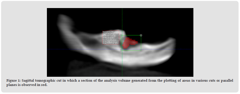
Both AL and AM were used in the form of gels made from stock solutions; for AM two concentrations of 20% (AM20) and 40% (AM40) were used. Control (C) was carboxymethylcellulose gel. The effect of the drugs was analyzed in male rats of the Wistar line weighing 120 ± 20 g, divided into 4 groups of 16 rats each. Animals were anesthetized with a ketamine/xylazine solution. Extraction of the lower right and left first molar was performed. Said alveoli were filled with gels prepared with the substances under study. The animals were treated according to universal standards of asepsis. At the end of the experiment, the animals were euthanized by intracardiac injection of potassium chloride, under general anesthesia. Determinations were made at the experimental times 0, 7, 15, 30 and 45 days. At all times, 3D DICOM images of rat jaws were taken (TC-CB Romexis – Planmeca; Radiology and Imaging Area of the UNC School of Dentistry). The radiographic densities (HU: Housnfield Units) corresponding to the dental extraction zone of the jaws were measured, delimiting analysis volumes with the tool provided by the software used in this study (Planmeca Romexis Viewer) (Figure 1). Data comparison was performed by variance analysis for two classification criteria (treatments: control, AL, AM20, and AM40; and treatment times: 0, 7, 15, 30, and 45 days). Parametric tests were used or not depending on the results obtained. Differences were considered significant if p<0.05.
Figure 1 Sagittal tomographic cut in which a section of the analysis volume generated from the plotting of areas in various cuts or parallel planes is observed in red.

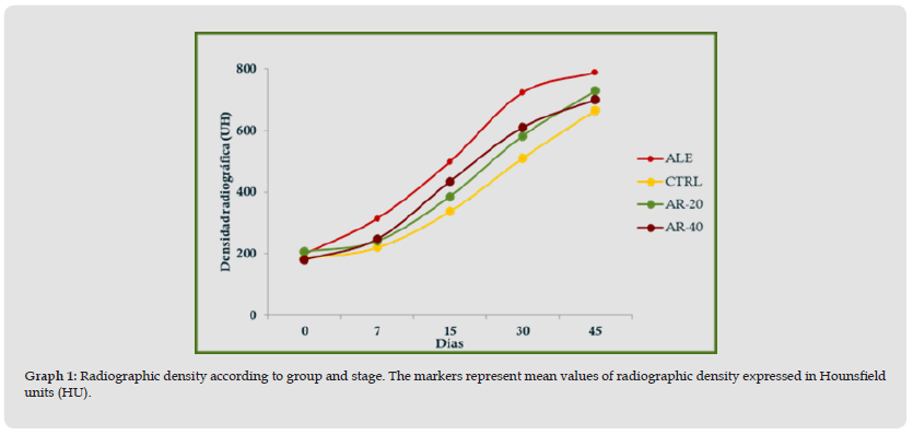
The densitometric values of central tendency (mean) and dispersion (standard deviation) registered in the three-dimensional spaces evaluated in each case were expressed, which coincided with the dental extraction zones for each group and time (Table 1). In order to visualize the changes experienced between stages according to group, the graph of means was made, in which it was observed that in global terms the values registered in each group only began to experience a visible growth after 15 days of extraction (Figure 1). When evaluating the density data using two-way ANOVA (group and stage), only the stage factor was significant (p<0.05), observing an increasing trend of density as a function of time, with significant differences between the later stages. (30 and 45 days) compared to the previous ones (initial, 7 and 15 days). Regarding the group contrast, although the differences were not significant (p>0.05), it is worth mentioning that the AL group registered the highest mean values in all post-treatment stages and that the two AM groups presented similar responses, with little apparent activity according to density in the early stages, and only after 30 days did, they show an appreciable densitometric increase compared to the control group (Graph 1).
Figure 2 Radiographic density according to group and stage. The markers represent mean values of radiographic density expressed in Hounsfield units (HU).

It should be remembered that in the dental field, osteonecrosis of the jaws related to medication is manifested as an adverse reaction due to the administration of bisphosphonates. For this reason, alternative routes of administration are needed, such as the local route or drug delivery systems capable of achieving efficient osteogenesis; in response, investigators have sought to find suitable carriers that can provide an osteoconductive matrix and impart the handling properties required for implantation at the repair site in order to improve AL loading and avoid side effects [5]. In this work, the local administration of alendronate proved to be an effective alternative to improve bone repair conditions in the post-extraction socket of the first lower molar in male rats.


