Impact Factor : 0.548
- NLM ID: 101723284
- OCoLC: 999826537
- LCCN: 2017202541
Frederico Keim1, Guilherme Constante Preis Sella2* and Mário Ferraz3
Received: December 20, 2022; Published: January 18, 2023
*Corresponding author: Guilherme Constante Preis Sella, R. das Azaléias, 476 - Conj. Hab. Inocente Vila Nova Júnior, Maringá - PR, Brazil
DOI: 10.26717/BJSTR.2023.48.007608
Traditionally, a scalpel blade number 11 or 15 is used to remove cartilaginous structures in rhinoplasty. However, this step can present some technical difficulty to achieve the exact modification desired; very often we overcorrect, undercorrect or end up having many irregularities. The use of electrosurgery in rhinoplasty is a technique that can be useful and very precise; so, the objective of this article is to describe a technique for cartilage refinement using electrosurgery. The use of electrosurgery is a quick, safe, and easily accessible instrument to remove irregularities and small amounts of cartilaginous structures in rhinoplasty.
Keywords: Rhinoplasty; Electrosurgery; Electrosection; Cartilaginous Structures
One of the main steps in rhinoplasty is to approach the cartilaginous and bone structures to modify and reshape them. The main action is to separate the soft tissue envelope from the osteocartilaginous structures and modify them to improve the functional and aesthetic aspect of the nose. Most of the structures are cartilaginous and changing them may not be so accurate. Usually, we use a scalpel blade number 11 or 15, however this step can present some technical difficulty to achieve the exact modification desired; very often we overcorrect, undercorrect or end up having many irregularities. Use of electrosection [1] is much easier because the removal of the desired portions may be done little by little. The objective of this article is to describe a technique for cartilage refinement using electrosurgery.
First, it is important to describe the correct terms of electrosurgery, that can be used to coagulate, cut, and ablate tissue and to produce hemostasis of blood vessels. Techniques of electrosurgery include electrosection (cutting), desiccation, fulguration, coagulation, and needle ablation [1]. Practically speaking, these terms describe the common operational modalities for electrosurgical systems, which can be more succinctly described as cutting, coagulation, and a combination of the two (“blend”). In a closed or open rhinoplasty, the nasal dorsum modification is usually the first stage, followed by the tip and the reduction of the alar base, if necessary. A regular dorsum is an essential condition for aesthetically pleasing nasal shape; there are several different techniques to reduce the dorsal hump including structured or preservation dorsal surgery [2-4]. If a structured technique is chosen, some authors recommend “en bloc” reduction of the cartilaginous dorsum, while others advocate the reduction of components, first separating the upper lateral cartilages from the nasal septum, in order to maintain the transverse portions of the upper lateral cartilages [5,6]. Regardless, the cartilaginous portion of the hump is traditionally cut with a 15 or 11 scalpel blade; afterwards, the reduction of the bony hump can be accomplished using rasp, osteotome, burr or piezotome [7]. Finally, lateral, medial and/or transverse osteotomies are performed by conventional osteotomes or Piezotome, and the reconstruction of the dorsum can occur with sutures and spreader grafts or flaps.
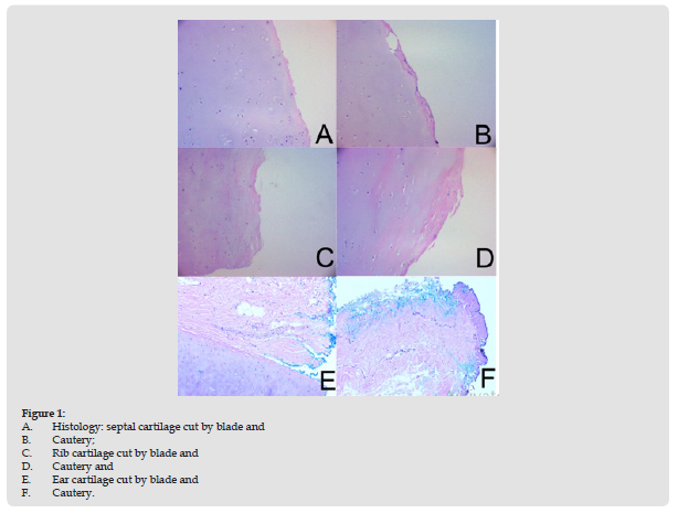
Another way to reduce nasal dorsal is through preservation rhinoplasty, a technique that does not cause the detachment of the upper lateral cartilage (ULC) from the nasal septum. In this type of surgery is necessary the septal preparation and transversal and lateral fractures to reduce nasal hump [3]. However, a preparation of the cartilaginous pyramid (like removing small irregularities) may be necessary without opening the nasal valve. Possible undesirable sequelae of dorsal hump reduction include long-term dorsal irregularities caused by irregular/over/under-resection of the osseocartilaginous dorsum, and many others complications [8]. The revision rate in rhinoplasty ranges from 0 to 15%2 in literature, and residual hump or irregularities in the dorsum correspond to a large part of these reviews. So, in the final step of the surgery, it is important to observe if small irregularities do not become visible. If small irregularities occur in the cartilaginous dorsum through traditional or preservation technique, they can be corrected with the cautious use of electrosection or also known as the cutting operational modality of electrosurgery system in the monopolar application [1]. It is a simple and easy method, available in almost any hospital, with proven safety and efficacy as long as the basic principles of electrosurgery are used. And the irregularities in the bony dorsum can be easily treated with powered drills or Piezoelectric. Minor irregularities in the lower cartilages can also be refined with the use of electrosection in the same way with great safety and precision – (Video). An interesting step for this use is after the domus sutures in which an excess of cartilage emerges in the medial region of the domus (like dog-ears), and a paradomal resection can be performed. We also did a histology study of the cautery, and there are not any differences between blade and cautery cut in septal, rib and ear cartilage – (Figure 1).
Figure 1 A. Histology: septal cartilage cut by blade and B. Cautery; C. Rib cartilage cut by blade and D. Cautery and E. Ear cartilage cut by blade and F. Cautery.

The use of electrosection is a quick, safe, and easily accessible instrument to remove irregularities and small amounts of cartilaginous structures that should be in the arsenal of every surgeon that performs a rhinoplasty.
All authors have made substantial contributions to the conception and design of the work; they has done the acquisition, analysis, interpretation, literature review and paper writing, analysis, paper review. All authors read and approved the final manuscript.
The authors declare that they have no conflict of interests.


