Impact Factor : 0.548
- NLM ID: 101723284
- OCoLC: 999826537
- LCCN: 2017202541
Vargas López, Antonio José1* and El Rubaidi Abdullah, Osama2
Received: November 29, 2022; Published: December 08, 2022
*Corresponding author: Antonio José Vargas López, Neurological Surgery Department, Hospital Universitario Torrecárdenas Avda. Donantes de Sangre s/n, Postal code: 04009, Almería, Spain
DOI: 10.26717/BJSTR.2022.47.007515
Background: To describe a case of bulky bilateral mirror meningiomas in an eighty-year-old female.
Case Description: We describe the case of an eighty-year-old woman with previous history of a one-year history of abulic depression and cognitive impairment. Neuroimage showed bilateral frontal masses sugestive of meningiomas. Surgical removal of both tumors was done in an unique stage. Postoperatively the patient presented symptomathic anemia that was succesfully treated. Afterwards the patient experienced neurological improvement with recovery of functional capabilities.
Conclusions: In presence of adequate clinical status surgical resection of symptomatic meningiomas in elderly patients should be considered.
Keywords: Mirror Meningioma; Meningioma; Ederly; Surgical Treatment
Abbreviations: ER: Emergency Room; MRi: Magnetic Resonance Image; CT: Computed Tomography; SSS: Superior Sagital Sinus; CSF: Cerebrospinal Fluid; WHO: World Health Organization; KPS: Karnofsky Performance Status
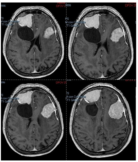
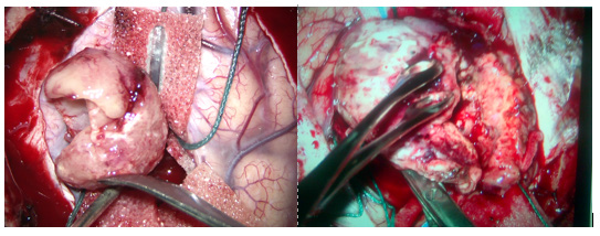
An eighty-year-old woman with a history of hypothyroidism and a one-year abulic depression and cognitive impairment consulted in ER after presenting syncope. No convulsions had been documented. She denied headache or nausea. Strenght was conserved in upper and lower limbs. Nevertheless she presented marked bradypsychia, time desorientation and gait apraxia. Blood tests were normal. CT head scan and brain MRi showed two extraaxial cotrast-enhancement frontal mirror masses of 45x40 mm in the left side and 40x35 mm in right side sugestive of convexity meningiomas (Figure 1). The right mass was associated to a Nauta type IV tumoral cyst of 40x35 mm. Both masses conditioned significative compression of frontal lobes collapsing frontal horns of lateral ventricles. Potential risks and benefits of intervention were discussed in clinical meeting and surgical resection in a single stage was planned. Bicoronal skin flap with left and right craniotomies sparing a bone bridge over SSS were performed. During disecction of dorsal aspect of right meingioma the cyst wall was opened observing normal CSF that was evacuated. Both tumors were resected without any incident (Figure 2). After intervention the patient presented mild confussion with no new neurological deficit. However, 48 hours after surgery she experienced hypoactive delirium with mixed dysphasia. CT scan ruled out significative complications (Figure 3). Postoperative blood test showed a decrease of Haemoglobin from 14 g/dl in preoperative test to 8g/dl. Red blood cell transfusion and adjustment in levothyroxine dosage were performed. Immediate ameloration of neurological status occurred. The patient experienced progressive recovery with marked improvement in gait and mental status compared to preoperative situation. Anatomopathological examination showed both WHO I meningiomas. The patient was discharged and after 1 year of follow-up she maintained independence for basic activities of daily living.
Figure 1: Brain MRi showed two frontal masses correspondig to meningiomas. Riht mass was associated to type IV cyst.

Figure 2: Intraoperative images showing circumpherencial dissection of extraaxial masses from brain parenchima.

We present a case of frontal mirror meningiomas in an eighty-year-old female. The size of the masses induced frontal lobes dysfunction with apathy and gait disturbance, mimicking depression, and cognitive impairment. No neuroimage had been performed until she was evaluated in ER. Aggressive management of these patients may be challenging due to the risk of increase clinical deterioration and death. Nevertheless, many authors have reported good outcomes of surgical treatment of meningiomas in elderly patients [1-4]. Preoperative KPS score >70 and no neurological déficits in presentation have been reported as major prognostic factor of meningioma surgery in elderly patients [5- 7]. Localization of the tumor, operative time and the ASA score have been also described as important prognostic factors for the mortality and neurological outcome in octogenarian patients [5- 7]. Different scales predicting clinical outcome have been also described [6,7]. In the case we present, management options were discussed in clinical meeting. Although presence of bilateral tumor could increase morbidity, the marked symptomatology and the practical abscence of medical comorbidities ruled out conservative treatment.
Despite the final good outcome, medical morbidities consisting in postoperative anemia and thyroid dysfunction presented as lifethreatening complications that could be corrected by geryatrician. This case ilustrates the importance of several aspects in elderly patients. First, neuroimage should be considered in evaluation of atypical depression and brief cognitive deterioration. In regard to the management of this condition, as many reports have shown, surgical treatment of meningiomas in elderly patients may be performed with good outcomes. Finally, the present case remarks on the importance of collaboration with medical departments in the attention of these patients. Cystic meningiomas have been classified in several categories as shown in Rengachary and Nauta descriptions [8,9]. Mirror meningiomas have been also previously reported [10,11]. Nevertheless, to the best of our knowledge the present case is the first one describing mirror meningiomas with a cystic one in an octogenarian patient being successfully treated.
In the presence of adequate clinical status surgical resection of symptomatic meningiomas in elderly patients should be considered. Multidisciplinary collaboration in the attention of these patient results crucial in order to achieve a good outcome.
None.
The authors have no conflicts of interest to declare.


