Impact Factor : 0.548
- NLM ID: 101723284
- OCoLC: 999826537
- LCCN: 2017202541
Youzhi Cai, Jiaqi Xu, Yijun Zhang and Chi Zhang*
Received: August 16, 2022; Published: September 06, 2022
*Corresponding author: Chi Zhang, Department of Orthopedics and Center for Sport Medicine, the First Affiliated Hospital, Zhejiang University School of Medicine, 79 Qingchun Road, Hangzhou, 310008, China; Tel: +86-571-87236848
DOI: 10.26717/BJSTR.2022.46.007283
Background: Arthroscopic reduction and fixation in ACL avulsion fracture would damage epiphysis, which is inapplicable to adolescents with persistent epiphysis.
Questions/purposes
1. Is arthroscopic “8” knotted fixation without bone tunnels in tibial-sided anterior cruciate ligament (ACL) avulsion fracture suitable for non-fusion-epiphysis patients?
2. Dose this novel method improve functional and radiographical outcomes?
Methods: Patients with tibial-sided ACL avulsion fracture who underwent onestep arthroscopic “8” knotted fixation without bone tunnels were indicated as included in our study, and between Jun 2014 and Dec 2016, 8 patients were enrolled in this study. All patients completed a follow-up of 1 year or more. Clinical outcomes were evaluated using IKDC and Lysholm scores at different follow-up points. CT scans were performed in all cases at 1 day, 3, 6 and 12 months postoperatively.
Results: Patients were followed up for at least 12 months, the average follow-up period was 14.6 months (range, 12-18 months). There were no intraoperative or postoperative complications. The mean IKDC score significantly increased from 30.8±5.1 preoperatively to 87.6±3.4 at 12 months after surgery (p<.001). The mean Lysholm score was statistically significantly improved from 37.3±5.9 preoperatively to 91.5±2.6 at 12 months after operation (p<.001). X-ray and CT scan showed fracture radiographically healing at 6 months follow-up.
Conclusion: One-step arthroscopic “8” knotted fixation without bone tunnels could provide secure treatment of tibial-sided ACL avulsion fracture in non-fusion- epiphysis patients regardless of fragment sizes and this novel method could be better suited to apply in non-fusion-epiphysis patients.
Level of evidence Level IV, therapeutic case series
Tibial-sided anterior cruciate ligament (ACL) avulsion fractures are relatively rare injuries, most frequently occurring in adolescents [1, 2]. Currently, this type of injury is generally treated with arthroscopic reduction and fixation with screws such as cannulate screws, staples, wires and nonabsorbable sutures [3-5], and the outcomes of arthroscopic treatment in children and adolescents were quite satisfactory. However, these surgical methods would damage epiphysis that may cause the premature closure of growth plate, which are inapplicable to adolescents with persistent epiphysis [6, 7]. The authors described a novel technique of one-step arthroscopic “8” knotted fixation without bone tunnels in the treatment of non-fusion-epiphysis patients for a tibial-sided ACL avulsion fracture and evaluated its short-term clinical efficacy.
Between June 2014 and December 2016, 8 childhood or adolescents with tibial-sided ACL avulsion fractures were treated at our institution. And all patients had obviously clinical symptoms: pain, swelling, giving way and locking, and pivot-shift test positive and Lachman test positive. Radiographic examinations were used to evaluate the injury on admission. Seven patients had type III fractures and one patient had type IV fracture, which were defined according to the classification of Meyers and McKeever. All 8 patients have intact ACL, without osteoarthritis or tibial and femoral fracture. Besides, all patients have non-fusion epiphysis. All patients were followed-up at least 12 months. Clinical outcomes were assessed using IKDC and Lysholm scores at 1, 3, 6, and 12 months after the operation. CT scans were also performed in all cases at 1 day, 3, 6 and 12 months postoperatively.
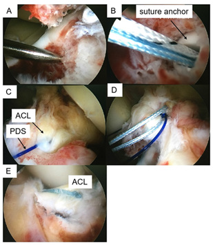
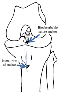
Patients were in a standard supine position with the knee at 90 degrees of flexion. Diagnostic arthroscopy was performed through the standard anterolateral portal and anteromedial portal, and the hemarthrosis was evacuated from the joint. The fracture was visualized, and the fracture bed on the underlying tibial cancellous bone was cleaned with a shaver and slightly deepened (Figure 1A). A pilot hole for the anchor was created with a 15.0 mm suture anchor punch (Bio-Corkscrew, Arthrex, Naples, FL, USA) angled at 45°in the sagittal plane with respect to the tibial plateau. The hole was located 5mm posterior to the fracture rim at 2 o’clock (10 o’clock in the left knee) (Figure 1B). While maintaining reduction with a probe through the anterolateral portal, the inferomedial part of the ACL was pieced with a suture hood loaded with NO.2 polydioxanone (PDS) through the anteromedial portal (Figure 1C). The PDS was advanced out through the hook and retrieved out from the media portal with a grasper. and the suture was passed through the base of the ACL to the anteromedial portal by pulling the PDS (Figure 1D). “8” knot-type was performed with the use of a self-locking sliding knots at the tip of the anchor to achieve appropriate tissue tension. Then, a 19.5 mm lateral-row of anchor (Bio-Corkscrew, Arthrex, Naples, FL, USA) was inserted angled at 90° in the sagittal plane located 10 mm inferior to the lateral side tibial tubercle to fix the knot (Figure 1E). Usually total one bioabsorbable suture anchor and one lateral-row of the anchor were used (Figure 2).
Figure 1: Technique for arthroscopic non-transosseous suture fixation.
a) Arthroscopic image showing a displaced tibial eminence fracture.
b) A suture anchor was inserted and tapped into the drill hole.
c) The inferomedial part of the ACL was pieced with a suture hook through the anteromedia porta.
d) The suture was passed through the ACL by pulling the PDS
e) After inserting a lateral row of anchor, the firmness of the fixation was checked using a probe.

Figure 2: Schematic drawing of left knee in anterior view, illustrating non-transosseous 2-point suture fixation technique.

Patients received the standard rehabilitation protocol: immobilization of the knee with a brace in full extension for 6-8 weeks, after which mobilization was started, below 90°of flexion until the third month and then, increasing the flexion of the knee 15° every week. Full range of motion and progressing weight bearing with brace unlocked were allowed at 3 months after surgery. If anatomic fracture healing was confirmed completely via CT scan at several months, the brace was discontinued, and return to sports was allowed at more than 4 months.
Data were analyzed using STATA version 12.0 software (College Station, TX, USA). IKDC and Lysholm scores were compared using independent-sample t-tests between pre-operation and different follow-up points postoperatively. The P value for determining statistical significance was set at <0.05.
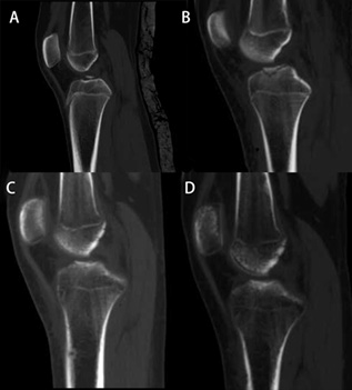
A total of 8 patients (5 males and 3 females) were included in the study, respectively. The right knee was affected in 4 patients and the left knee was affected in 4 patients. Mean age of patients at the time of trauma was 12.3 years (range, 9-14 years). Seven patients (87.5%) had type III fractures and one patient had type IV fracture (comminuted fracture). The mean operative time was 77.7 minutes (range, 62-90 minutes), and the mean bleeding volume was 9.3 ml (range, 5-15 ml). And four patients underwent injured meniscus suturing simultaneously. All patients underwent at least 12 months of follow-up, the mean follow-up period was 14.6 months (range, 12-18 months). The demographic data of selected patients was shown in (Table 1) There were no intraoperative or postoperative complications, such as infection, synovitis, or synarthrophysis in all patients. CT scan (Figure 3) showed that all 8 patients had an anatomical reduction of fracture at 1 week after the operation.
Table 1: Demographic data of patient population.

Note: *Type of fractures were defined according to the classification of Meyers and McKeever.
Table 2: Preoperative and follow-up outcome scores for all patients.

Note: *Knee clinical scores at 12 months follow-up compared with scores preoperatively
Figure 3:
a) Lateral CT scan showed ACL tibial avulsion fracture preoperatively.
b) Lateral CT scan showed anatomical reduction of fracture at 1 day after operation.
c) Lateral CT scan showed the fragment healing at 3 months follow-up.
d) At 12 months follow-up, lateral CT scan showed the fragment healed superiorly with persistent epiphysis.

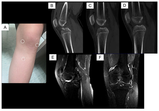
Figure 4: A typical case of ACL tibial avulsion fracture (female, 12 years old).
a) Three mini-portals on left knee on gross view postoperatively.
b) Lateral CT scan showed ACL tibial avulsion fracture.
c) (C, D) Lateral CT scan showed an anatomical reduction of fracture 1 month and 1 year after the operation.
d) (E, F) MRI scan showed the fragment healing at 24 months follow-up.

And 3 months after the operation, CT showed fracture healing and at 12 months follow-up, the fragment healed superiorly with persistent epiphysis in all patients, and all patients had stable Lachman and negative pivot shift tests. IKDC score improved from 30.8±5.1 preoperatively to 87.6±3.4 at 12 months postoperatively (p<.001). The Lysholm scores also significantly increased from37.3±5.9 preoperatively to 91.5±2.6 at 12 months followup (p<.001). Subjective evaluation development over time by the described scores was shown in (Table 2). Statistical analyses of clinical scores were performed with regard to the different followup points. The IKDC and Lysholm score showed a significant increase comparing the different follow-up points (1, 3, 6 and 12 months) with the preoperative situation, especially at 6 months follow-up, the functional scores significantly were greatly improved (p<.001). A typical case of ACL tibial avulsion fracture was showed in figure 4. On the other head, the data on the height of all patients before and 1 year after operation (Table 3) was collected, and this technique did not affect growth based on the normal height scale.
Table 3: Height of all patients pre-operation and at final follow-up

Note: *Knee clinical scores at 12 months follow-up compared with scores preoperatively
This prospective study firstly described a novel technique of one-step arthroscopic “8” knotted fixation without bone tunnels in non-fusion-epiphysis patients who suffered tibial-sided ACL avulsion fractures using bioabsorbable suture anchors. The clinical efficacy was satisfactory at 12 months follow-up with fracture radiographically healing at 6 months follow-up. This operation had the advantages of promoting bone healing and moulding with no damage to the non-fusion epiphysis of patients. Thus we answered the following questions:
1. Is arthroscopic “8” knotted fixation without bone tunnels in tibial-sided anterior cruciate ligament (ACL) avulsion fracture suitable for non-fusion-epiphysis patients?
2. Dose this novel method improve functional and radiographical outcomes? Tibial-sided ACL avulsion fractures result in anterior knee instability and occasionally in loss of knee extension. Surgical treatment is recommended for all Meyer- McKeever classification except type I. A wide variety of fixation methods have been used to fix tibial-side ACL avulsion fractures including screw, steel wire, band wire, PDS suture and suture anchor. Fixation with screws with the advantage of direct reduction and compression of the fracture fragment was the most common method [8]. However, operation in the epiphyseal region would probably interfere with the growth of epiphysis, especially in childhood or adolescence. Besides, screw fixation destroyed the collagenous fibers of ACL attachment and was not appropriate for the small or comminuted fragment [9]. In addition, using steel wire or band wire was so complicated that needed two bony tunnels drilled from the proximal tibial cortex which may destroy the epiphysis even using 1.0 mm-sized K-wire [10]. By the way?, there may necessitate implant removal after fracture healing [9, 11, 12]. Besides, simply using PDS suture or suture anchors does not provide a direct reduction of the fracture fragment, although suture anchors could rely on a tension band effect from force applied to the ACL [13].
However, the limitations could be overcome using one-step arthroscopic “8” knotted fixation. First, “8” knotted fixation is not only suitable for large fracture fragments but also reliable for small or comminuted fragments. Second, previous researches have demonstrated that Wire suture is superior to single screw fixation in biomechanical testing [14,15]. In addition, Anderson et al [16] also showed that Wire suture tired over a suture button could improve fixation when compared with a single screw. In addition, the double-row suture technique in rotator cuff repair could provide a great contact area, as well as biomechanical advantages [17]. So this “8” knotted fixation technique achieved in one step with only one suture anchor may provide strong biomechanical strength for tibial-sided ACL avulsion fracture. Besides, this one-step technique further shortened the time of operation and decreased bleeding. Besides, there is no need to take out the suture anchors for their degradability. Based on the surgical benefits of this technique, we used one-step arthroscopic “8” knotted fixation without bone tunnels in non-fusion-epiphysis patients to fix the ACL avulsion fracture, and achieved good clinical results with anatomical reduction intraoperatively and radiographically healing postoperatively at 6 months after the operation. Consequently, this technique may be reliable for its biomechanical strength and simplification. In addition, there is no need to drill tibial tunnels through osteoepiphysis, and completely avoid damaging the epiphyseal plate during internal and external anchors insertion.
We collected the data on the height of all patients before and 1 year after operation, which indicated that this technique did not affect growth based on the normal height scale. The rehabilitation protocol tends to advocate a more conservative approach with longer periods of non-weight bearing, bracing and a delayed return to sport in comparison with more functional-based adult protocols [18, 19]. Furthermore, there seems to be a consensus on the use of a knee brace during sports activities, although no solid studies have been published on the area of bracing and ACL injuries in children [20]. In this study, the brace should be kept on being used until the fracture heals radiographically. As a result, the clinical scores at 1 and 3 months were not particularly satisfactory because of the conservative rehabilitation with the brace. Whileonce removed the brace at 4 months or more postoperatively, the patients underwent positive rehabilitation, consequently, the clinical scores were significantly improved than 1 and 3 months after the operation. The limitations of the present study are: first, we did not investigate the clinical scores and radiographical results at long-term follow-up, although this study showed satisfactory short-term results; second, this was a small initial case series without comparative studies, we cannot conclusively show the effectiveness of this technique. And further randomized controlled studies are desired to demonstrate the efficacy of this novel technique. Besides, it should be emphasized that no biomechanical study was conducted to determine fixation strength during this study.
The technique of one-step arthroscopic “8” knotted fixation without bone tunnels in ACL avulsion fracture was significantly effective in terms of CT scan and clinical outcomes in nonfusion- epiphysis patients. The use of arthroscopic fixation has an obvious advantage in that the osteoepiphysis could be protected during arthroscopy. This novel method with no need to remove the bioabsorbable anchors may be less invasive than traditional treatment. To conclusively show the effectiveness of this treatment requires comparative studies, especially with an established traditional arthroscopic procedure.
We thank all the authors for contributing to this study. This work was supported by the National Natural Science Foundation of China (82072398, 82002320, 81201395, 81171703) and Natural Science Foundation of Zhejiang Province (LQ21H060005).


