Impact Factor : 0.548
- NLM ID: 101723284
- OCoLC: 999826537
- LCCN: 2017202541
Iara Cláudia Vasconcelos1 and Paolo Ruggero Errante1,2*
Received: August 10, 2022; Published: September 01, 2022
*Corresponding author: Paolo Ruggero Errante, Department of Pharmacology, Universidade Federal de São Paulo, Escola Paulista de Medicina, São Paulo, Brazil
DOI: 10.26717/BJSTR.2022.45.007272
Multilobular bone tumor, also known as multilobular osteochondrosarcoma or chondroma rodens, it is a slow growing, locally invasive, malignant tumor capable of compressing and invading adjacent tissue. Its occurrence is higher in the bones of the skull. Clinical signs depend on the location of the tumor and are usually related to compression of adjacent structures. This report describes a case of multilobular bone tumor in an eight-year-old male Brazilian crossbreed dog, with progressive growth in a region overlapping the topography of the right parietal and occipital bone cranium. After performing a cranial radiography, computed tomography evaluation, laboratory investigation and histopathology which confirmation of a multilobular bone tumor, the animal was euthanized in function of severe neurological symptoms. The histopathology associated with imaging exams allowed the establishment of a diagnosis of multilocular bone tumor, a neoplasm rarely described in the Brazilian veterinary clinic of dogs.
Keywords: Multilobular Bone Tumor; Multilobular Osteochondrosarcoma; Bone Tumor; Dogs
The multilocular bone tumor is a rare, slow growing, locally invasive bone neoplasm and with high recurrence rate, occurring most frequently in the bones of the skull of dogs [1- 3]. The multilobular bone tumor has different names such as calcifying aponeurotic fibroma, juvenile aponeurotic fibroma, chondroma rodens, multilocular osteoma and multilocular osteochondrosarcoma [4]. In dogs it occurs more frequently in medium or large breeds and rarely in giant and small breeds, being more common in middle-aged to elderly animals [5]. This type of tumor usually presents as a firm and immobile mass with a nodular appearance on the surface of the skull bones [6]. In function of location the tumor can lead to the manifestation of different clinical signs and symptoms in affected dogs that include exophthalmos and disfigurement of the face and head due to a presence of protruding tumor mass, difficulty in chewing, obstruction of the sinuses and neurological signs [7]. The diagnosis of the tumor can be defined through imaging tests, including radiography, magnetic resonance imaging and computed tomography [8,9]. The histological evaluation is important to the diagnosis definition, and the histological alterations include the presence of a multilobular mass containing many lobules, with lobules containing welldefined osteoid and cartilage separated by fibrovascular septa [5]. These lobules have a trilaminar appearance formed by islands of bone or mineralized cartilage circumscribed by ovoid or elongated cells, delimited by peripheral areas of fibrosis [1,2,4,5].
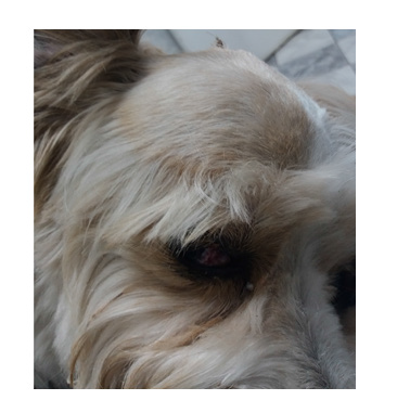
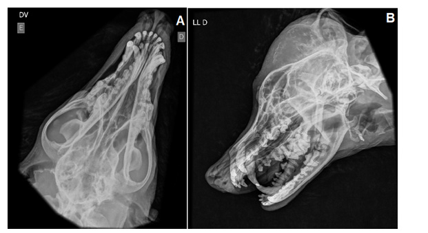
An 8-year-old male, Brazilian crossbreed dog, mediumsized, was seen at a private clinic in São Paulo, with a history of increased cranial volume superimposed on the topography of the parietal bone to the right occipital bone (Figure 1). According to the owner, the growth was observed 6 months ago before the clinical consult of dog. A radiographic examination of the skull and chest computed tomography of the skull and chest, electrocardiogram, echocardiography and laboratory tests such as blood count and serum biochemistry with evaluation of serum levels of alanine aminotransferase, alkaline phosphatase, urea, creatinine, albumin and total proteins were requested. The radiographic examination of the skull was performed in the right laterolateral, right oblique laterolateral and dorsoventral views, which revealed marked bone proliferation of high radiopacity, with an irregular and amorphous aspect superimposed on the topography from the parietal region to the occipital right region (Figures 2A & 2B). Three-view chest radiographs were taken to assess whether metastatic disease was present, with results within normal limits. In the tomographic exam, images were obtained in the transverse plane before and after intravenous contrast, with multiplanar reconstructions after the examination. The presence of amorphous bone proliferation was observed, with a format tending to round, multilobular, delimited by a thin layer of bone tissue forming septations that delimit hypoattenuated central areas. This area was attuned to the right occipital, temporal, parietal, frontal and lacrimal bones, as well as the dorsal portion of the left occipital, parietal and frontal bones (Figures 3A-3C).
Figure 1: Macroscopic aspect of the increase in cranial volume overlaid on the topography of the parietal bone up to the right occipital bone in Brazilian crossbreed dog with 8-year-old. Font: Errante (2022).

Figure 2: Radiographic examination of the skull.
A. Ventrodorsal cranial incidence.
B. Right laterolateral cranial incidence. In both projections, the presence of a mass tending to round is observed, constituted by irregular and amorphous bone proliferation superimposed on the topography of the right cranial region since the parietal to the occipital bone. Font: Errante (2022).

Figure 3:
A. Computed tomography in transverse plane right, with the presence of prominent multilobulated bone proliferation, with regular margins and defined limits, affecting since the parietal region to the right occipital region.
B. Computed tomography in axial plane, with the presence of multilobulated bone proliferation with involvement of parietal and occipital right regions.
C. Computed tomography in transverse left, with the presence of multilobular bone proliferation under parietal right region with dissemination to parietal left region. Font: Errante (2022).

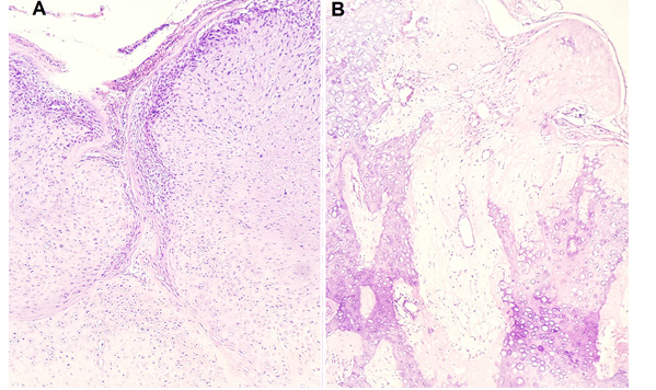
Figure 4:
A. Mesenchymal proliferation with expansive growth, accompanied by lobular outlines outlined by bundles of spindle cells. Inside the lobules, presence of an atypical cartilaginous component and foci with immature osteoid matrix (Hematoxylin/Eosin staining, 4x magnification).
B. Presence of fusiform or rounded tumor cells containing rounded to oval nuclei or slightly irregular (Hematoxylin/Eosin staining, 10x magnification). Font: Errante (2022).

The electrocardiogram and echocardiogram showed results within the normal limits regarding electrical conduction, anatomical and hemodynamic aspects. Laboratory hematological evaluations showed almost all parameters within the reference limits for the species, with the exception of alkaline phosphatase (1155.00 U/L), which was above the reference limits (normal values between 20 to 156 U/L). In the histopathology, the microscopic examination of the biopsied samples revealed the presence of mesenchymal proliferation with expansive growth, stretches forming lobular outlines delineated by bundles of spindle cells. Inside the lobes, an atypical cartilaginous component, areas of ossification and some foci of immature osteoid matrix were observed. Tumor cells presented with a fusiform or rounded appearance, with rounded to oval nuclei or slightly irregular (Figures 4A & 4B). Based on imaging and histopathology findings, the diagnosis of multilobular bone tumor was established. Due to the appearance of neurological alterations in the form of convulsive crises and the impossibility of surgical removal of the neoplasm with wide margins, euthanasia of the animal was requested by the tutor.
Multilobular bone tumor is an uncommon bone neoplasm, most frequently described in the skull of medium to large breed dogs, with absence of sexual predisposition [5]. In this case report we describe its occurrence in an 8-year-old mixed breed dog, medium size, located in the parietal region to the right occipital region of skull. This neoplasm presents as a firm, palpable mass with symmetrical and eccentric growth, usually over the bones of the head [1,2,5,6]. Clinical manifestations and behavioral changes arise due to compression of adjacent structures [5,10], a phenomenon described by the animal’s owner in the form of convulsive crises. In this type of neoplasm, an increase in serum levels of alkaline phosphatase may occur due to the release of bone isoenzyme in cases of reactive lesions [11], a finding verified in our report. By performing imaging tests such as radiography and magnetic resonance, it was possible to observe the location of the bone tumor since the parietal region to the right occipital region, with dissemination to the parietal left of skull. These imaging tests were essential for establishing a diagnosis and providing the owner with guidance on the animal’s prognosis, since the location of the neoplasm made aggressive surgical excision with a wide margin impossible [12]. Histologically, a mesenchymal proliferation was observed with stretches forming lobular outlines delineated by bundles of fusiform cells. Inside the lobules, an atypical cartilaginous component and foci with immature osteoid matrix were visualized, containing tumor cells with a fusiform or rounded appearance, with rounded to oval nuclei, as described in the literature [4,5,13,14]. After the diagnostic confirmation of a multilobular bone tumor, the owner was instructed on the patient’s prognosis. Due to the development of neurological symptoms and the impossibility of surgical removal, the animal’s owner opted for euthanasia.
The association between the clinical signs, imaging tests and histopathology allowed the establishment of the diagnosis of a multilobular bone tumor in an 8-year-old male Brazilian crossbred dog.


