 Research Article
Abstract
This work was conducted to explore the potential for dietary administration of Nano-selenium (Nano-Se) versus bulk selenium (sodium selenite, SSE) in New Zealand White (NZW) rabbit and its effects on growth efficiency, hematological parameters and immune responses. Green synthesis of Se NPs was achieved using the reducing power of iron present in the fenugreek (Trigonella foenum-graecum L ) seed extract. Sixty female rabbits (8 wk old) were grouped randomly into 5 groups; 6 rabbits each in two replicates, Se was added to the ration at two concentrations (0.1, 0.3 mg / kg). Feeding was continued for two months, then growth parameters were investigated. Rabbits were experimentally infected with S. aureus bacteria to investigate the effect of Se in both forms on immune system
Results from SEM image of Nano-Se has shown that the size of the particles is ˃50 nm in diameter so that they are converted into nano se. Growth rates were increased in rabbits with Nano-Se feed additive compared to SSe and control. Hematological parameters showed that nano se increased WBC counts, haemoglobin levels, RBCs and neutrophils but decreased lymphocyte counts relative to bulk se.. Lysozymes displayed no clear pattern with nano / bulk se, the detected nitric oxide was higher in nano se-fed rabbits compared to control and bulk se groups. The cytokines genes expression of the rabbits innate immune response to infection (IL 6, IL 1β and TNF-α) revealed that nano se (0.3mg/kg) increased these genes expression than all other groups. It was clear from the comet assay that there was no definite trend can be obtained to say that nano se has a risk effect on the DNA of the rabbit. It can be concluded that Nano-se supplementation has induced positive changes in blood constituents which have improved the growth performance and the immune function of growing NZW rabbits more than SSe.
Keywords: Nano-Selenium; Sodium Selenite; Growth; Immunity; Rabbits
Abbreviations: Nano-Se: Nano-Se; NZW: New Zealand White; IL-1β: Interleukin 1beta; TNF-Α α: Tumor Necrosis Factor; Se NPs: Selenium Nanoparticles; SEM: Scanning Electron Microscope; BW: Body weight; HK: House Keeping; WG: Weight Gain; NO: Nitric Oxide; APPs: Acute Phase Proteins; OECD: Organization for Economic Co-Operation and Development; SCGE: Single Cell Gel Electrophoresis
Introduction
Since rabbit production makes up only a very small percentage
of the total animal production in the world, there is little
information available or research performed on the nutrition of the
rabbit. Rabbit meat may play a great role in man nutrition. Rabbit
efficiency in producing meat compares favorably with most other
domesticate animals and may play a significant role in solving a
part of meat shorting in Egypt especially during poultry crisis as
bird flu. Animals feed supplementation of minerals, some elements
and vitamins to ration promotes more rapid growth (e.g. growth
promoters), maintain health or stimulate an immune response and
disease resistance. Selenium is an essential trace element important
as animals feed additive for many physiological processes,
especially for immune and defense functions, as well as metabolism
of thyroid hormones. The most important action of selenium
biological functions comes from several specific seleno-proteins,
some of which are involved in thyroid hormone metabolism, while
others play an antioxidant defense role. Selenium is also necessary
for immune function regulation and has a vital role in non-specific
immune response [1,2]. The weakened immune system is a result
of Se deficiency [3].
Application of Nanotechnology in the agri-food sector potentially
offers huge benefits for both consumers and manufacturers in a wide range of applications. Many of these benefits result from the
ultrafine dimensions of Nano-particles, which enable them to reach
new locations in the body, and thus can perform their biological
functions more efficiently [4]. According to many literatures,
consumers are more concerned about food produced by the
application of Nanotechnology [5]. Nano-Se has attracted more
attention because of its high bioavailability, high catalytic efficiency,
strong adsorbing ability and low toxicity compared with selenite
in rats (Jia, et al.), mice [6] and goats [7]. The immune system
provides body defense against invading microorganisms as well
as being importantly involved in tissue repair after injury. Immune
responses can be divided into nonspecific (or innate) and specific
(or acquired) immunity [8]. Non-specific immunity exists from
birth and operates in a generalized fashion without regard to the
exact identity of the pathogen. It serves as the body’s first line of
defense against invasion.
There are a wide variety of innate immune defenses, ranging from anatomic (e.g., the integrity of the skin; tears that wash out bacteria), to physiological (e.g., lysozymes in mucus cleave bacterial cell walls; acid in the stomach kills pathogens), to phagocytic (e.g., macrophages engulf and destro bacteria) [9]. Both non-specific and specific immune responses are regulated by mediators such as cytokines. Cytokines are a diverse group of non- antibody proteins that act as mediators between cells. They were initially identified as products of immune cells that act as mediators and regulators of immune processes, but many cytokines are now known to be produced by cells other than immune cells and they can have effects on non-immune cells as well. Cytokines are currently being used clinically as biological response modifiers for the treatment of various disorders. Molecular understanding of signaling mechanisms orchestrating the immune responses is required to define new targets for future treatments of bacterial infections [10]. Staphylococcus aureus (S. aureus) is an adaptive opportunistic pathogen, capable to persist and replicate under various conditions. This microbial species causes a wide range of diseases in both men and animals [11].
In rabbits, problems of staphylococcosis arise when S. aureus bacteria infect small dermal lesions and invade subcutaneous tissue [12], provoking a number of lesions including pododermatitis, visceral abscesses and mastitis [13]. Occasionally, abscesses in internal organs, most commonly lungs, liver and uterus, are observed [14]. This research work studies the evaluation of Nano selenium as a feed additive in enhancing the processes of growth and immunity in rabbit (Oryctolagus cuniculus). Growth will be evaluated through growth parameters and measurements. Evaluation of immunity will be carried out based on molecular studies by examining some immune genes expression (interleukin 6 (IL6), interleukin 1beta (IL-1β) and tumor necrosis factor (TNF-Α α)) as a proinflammatory cytokines after Staphylococcus aureus experimental infection (Challenge) using the high technology of real-time PCR (qPCR). Immunity will be assessed also on a biochemical bases exemplified by nitric oxide and lysozymes activity measurements . Finally, comet assay will be carried out to assess if there is a hazardous effects of using nano selenium in comparison to bulk selenium on rabbit tissues DNA?
Materials and Methods
Ethical Approval and Place of Experimental Work
Rabbits were handled according to the EU Directive 86/609/ EEC on the protection of animals used for experimental and other scientific purposes. The experimental work of the present study was carried out at the Animal Reproduction Research Institute, Giza, Egypt.
Experimental Animals, Batteries , Feed and Management
Sixty female White New Zealand rabbits were purchased from El-fayoum farm, 8 weeks old, with average weight of 1 to 1.5 kgs and housed in batteries in a special lab. The rabbit room was naturally ventilated by wired windows and provided with sided fans. Each cage had its feeder and automatic nipple drinker, feed and water were offered. All animals were kept under the same managerial, hygienic and environmental conditions and were maintained and treated according to the accepted standards of animals treatment (Figure 1). Rabbits were vaccinated with viral immunization (SERVAC RHDV Vaccine) which was given by Pathology department, Animal Reproduction Research Institute, Giza, Egypt.
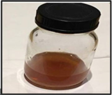
Figure 1: Red colour of Nano-selenium.
Feed and Feed Supplements
The experiment was conducted, and ration was purchased with factorial arrangement of treatments, being selenium additive-free rabbit ration. Ingredients and chemical analyses of the basal diet are presented in Table 1 following NRC (1977) recommendations.
Selenium Additive
In the present study, selenium bulk (Sodium Selenite ) was supplied by the Egyptian National Research Center. The use of Green synthesis to generate selenium Nanoparticles was demonstrated so, the procedure is free from the drawbacks of using any chemicals. Green synthesis of selenium Nanoparticles (Se NPs) was achieved according to Ramamurthy, et al. [15], by a simple biological procedure using the reducing power of iron present in the fenugreek (Trigonella foenum-graecum L, Fabaceae family ) seed extract. The plant is cultivated worldwide as a semi-arid crop (Table 1).
Table 1: Ingredients and chemical composition of the basal diets.
Fenugreek seeds were purchased from a super market then washed well and left overnight to dry, dried seeds were crushed well and turned into power, five grams of the powder were added to 500 ml distilled water (DW) and left for 24 hrs to obtain extract. Filtration of the solution was carried out by using filter paper to obtain a pure extract. A mixture of 0.02 gm of selenium + 2ml ethanol + 1ml ascorbic acid was prepared and two ml of the fenugreek extract were added to the mixture prepared. The solution was shacked well then placed in a dark bottle at room temp for 24hrs. After 24hrs, the extract was turned from white into red color (Figure 1) and selenium was expected to be converted into Nano selenium. Chetosane added to Nanoselenium as stabilizer (Figure 1).
Scanning Electron Microscope Analysis
Scanning analysis using scanning electron microscope (SEM); JEM-2100) was carried out in the National Research Centre, images were taken for the analysis of size and shape of SeNPs. The size of selenium Nanoparticles was ˂50 nm.
Experimental Design
Selenium was added to the ration in the designed rates, Rabbits (no. 60) were housed in grow-out batteries with access to water and commercial feed, they were randomly divided into 5 groups (Control and 4 treatments). Se was added to the ration in two concentrations ( 0.1 and.3 mg/kg). Feeding was continued for two months.
Growth Parameters Estimation
Body weight (BW) was measured at the beginning of the
experiment (8th week of age).Growth performance was calculated
as weight gain through an equations:
Weight gain (WG %) = (final weight - initial weight) × 100/
initial weight
Challenge Test
To the evaluate the immunity function in rabbits after feeding for 2 months on bulk and Nano-forms of selenium supplemented feed, a challenge test was carried out through Staphylococcus aureus experimental bacterial infection in rabbits and examination of some immune genes expression.
Staphylococcus aureus
Staphylococcus aureus was supplied by Microbiology department of animal reproduction institute. The bacteria cultured and the bacterial density was determined by McFarland’s tube. The strain of S. aureus was grown in 250 ml of brain heart infusion broth. Heat shock stress conditions were imposed as the culture broth was incubated at 25, 37 and 50 oC respectively. At each temperature, samples were taken at intervals after heat shock, and crude cell extracts were prepared.
Experimental Infection
Rabbit groups were immunized by experimental infection with sub lethal pathogenic bacteria Staphylococcus aureus (8×10⁸ CFU).
Clinical Investigations
Rabbits were clinically investigated for disease symptoms and investigated for the internal organs and tissue lesions.
Samples Collection
Blood Sampling and Analysis of Some Immune Response: Three days after the experimental infection of different rabbit groups under investigation, blood samples were collected from the marginal ear vein of the immunized rabbits into in plain sterile glass tubes; EDTA supplemented for blood count. and free tubes for serum analysis. Blood samples for serum analysis were left to clot, then centrifuged (4000 rpm) for 10 min; the clear serum was collected and stored at –20°C until the subsequent assessment of cellular (nitric oxide and lysozymes).The animals were then euthanized and liver and spleen samples were collected and kept in RNA latter and stored at -80°C for further RNA extraction and Real- Time PCR analysis and immune genes expression quantification.
Methods of Analysis
Estimation of Serum Nitric Oxide and Lysozymes Levels: Measurement of nitric oxide was assessed according to the assay described by Ramadan, et al. [16].
Molecular studies (IL6, IL1-β and TNF-Α Genes expression): Gene expression was assessed using Real-Time PCR, RNA Extraction was carried out using Gene-Jet RNA Purification Kit following the manufacture’s recommended protocol, cDNA Synthesis: using Maxime RT PreMix Kit and Polymerase Chain Reaction (Real-time PCR) was carried out using TaqMan Universal Master. NCBI website (http://www.ncbi.nlm.nih.gov/) was used for searching for the target genes; interleukin 6 (IL 6), interleukin 1-β (IL1-β) and tumor nicrosis factor alpha (TNF-α) sequences and the house keeping (HK) gene (rabbit GAPDH). Primers and probe were designed using NCBI Blast (http://blast.ncbi.nlm.nih.gov/Blast.cgi) and GenScript (http://www.genscript.com/) (Table 2). Rotor-Gene Q - QIAGEN instrument was used with 36 wells using Qiagen PCR Tubes for quantification of gene expression in both target and housekeeping genes (Table 2).
Table 2: Illustrates the sequences of both the target and housekeeping genes.
F denotes to a forward, R denotes to reverse. L denotes to forward primer, R denotes to reverse primer, P denotes to Probe. Probes are modified with 5’Fam - 3’Tamra as (reporter- quencher).
Assessment of Genotoxicity in Rabbit Tissues by the Single Cell Gel Electrophoresis (SCGE)/ Comet Assay: The comet assay was carried out according to the technique described by Singh, et al. [17], a protocol that can be used to detect DNA damage.
Statistical Analysis
The obtained data in the present study were statistically analyzed using the computer program (SPSS version 15 for Windows), and comparison were made using one-way ANOVA.
Results
The scanning electron microscope (SEM) image of Nano-Se is shown in Figure 2. It can be seen that the particle sizes is ˃ 50 nm in diameter, they were spherical in shape and there were some agglomerations among the NPs.
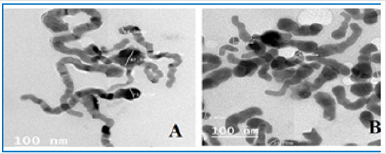
Figure 2: TEM image of Selenium Nanoparticles with diameters ˂ 50 nm; A: 14.6, 42.34 and 23.62 nm, B: 11.06, 20.23 and 18.87nm.
Rabbit Mortality Rates
No mortalities were recorded along the course of the experiment in all the control and the challenged groups.
Clinical Investigation after the Experimental Infection
Experimentally infected rabbits showed some illness exhaustion, and lack of appetite for food, examination of the internal organs revealed cases of lung hematoma and some hemorrhagic patches in 40% of the cases. Macroscopic examination revealed deeply red consolidated area in the lungs of the infected rabbits (Figure 3).
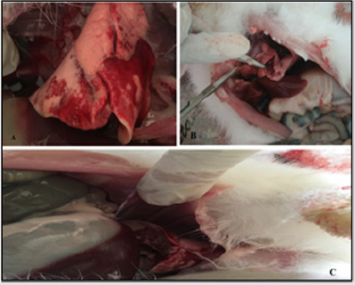
Figure 3:
A. Lung hematoma,
B. Haemorrhagic pneumonia and
C. red consolidated areas.
Growth Performance in Rabbits
The effects of feeding diets supplemented with selenium and Nano-Selenium for two months on weight gain in rabbits are presented in Figure 4. The effects of dietary Se supplementation on the growth performance of NZW rabbits showed that, among the tested groups of conventional and Nano- Se supplementation, it was noticed that; the weight gain of rabbits increased than the control group which was fed on ration without any supplementation. There was a positive relationship between the increase in the concentration of Se and the weight gain in groups fed on Se supplemented feed (Either bulk selenium or Nano selenium) with observation that Nano-Se supplementation enhanced the growth rates more than bulk selenium in respect to the two concentrations (0.1 and 0.3mg/kg.) with observation that 0.3 mg/kg Nano-Se enhanced the growth rates better than the concentration of 0.1 mg/ kg (Figure 4 & Table 3).
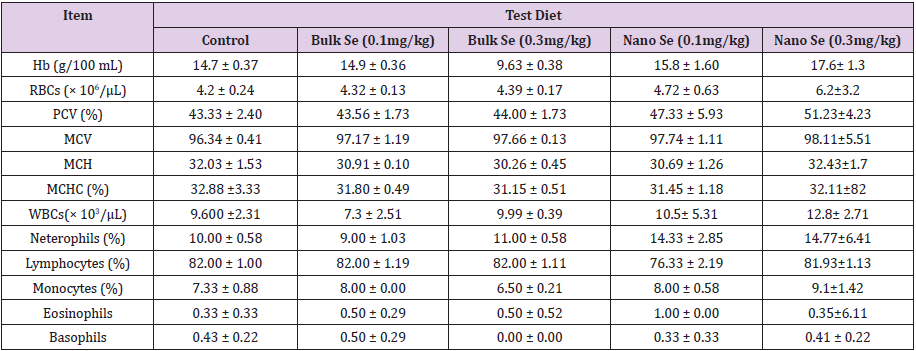
Table 3: Blood hematological parameters of New Zealand (NWZ) rabbit groups fed with test diets for 8 weeks.
Values expressed as means ± SE (n = 3). Different superscript letters indicate significant differences for each pair comparison.
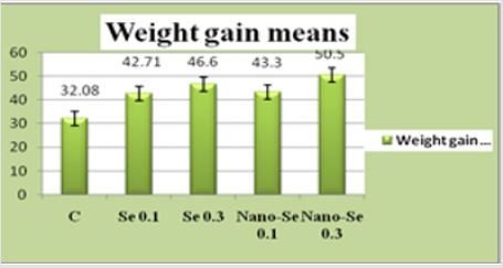
Figure 4: The weight gain in New Zealand (NWZ) rabbits after two months feeding on diets supplemented with selenium (0.1 mg/kg. and 0.3mg/kg.) and Nano-Selenium (0.1 mg/kg. and 0.3mg/kg.). Values are expressed as mean ± SE.
Blood Hematological Parameters of Tested New Zealand (NWZ) Rabbit Groups
Table 3 illustrates the effect of utilizing different Se forms (Nano and bulk) as a feed additive in the New Zealand (NWZ) rabbit. In the present study, blood hematological parameters was found to be affected by Se additive where nano se increased the WBC counts, hemoglobin levels, RBCs and neutrophils values and some decrease in the Lymphocytes count when compared to bulk se.
Lysozymes Analysis and Nitric oxide Production Analysis
Data of Lysozymes and nitric oxide production in the present study is illustrated in Figure 5. It was noticed that no specific or regular form of immune response was observed in the form of lysozyme using the feed additives of selenium, in the two forms of nano and bulk Se and different concentrations, where it was clear that immunized rabbit groups that were fed on Nano selenium produced higher increase in the mean values of serum nitric oxide production three days post vaccination (72.53 & 70.05 μg/ml) for Nano selenium additives concentrations (0.1 mg/kg and 0.3 mg/ kg, respectively) in comparison with vaccinated non treated control group (35.17 μg/ml) and immunized groups that were fed on bulk selenium (44.98 and 46.4 μg/ml for bulk selenium additives concentrations (0.1 mg/kg and 0.3 mg/kg, respectively) (Figure 5).
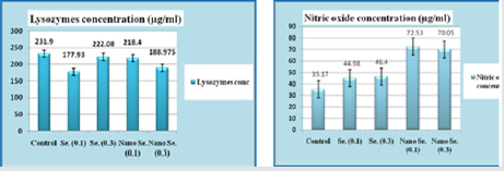
Figure 5: Blood immune responses of lysozyme (μg/ ml) and Nitric oxide (μg/ml) in experimentally infected New Zealand (NWZ)rabbit groups (No.=30) 3 days post infection with S. arues. Values are expressed as mean ± SE.
Expression of IL6, IL1-β and TNF-Α Genes Response to the Bacterial Infection
Real time PCR results were analyzed, for all samples, dissociation curves were acquired for quality control purposes. In addition, amplification products were visualized by agarose gel electrophoresis. For gene expression quantification, we used the comparative Ct method. First, gene expression levels for each sample were normalized to the expression level of the housekeeping gene encoding GAPDH within a given sample (ΔCt); the difference between the treated groups (Fed on selenium bulk and Nano) compared to the control (feeding without any selenium supplementation) was used to determine the ΔΔCT. The log2 ΔΔCT) gave the relative fold change in the gene expression of the test versus the control condition. The relative changes in mRNA transcript levels for IL6, IL 1β and TNF-α were presented in Figure 6. The results clearly demonstrated that the mRNA expression of the three genes under investigation was increased in tissues of rabbits fed on ration supplemented with Nano Se in comparison to rabbits fed on feed supplemented with bulk selenium within the same concentration of selenium used either 0.1 or 0.3 mg/kg, but the values were comparable between the 0.1 mg/kg Nano Se group and the 0.3 mg/kg bulk se group. All Se treated groups revealed better expression of studied genes when compared to the control group with observation that groups fed on Nano Se gave higher expressions over the other groups (Figure 6).
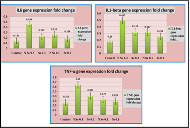
Figure 6: IL 6, IL 1β and TNF-α genes expression in experimentally infected New Zealand (NWZ) rabbit groups (No.=30) 3 days post infection with S. arues. Values are expressed as mean ± SE.
Comet Assay Data Analysis
Comet assay or single cell gel electrophoresis has been shown to be a simple and sensitive genotoxic approach for evaluating DNA damage (Frenzilli, et al.). In this study, comet assay was carried out to assess the hazard effects on DNA after using Nano selenium in comparison to bulk selenium. Results indicated that there are a rate of comet cells among different rabbit groups (Figure 7) ranged between 13.15% and 17.08% with a comparable values (Figure 8). There is an observation that the percent of comet cells was higher in rabbits group which were fed on ration supplemented with Nano selenium (Concentration = 0.1 mg/kg feed) but still near the rate in other groups even the control (Figure 8 & Table 4). Concerning the tail length and DNA percent in comet tails, Table 4 illustrates that the tail length was lower in comet cells of rabbits fed on feed supplemented with se than the control which had a feed without se. Moreover, the shortest tail length appeared in cells of animals fed on nano se concentration (0.3mg/kg). The DNA percent in the tail showed irregular image and had no specific trend.
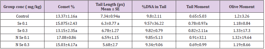
Table 4: The effects of selenium feed additive on rabbits Genotoxicity using comet assay (mean ± S.E): Comet data analysis.
Data are shown as mean ± SD. 1px is equal to 0.24μm for unit conversion.
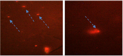
Figure 7: Comet cells present in different rabbit groups.
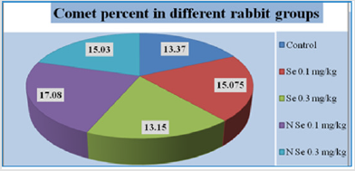
Figure 8: Comet cells percent in different New Zealand (NWZ) rabbit groups.
Discussion
Se NPs were synthesized from sodium selenite by a simple and
eco-friendly approach using fenugreek plant extract as an effective
reducing and stabilizing agent. On addition of fenugreek extract,
the sodium selenite solution changed to yellow within a minute
and attained to red colour within few minutes. This indicates
synthesis of Se NPs [18], also SEM indicated the nano particles of
se. Concerning the mortality rate, no deaths of rabbits occurred
during the duration of this study. The results indicated positive
effects of using nano-selenium supplementation to enhance
growth performance of growing NZW rabbits more than those of
sodium selenite or bulk selenium, this result is consistent with a
previous results of Emara, et al. [19], where they found that dietary
supplementation of nSe revealed heaviest live body weight and
highest WG and the lowest mortality rate when compared to the
control and bulk Se diets, suggesting a better absorption and higher
bioavailability of nano-Se. However, previous studies reported that
dietary supplementation of Se at 0.1 ppm produced an improvement
in fetal and birth weight of offspring in rabbit does, but the level of
0.3 ppm was not effective [20].
Ebeid, et al. [21] reported that feeding male California rabbits
on diets supplemented with organic Se (0.15 or 0.30 ppm) resulted
in an increase in live body weight and daily weight gain compared
with their control counterparts and these results are consistent
with the results of this work. The different physiological effects
of Nano-Se and sodium selenite may be attributed to the different
absorption process and metabolic pathways. It has been reported
that the superior performance of nanoparticles may be attributed to their smaller particle size and larger surface area, increased
mucosal permeability, improved intestinal absorption and tissue
depositions, while nanoparticles show new transport and uptake
characteristics and exhibit higher absorption efficiencies [22].
Mohapatra, et al. [23] found that feeding diets supplemented with
nano Se up to 0.30 mg/k g from 9 to 20 weeks of age positively
affected feed conversion ratio of pullets but when the level of added
nano Se reached 0.60 mg/kg FCR was negatively affected, this in
part is matching our results where, nano Se raised the growth rates
but was positively consistent with the increase of concentration.
The improvement in the performance of rabbits due to Se
supplementation may be due to that decreases the oxidative stress
and helps in the scavenging of ROS from the tissues, because Se
is a cofactor for many antioxidant enzymes such as glutathione
peroxidase and thioredoxin reductase [24]. However, data of this
work in investigating growth rates resulted from feeding on nano
Se feed additive was inconsistent with the study of Dokoupilova, et
al. [25] who noted that added Se (Se-enriched yeast) did not affect
FCR of growing rabbits. Also, Dorra, et al. [26] found that addition
of nano- Se had no significant effect on body weight gain (WG) of
rabbits at all age intervals and the whole experimental period. In
general, these differences in results and opinions could be due to the
species-specific differences or the differences in diet contents and
the nano material types and levels [27]. The contradictory findings
of different scientific literature studies on the responsiveness of
growing animals to dietary supplementation with nano selenium
may also be attributed to a variety of factors such as species,
genotype, age, dietary composition (amino acid profile), length of
analysis, nano selenium processing, and dose used.
Three days after the experimental infection with S. aureus,
external clinical investigations showed have showed some illness
symptoms such as lethargy, fatigue, and lack of appetite for food in
infected animals that are susceptible to S. aureus Bacterial disease.
Examination of the internal organs revealed cases of lung hematoma
and some hemorrhagic patches, macroscopic examination revealed
deeply red consolidated areas in the lungs of the infected rabbits.
Rabbits are important natural hosts of S. aureus, . The strains
in rabbits can be divided into low virulence and high virulence.
Infection of low-virulence strains is limited to a few birds, while
infection of high-virulence strains indicates an outbreak spread
through the entire flock causing major economic losses [28]. S.
aureus poses a major threat to rabbit farming. Manifestations
of infected rabbits include subcutaneous abscesses, mastitis
and pododermatitis [29]. At the end of August 2017, a serious
respiratory disease broke out in a rabbit farm with about 1,000
adult female rabbits in the Fujian Province of south-eastern China,
killing approximately 1,000 rabbits in a four-week timeframe.
Infected rabbits had clinical symptoms of weight loss, coughing
and purulent nasal discharge, haemorrhage in trachea and purulent
pneumonia was found in dead rabbits [30], These results are consistent with our findings in the concern. Blood hematological
parameters was found to be affected by Se addition where nano se
increased the WBC counts, hemoglobin levels, RBCs and neutrophils
values if compared to bulk se. When comparing the effect of SeNPs
(red selenium) (orally 1 mg•kg-1 BW) and inorganic selenium
(orally 1 mg Se•kg-1 BW) on the antioxidant function of neutrophils
and haematological parameters in sheep for 30 days, it was found
that thiobarbituric acid-reactive levels were significantly higher
in both groups than in control animals on the 30th day, in contrast
to the predicted decline. There were no major variations between
the amount of packed cells and the number of red blood cells in
the experimental and control groups. The white blood cell count in
the Nano-Se group showed a substantial increase on the 20th and
30th days and in the Na2SeO3 group on the 20th day compared to the
control group. There was also a substantial rise in neutrophil count
and a large decrease in lymphocyte count on day 10 in the Nano-
Se group compared to the second experimental group and control
group and on days 20 and 30 in all experimental groups compared
to the control group [31].
Differences between studies can be explained as the fact that
combined synergistic effects of additives might be different from
individual effects. Selenium has been shown to be commonly
concentrated in organs of immune response such as liver, lymph
nodes, and spleen [32]. To rise immunogens; treatment with
Se may increase the production of antibodies and broaden
complementary responses via various mechanisms that include
different selenoproteins. Nitric oxide (NO) is an important molecule
which is a general characteristic of activated macrophages [33].
Nitric oxide (NO) initially described as a physiological mediator of
endothelial cell relaxation. It is an intercellular messenger and has
been recognized as one of the most versatile players in the immune
system. Immediately after engulfment of the bacteria, inducible
nitric oxide synthase enzyme is expressed in the phagocytic cells
(Antoine, et al.) and the levels of NO sharply increase. In the current
study dietary administration of nano Se to immunized rabbits
resulted in a substantial increase in mean values of serum nitric
oxide production, approximately two-fold and approximately 1.6-
fold, compared to rabbits fed on feed supplemented with bulk
selenium two days post-immunization.
The current NO results are in opposition to those of Abd-Allah,
et al. [34], who recorded no differences in NO level due to Nano-Se,
although ours were significantly high (72.53μg/ml and 70.05 μg/
ml for the concentrations 0.1 mg/kg & 0.3mg/kg respectively) and
about twice the effect of the bulk Se (44.98 μg/ml & 46.4 μg/ml for
the concentrations 0.1 mg/kg &0.3 mg/kg respectively) and to the
control(35.17 μg/ml). Lysozymes is an active molecule in the defense
system of the organisms which is secreted by neutrophils and
phagocytes. Lysozyme is responsible for lysing pathogenic bacteria
with the key feature of pathogen restriction by targeting and disrupting
the glycoside bonding of the peptidoglycan substrate [35].
Lysozyme activity is known to be part of a non-specific immune response and is found in leukocytes. It is a natural enzyme that acts
as an antimicrobial and has had an immune modulating effect. It
serves as a non-specific defensive mechanism, representing the behaviour
of macrophages [36]. The activation of phagocytic activity
would therefore be reflected in the activity of the serum lysozyme.
The present study has revealed that, dietary administration
of Se additive for 60 successive days, either nano or bulk showed
revealed no specific trend of elevating of the values of lysozymes,
moreover the control had higher values in all groups. Carroll, et al.
[37] in their study reported that, Although normal rabbit serum
and plasma serum also contain identical (though reduced) levels of
lysozyme, they differ significantly in their bactericidal ability. These
results clearly indicate that the predominant antibacterial element
in rabbit serum is not lysozymes and that the exact source of serum
bactericide in these animals is not the same. They continued to
indicate this explanation as Further evidence that lysozyme is
not the primary bactericidal agent in rabbit serum comes from an
examination of the purified enzyme in the standard killing assay.
When tested at concentrations equivalent to those in 2 to 5% serum
or plasma, rabbit lysozyme exhibits little bactericidal activity.
In another study of Weksler, et al. [38] they said that, differences
observed for rabbit serum and plasma serum indicate that activity
is “released” from cellular elements during coagulation or immune
injury, and several investigators have presented evidence suggesting
that platelets are the source of rabbit serum bactericides.
The role of lysozyme in host defense against infection
remains relatively elusive although elevated circulating lysozyme
concentrations were evidenced after antigen stimulation [39]. In
the same way, Pawlikowska, et al. [40] reported that lysozymes
concentrations increased before the efficient production of specific
antibodies. Georgieva, et al. [41] in their study to evaluate the
changes in the concentrations of major blood proteins associated
with experimental E. coli infection in New Zealand rabbits revealed
that E. coli infected rabbits exhibited considerable increases of the
serum lysozymes concentrations on days 11 and 18, confirming the
role of this protein in the innate immunity and that lysozyme can
be considered as a late major positive acute phase proteins (APPs)
with its low values, data from our work is consistent with those
studies and it could be proved that the low values of lysozymes
in this study may be due to that lysozymes is a late major positive
acute phase proteins (APPs) and there are other immune elements
that had the main role as a bactericidal agent.
The relative changes in mRNA transcript levels for some innate
immune genes exemplified by IL6, IL1β and TNF-α were analyzed.
Results clearly demonstrated that the mRNA expression of these
genes was unregulated or increased in spleen and liver of the
rabbits fed on nano Se more than bulk organic Se when compared
with the control group. These results revealed feeding on nano Se
supplemented diet, at 0.3mg/kg showed the highest expression
status compared with the other protocols. Such amelioration of proinflamatory cytokines expression as an immune response to
bacterial infection can be ascribed to the activation effect of Se on
signaling pathways that react with the signal transduction of Tolllike
receptors in monocytes and thereby stimulate the secretion
of pro-inflammatory cytokines [42]. In parallel with our result,
Schnupf, et al. [43] reported that, during the natural infection,
Shigella infection of rabbit ileal loops led to a large increase in
mRNA abundance for the pro-inflammatory cytokines IL-1b, TNFa,
IL-6, IL-4, IFN-c and IL-8.
Using nano ZnO as a feed additive in rabbits, they said that
the administration of zinc nanoparticles to growing rabbits as
an alternative to conventional zinc sources could elicit favorable
influences on growth performance, serum parameters and splenic
expression of IL6. Also, a study conducted by Sundaresan, et al.
[44] reported that the expression of IL6 in the ovary and oviduct
of hens was up regulated significantly during zinc-induced molting.
Experimental infection with a sub-lethal dose of Staphylococcus
aureus bacteria caused increased expression of IL1-β after two
days of the experimental infection. Raw real-time threshold cycle
(Ct) data showed an improvement in IL1-β expression in rabbit
groups supplemented with nano Se in their feed more than in the
bulk selenium and the control groups. Also, the nano selenium
concentration 0.3 mg/kg revealed higher value more than 0.1 mg/
kg. In line with the result obtained from this study concerning the
higher rate of NO in case of dietary supplementation of nano Se,
iNOS is involved in immune response and is regulated by cytokines
and determined primarily by the novo synthesis and stability of
iNOS mRNA, the expression of interleukins was found to be higher
also in case of utilizing the nano Se, this may indicate the same
trend of the proinflamatory cytokins and the NO production [45].
Genotoxicity endpoints are crucial in assessing the safety
of chemicals. The Organization for Economic Co-operation and
Development (OECD) has published guidelines for several validated
and standardized in vitro and in vivo methods including genotoxicity
assays covering different endpoints. It is clear that the methods and
standard procedures used to identify the toxicity of chemicals may
not be entirely sufficient for the safety evaluation of NMs. NMs can
interfere with test components or detection systems for standard
toxicity tests [46]. In 2006, the OECD reviewed the Manufactured
Nanomaterials (WPMN) with the goal of updating the OECD
guidelines on genotoxicity and evaluating their suitability for NMs.
Magdolenova, et al. [47] analyzed the genotoxicity methods used
in 112 papers published between 2000 and 2012 on the possible
genotoxicity of NMs. Similarly, Azqueta, et al. [48] reviewed 102
papers evaluating the genotoxicity of NMs with possible use in
medicinal products. Based on the findings of both reviews, where
the authors of this paper were personally involved, the comet assay
and the micronucleus assay are the most commonly used in vitro
and in vivo techniques, the comet assay being the most commonly
used in vitro and the micronucleus assay in vivo studies.
In this work, assessment of genotoxicity by the single cell gel
electrophoresis (SCGE)/ comet assay was carried out to assess if
there is any hazard effect in using nano se supplement. Dietary
supplementation of nano se versus bulk organic se for rabbits
produced comparable rates of comet cells like the control after 60
days post immunization. With regard to tail length and DNA percent
in comet tails, Table 4 shows that the tail length in comet cells of
rabbits fed on feed complemented with se was lower than the
control which had se-free feed. In addition, the shortest tail length
was observed in cells of animal that fed on nano-se (concentration
0.3 mg / kg). The DNA percentage in the tail displayed an unusual
picture and had no clear pattern. As SeNPs have been shown to be a
promising agent to regulate chronic toxicity caused by exposure to
heavy metals. Prasad, et l. [49] studied the effects of T-synthesizing
SeNPs. Arjuna leaf extract on human lymphocytes treated with
arsenite (As+III) [50-55].
Studies of cell viability using MTT assay and DNA damage using
comet assay showed the protective effect of SeNPs against As+IIIinduced
cell death and DNA damage. This approach may be used in
the future to mitigate ROS-induced arsenic-mediated toxic hazards,
especially in areas with arsenic-contaminated groundwater and
the prevalence of arsenicosis. However, if nano-selenium has been
used as a protective agent against induced cell death and DNA
damaging materials, it is a fact that it does not have the character
of causing cell death or DNA damage. Finally, We may infer that
Nano-se supplementation is capable of causing positive changes
in the blood composition, making it possible to boost the growth
efficiency of growing NZW rabbits and improve its immunity more
than supplementation with bulk Se [56-60].
Conclusion
From this study it could be mentioned that administration of Se nanoparticles to growing rabbits as an alternative to conventional Se sources could improve growth performance, immune system function utilized by some hematological parameters, serum No and expression of some proinflamatory cytokines like IL 1-β, IL6 and TNF-α. From the economic point of view 0.3 mg/kg of Se nanoparticles is the optimal concentration for use. Comet assay revealed that using nano selenium showed no hazardous effect on liver and spleen cells, but still a great efforts in research is needed to improve our knowledge of SeNPs involvement in the functionality of organs and the potential role of SeNPs in unwanted disorders.
Conflict of Interest
Authors hereby that there is no conflict of interest concerning this work.
Acknowledgement
Authors thank the Animal Reproduction Research Institute for the financial and technical support to carry out this work. Thanks also to prof. Iman Abumourad for her support in the molecular work.
References
- Dercksen DP, Counotte GH, Hazebroek MK, Arts W, Van RT (2007) Selenium requirements of dairy goats. Tijdschr. Diergeneeskd 132(12): 468-471.
- Köhrle J, Gartner R (2009) Selenium and thyroid. Best Pract Res Clin En 23(6): 815-827.
- Effraimidis G, Wiersinga M (2014) Mechanisms in endocrinology: Autoimmune thyroid disease: Old and new players. Eur J Endocrinol 170(6): 241-252.
- Kaiser H (2007) Nanotechnology in food , food processing . Agriculture , packaging and consumption. State of science , Technologies , Markets , Application and Developments to 2015 and 2040.
- Siegrist M, Stampfli N, Kastenhol H, Keller C (2008) Perceived risks and perceived benefits of different nanotechnology foods and nanotechnology food packaging. Appetite 51(2): 283-290.
- Wang Z, Cerrate S, Coto C, Yan F, Waldroup PW (2007) Use of constant or increasing levels of distillers dried grains with solubles (DDGS) in broiler diets. Int J Poult Sci 6(7): 501-507.
- Shi L, Xun W, Yue W, Zhang C, Ren Y, et al. (2011) Effect of elemental nano-selenium on feed digestibility, rumen fermentation, and purine derivatives in sheep. Animal Feed Science and Technology. 163(2-4): 136-42.
- Maier SF, Kalman BA, Grahn RE (1994) Chlordiazepoxide microinjected into theregion of the dorsal raphe nucleus eliminates the interference with escaperesponding produced by inescapable shock whether administered beforeinescapable shock or escape testing. Behav Neurosci 108(1): 121-130.
- Kuby J (1992) Immunology. WH Freeman, New York, pp. 304-306.
- Boekel J, Kallskog O, Ryden Aulin M, Rhen M, Richter Dahlfors A (2011) Comparative tissue transcriptomics reveal prompt inter-organ communication in response to local bacterial kidney infection. BMC Genomics 12: 123.
- Cucarella C, Tormo MA, Ubeda C, Trotonda MP, Monzon M, et al. (2004) Role of biofilm-associated protein bap in the pathogenesis of bovine Staphylococcus aureus. Infect Immun 72(4): 2177-2185.
- Hagen KW (1963) Disseminated staphylococcic infection in young domestic rabbits. J Am Vet Med Assoc 142: 1421-1422.
- Parsonnet J, Gillis ZA, Richter AG, Pier GB (1987) A rabbit model of toxic shock syndrome that uses a constant, subcutaneous infusion of toxic shock syndrome toxin 1. Infection and Immunity 55(5): 1070-1076.
- Bisnoa L, Stevens DL (1996) Streptococcal infections of skin and soft tissues. New Engl J Med 334(4): 240-246.
- Ramamurthy CH, Sampath KS, Arunkumar P (2013) Green synthesis and characterization of selenium nanoparticles and its augmented cytotoxicity with doxorubicin on cancer cells. Bioprocess Biosyst Eng 36(8): 1131-1139.
- Ramadan AA, Attia ERH (2003) Natural killing molecules in cervical mucus of buffaloes during estrous cycle. 7th Sci Cong Egyptian Society for cattle Diseases. Assuit, Egypt.
- Singh NP, Mc Coy MT, Tice RR, Schneider EL (1988) A simple technique for quantitation of low levels of DNA damage in individual cells. EXP Cell Res 175(1): 184-191.
- Thangavelu S, Periyakali SB, Subramaniyam K (2017) Green synthesis of selenium nanoparticles from sodium selenite using garlic extract and its enrichment on Artemia nauplii to feed the freshwater prawn Macrobrachium rosenbergii post- larvae. Res J Chem Environ 21(10): 1-12.
- Emara SS, El Zaher HM, Michael MI, Eid SY (2019) Comparative Effects of Nano-Selenium and Sodium Selenite Supplementation on Blood Biochemical Changes in Relation to Growth Performance of Growing New Zealand White Rabbits. Arab J of Nuc Sci Applic 52(4): 1-14.
- Strucklec M, Dermelj M, Stibilij V, Rajh I (1994) The effect of selenium added to feed stuffs on its content in tissues and on growth of rabbits . Krmiva 36(3): 117-123.
- Ebeid T, Suzuki T, Sugiyama T (2012) High ambient temperature influences eggshell quality and calbindin-D28k localization of eggshell gland and all intestinal segments of laying hens. Poult Sci 91(9): 2282 - 2287.
- Liao H, Liu D, Loi R (2010) Looking at both sides of the social exchange coin: a social cognitive perspective on the joint effects of relationship quality and differentiation on creativity. Academy of Management Journal 53(5): 1090-1109.
- Mohapatra S, Sargaonkar A, Labhasetwar PK (2014) Distribution network assessment using EPANET for intermittent and continuous water supply Water Resour. Manag 28(11): 3745-3759.
- El Batal AI, Abou Zaid O, Noaman E, ES Ismail (2012). Promising antitumor activity of fermented wheat germ extract in combination with selenium nano-particles. Int J Pharm Health Care 2(6): 1-22.
- Dokoupilová A, Marounek M, Skřivanová V, Březina P (2007) Selenium content in tissues and meat quality in rabbits fed selenium yeast. Czech J Anim Sci 52: 165-169.
- Dorra TMI, Gihan ME, Hayam MA, Rana HEE (2014) Effect of dietary supplementation with Nano-- Selenium or glutamine on growth performance and carcas characteristics of growing rabbits fed diets containing two crude protein levels. J Anim Poul Prod, Mans. Univ 5(9): 549-562.
- Fardos AMH, Rania M, Iman E (2017) Growth Performance, Serum Biochemical, Economic Evaluation and IL6 Gene Expression in Growing Rabbits Fed Diets Supplemented with Zinc Nanoparticles. Zag Vet J 45(3): 238-249.
- Hermans K, Devriese LA, Haesebrouck F (2003) Rabbit staphylococcosis: difficult solutions for serious problems. Vet Microbiol 91(1): 57-64.
- Vancraeynest D, Haesebrouck F, Deplano A, Denis O, Godard C, et al. (2006) International dissemination of a high virulence rabbit Staphylococcus aureus clone. J Vet Med B 53(9): 418-422.
- Wang J, Sang L, Chen Y, Sun S, Chen D, et al. (2019) Characterization of Staphylococcus aureus strain causing sever respiratort disease in rabbits. World Rabbit Sci 27(1): 41-48.
- Sadeghian S, Kojouri GA, Mohebbi A (2012) Nanoparticles of selenium as species with stronger physiological effects in sheep in comparison with sodium selenite. Biol Trace Elem Res 146(3): 302-308.
- Dickson RC, Tomlinson RH (1967) Selenium in blood and human tissues (1967) Clinica Chimica Acta 6(2): 311-321.
- Stuehr DJ, Marletta MA (1987) Synthesis of Nitrite and Nitrate in Murine Macrophage Cell Lines. Cancer Res 47(21): 5590-5594.
- Abd Allah S, Hashem KS (2015) Selenium nanoparticles increase the testicular antioxidant activity and spermatogenesis in male rats as compared to ordinary selenium. Int J Adv Res 3: 792-802.
- Saurabh S, Sahoo PK (2008) Lysozyme: an important defense molecule of fish innate immune system. Aquac Res 39(3): 223-239.
- El Sayed MG, Manal BM (2007) The immunomodulatory effects of amoxicillin and florfenicol in buffalo after vaccination with FMD vaccine. 5th Int Sci Conf, Mansoura 69: 801-817.
- Carroll SF, Martinez RJ (1979) Role of Rabbit Lysozyme in In Vitro Serum and Plasma Serum Bactericidal Reactions Against Bacillus subtilis. Infection and 25(3): 810-819.
- Weksler BB, RL Nachman (1971) Rabbit platelet bactericidal protein. J Exp Med 134(5): 1114-1130.
- Sotirov L, Semerdjiev V, Maslev T, Draganov B (2007) Breed-related differences in blood lysozyme concentration and complement activity in cows in Bulgaria. Rev Med Vet 158(5): 239-243.
- Pawlikowska M, Deptula W (2003) Non-specific humoral immunity in rabbits immunized with Chlamydia psittaci - Gocaltovo strain. Pol J Vet Sci 6(3Suppl 1): 31-33.
- Georgieva RN, Iliev IN, Chipeva VA, Dimitonova SP, Samelis J, et al. (2008) Identification and in vitro characterisation ofLactobacillusplantarumstrains from artisanal Bulgarian white brined cheeses.Journal of Basic Microbiology 48(4): 234-244.
- Haase H, Rink L (2007) Signal transduction in monocytes: the role of zinc ions. Biometals 20(3-4): 579-585.
- Schnupf P, Sansonetti PJ (2012) Quantitative RT-PCR profiling of the rabbit immune response: assessment of acute Shigella flexneri infection. PLoS One 7(6): e36446.
- Sundaresan NR, Anish D, Sastry KV, Saxena VK, Nagarajan K, et al. (2008) High doses of dietary zinc induce cytokines, chemokines, and apoptosis in reproductive tissues during regression. Cell Tissue Res 332(3): 543-554.
- Pautz A, Julia A, Hahn S, Nowag S, Voss C, et al. (2010) Regulation of the Expression of Inducible Nitric Oxide Synthase. Nitric Oxide 23(2): 75-93.
- Guadagnini R, Halamoda Kenzaoui B, Cartwright L, Pojana G, Magdolenova Z, et al. (2015) Toxicity screenings of nanomaterials: challenges due to interference with assay processes and components of classic in vitro tests. Nanotoxicology 9(Suppl 1): 13-24.
- Magdolenova Z, Collins A, Kumar A, Dhawan A, Stone V, et al. (2014) Mechanisms of genotoxicity. A review of in vitro and in vivo studies with engineered nanoparticles. Nanotoxicology 8(3): 233-278.
- Azqueta A, Arbillaga L, López de Cerain A (2014) “Genotoxicity of nanoparticles. A,” in Nanomedicine: Current View, Present and Future Main Regulatory, (Eds.). VB Sutariya, Y Pathak, Taylor and Francis, Boca Raton, FL, USA, pp. 353-363.
- Prasad KS, Selvaraj K (2014) Biogenic synthesis of selenium nanoparticles and their effect on As (III)-induced toxicity on human lymphocytes. Biol Trace Elem Res 157(3): 275-283.
- Antony J Prabhu P, Schrama JW, Kaushik SJ (2016) Mineral requirements of fish: a systematic review. Rev Aquac 8 (2): 172-219.
- Henson SE, Nichols TC, Holers VM, Karp DR (1999) The ectoenzyme γ-glutamyl transpeptidase regulates antiproliferative effects of S-nitrosoglutathione on human T and B lymphocytes. J. Immunol 163(4): 1845-1852.
- Quintana M, Haro Poniatowski, E Morales J, Batina N (2002) Synthesis of selenium nanoparticles by pulsed laser ablation. Appl Surf Sci 195(1-4): 175-186.
- Rajendran D, Thulasi A, Jash S, Selvaraju S, Rao SBN (2013) Synthesis and application of nano minerals in livestock industry. Animal Nutrition and Reproductive Physiology (Recent Concepts). Satish Serial Publishing House, Delhi, India, pp. 517-530.
- Sauvaire Y, Baissac Y, Leconte O, Petit P, Ribes G (1996) Steroid saponins from fenugreek and some of their biological properties. Adv Exp Med Biol 405: 37-46.
- Tan PLJ, Farmiloe S, Yeoman S, Watson JD (1990) Expression of the interleukin 6 gene in rheumatoid synovial fibroblasts. J Rheumatol 17(12): 1608-1612.
- Torres SK, Campos VL, León CG, Rodríguez L lamazares SM, Rojas SM, et al. (2012) Biosynthesis of selenium nanoparticles by Pantoea agglomerans and their antioxidant activity. J Nanopart Res 14(11): 1236.
- Turner NA, Sharma Kuinkel BK, Maskarinec SA, Eichenberger EM, Shah PP, et al. (2019) Methicillin-resistant Staphylococcus aureus: an overview of basic and clinical research. Nat Rev Microbiol 17(4): 203-218.
- Van Snick J (1990) Interleukin-6: an overview. Ann Rev Immunol 8: 253-278.
- Ventura M, Melo M, Carrilho F (2017) Selenium and Thyroid Disease: From Pathophysiology to Treatment. International Journal of Endocrinology 2017: 1297658.
- Vilcek J, Lee TH (1991) Tumor necrosis factor. New insights into the molecular mechanisms of its multiple actions. J Biol Chem 266(12): 7313-7316.
