Impact Factor : 0.548
- NLM ID: 101723284
- OCoLC: 999826537
- LCCN: 2017202541
Xingyu Lv1,2, Guangyi Jiang1,2, Qiang Hong3, Yifan Wang1,2, Xinjie Zhang1,2, Jisheng Lv4 and Xiujun Cai*1,2
Received:January 30, 2019; Published: February 07, 2019
*Corresponding author: Xiujun Cai, Department of General Surgery, Sir Run Run Shaw Hospital, School of Medicine, Zhejiang University, Hangzhou, 310016, Zhejiang Province, China
DOI: 10.26717/BJSTR.2019.14.002517
Background: Multiple primary neoplasms (MPNs) are of rare occurrence. The majority of MPNs are double primary neoplasms while triple and quadruple primary neoplasms are extremely rare. The clinical diagnosis of MPNs relies on pathological examination and pathogenesis is still unclear.
Case Presentation: Here, we report a rare case of a 78-year-old woman diagnosed with simultaneous triple primary neoplasms in the ascending colon, the transverse colon and the appendix. The patient presented with lower abdominal pain in the right upper quadrant of 2 months duration. The serum carcinoembryonic antigen (CEA) level (185.83 ug/L) was significantly higher than the normal range (0-5 ug/L). An abdominal computed tomography (CT) scan with intravenous contrast scan showed multiple soft tissue masses in the hepatic flexure of the ascending colon and transverse colon, and a 2.8 × 2.8 cm soft tissue mass in the right ileocecal appendix. She underwent a laparoscopic radical extended right hemicolectomy. The patient has been seen in follow-up every 3 months, and there has been no recurrence after 16-months. We explore the etiology and discuss the treatment and prognosis of MPNs.
Conclusions: Simultaneous triple primary neoplasms are uncommon cases. Image examination and pathological evaluation are pivotal for diagnosis. Although the pathogenesis of MPNs is unclear, we believe that poor dietary habits were the largest contributor in this case. Surgery-based comprehensive therapy is the recommended therapy for simultaneous multiple primary neoplasms.
Keywords: Case Report; Multiple Primary Neoplasms; Primary Colon Cancer
Abbrevations: MPNs: Multiple Primary Neoplasms; CT: Computed Tomography; CEA: Carcinoembryonic Antigen
Multiple primary neoplasms (MPNs) were not recognized until the 1890s [1]. MPNs were first defined by Warren and Gates in 1932 [2]. The diagnostic criteria for MPNs are as follows:
a) at least two neoplasms are detected in an individual patient;
b) each neoplasm has its identified pathology;
c) the possibility of one being a metastasis of the other must be ruled out.
In 1961, Moertel [3] proposed that if MPNs are diagnosed within 6 months, they are simultaneous MPNs; if MPNs are diagnosed with a time interval more than 6 months, they are metachronous MPNs. Today, the diagnosis of MPNs has become more precise due to the development of imaging equipment and pathology examinations [4]. The incidence of MPNs in cancer patients is estimated to be 0.5% to 10% [5]. The majority of MPNs are metachronous double primary neoplasms, while simultaneous triple and quadruple primary neoplasms are much rarer [6]. In this report, we provide a rare case of simultaneous triple primary adenocarcinomas of the ascending colon, transverse colon, and appendix.
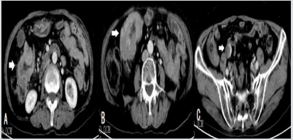
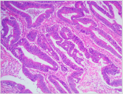
TA 78-year-old female presented with lower abdominal pain in the right upper quadrant of 2 months duration without remarkable previous disease histories. The abdominal pain was paroxysmal with nausea and vomiting and the pain relieved spontaneously. The serum carcinoembryonic antigen (CEA) level (185.83 ug/L) was significantly higher than the normal range (0-5 ug/L). No member in her family was ever diagnosed with neoplasm. The patient reported a 5 kg weight loss in the past three months. Physical examination revealed pain on palpation of the right upper abdomen. An abdominal computed tomography (CT) scan with intravenous contrast scan (Figure 1) showed multiple soft tissue masses in the hepatic flexure of the ascending colon and transverse colon, and a 2.8 × 2.8 cm soft tissue mass in the right ileocecal appendix. A large mass was found in her transverse colon by enteroscopic examination (Figure 2). Colonoscopy and biopsy revealed a tubulovillous adenoma in the transverse colon (Figure 3).
Figure 1: Abdominal contrast-enhanced CT scan CT reveals marked bowel-wall thickening of the ascending colon (A) and the transverse colon (B), and the appendix (C) is markedly enlarged.

Figure 3: Histopathology of colonoscopy samples shows villous-tubular adenomas in the transverse colon. (magnification × 200).

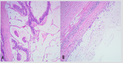
After multidisciplinary discussion, a laparoscopic radical extended right hemicolectomy was performed. Surgical findings (Figure 4) showed a 6 × 5 cm mass in the hepatic flexure of the ascending colon and a 4 × 4 cm mass in the hepatic flexure of the transverse colon. The two masses, leading to incomplete intestinal obstruction, were 4 cm apart. Postoperative histopathology of the specimen from the ascending colon (Figure 5A) revealed a moderately differentiated adenocarcinoma infiltrating the intestinal wall. Moreover, the mass from the appendix was diagnosed as a low-grade appendiceal mucinous neoplasm (Figure 5B). The postoperative course was uneventful. The patient refused to receive postoperative chemotherapy, radiotherapy, immunotherapy or targeted therapy. The patient has been seen in follow-up every 3 months, and there has been no recurrence after 16-months.
Figure 4: The resected specimens. Specimens from the hepatic flexure of the transverse colon (A), the ascending colon (B), and the appendix (C).


With the medical technology and equipment evolution, an increasing number of multiple primary neoplasms are being reported. However, MPNs are still rare, accounting for about 0.5% to 10% of the incidence of malignant tumors [5]. The etiology of MPNs is still unclear and is generally considered to be the result of multiple carcinogenic factors. Possible etiologies include genetic defects [7,8], medical treatment-related factors [9], environmental factors [10], and unhealthy living habits [11]. Pickled foods contain N-nitrosodimethylamine and other volatile N-nitroso compounds that show mutagenicity and carcinogenicity [12-14]. In this case, the patient had a diet high in salty and pickled foods. She denied using tobacco or alcohol and being exposed to polluted environment. The patient had no history of exposure to ionizing radiation or having undergone chemotherapy. No member in her family was ever diagnosed with neoplasm. Therefore, we suspect that dietary factors are the largest contributor to her MPNs. The treatment of multiple primary neoplasms is fundamentally different from that of metastatic and recurrent cancers [15]. Metastatic and recurrent cancers are mainly treated with nonsurgical palliative care. However, MPNs are mainly treated with surgery-based comprehensive therapy (radical surgery combined with chemotherapy or radiotherapy).
For metachronous MPNs, appropriate treatment options can be selected according to the characteristics of each primary tumor, whereas treatment for simultaneous MPNs must be prioritized for those tumors that have poor prognosis, are directly life-threatening, or have obvious symptoms. The patients who accepted surgerybased comprehensive therapy had a longer survival time than the patients who accepted surgery alone [6]. In the present case, the patient was diagnosed with simultaneous triple primary neoplasms of the ascending colon, transverse colon, and appendix but refused to receive postoperative adjuvant therapy. Therefore, laparoscopic radical extended right hemicolectomy was performed. The prognosis of MPNs is better than that of recurrent and metastatic cancers [16]. In summary, to our knowledge, simultaneous triple primary neoplasms have an extremely low occurrence [17]. The exact etiology of MPNs is still unknown and we suspect that in this case, a diet high in salty and pickled foods was a major contributing factor. Surgery-based comprehensive therapy is the recommended therapy for simultaneous multiple primary neoplasms. However, exact etiologic origin of such MPNs needs to be explored further.
This study was approved by the Ethical Review Committee of Guangfu Hospital (Jinhua, Zhejiang, China). The patient signed the informed consent form.
Written informed consent was obtained from the participant for publication of this case report.
All patient data and clinical images are contained in the medical files of Guangfu Hospital in Jinhua. Data is not publicly available due to patient confidentiality concerns but is available from the corresponding author on reasonable request.
All authors participated in the management of the patient in this case report. XYL and GYJ wrote the manuscript; QH and JSL took care of the patient and performed the operation; GYJ, XJZ and YFW revised the manuscript. XJC supervised the entire process. All authors read and approved the final manuscript.


