Impact Factor : 0.548
- NLM ID: 101723284
- OCoLC: 999826537
- LCCN: 2017202541
Yu Wen Hung1,2, Cheng Sheng Tsung3, Ching Feng Chiu4, Chien Chao Chiu5, Hsuan Wen Chiu6, Yu Hsing Lin2, Wei Huang Tsai7 and Shao Wen Hung*2,5
Received: January 21, 2019; Published: January 28, 2019
*Corresponding author: Shao Wen Hung, Division of Animal Industry, Animal Technology Laboratories, Agricultural Technology Research Institute, Xiangshan, Taiwan
DOI: 10.26717/BJSTR.2019.13.002443
Colon cancer is very important cancer worldwide. Although many therapies and therapeutic strategies are applied in the patients with cancer, however, the deaths of patients with cancer maintain in the top of the dead people worldwide. The visible light at a wavelength corresponding to the drug absorption activates these agents and induces highly cytotoxic products was verified. Application of the visible light at a wavelength is a potential for cancer therapy. In this study, in vitro and in vivo data were demonstrated that LED light at 450 nm induces highly cytotoxic ability and suppresses the growth of colon cancer in the CT-26 bearing mice. Taken together, LED light at 450 nm has the potential to be developed as a new therapeutic method for colon cancer in the future
Keywords: Colon cancer; In vitro; In vivo; LED light; Phototoxicity
Cancer may be viewed as a disease that progresses through multiple stages that involve initiation, promotion, progression, and metastasis [1]. Colon cancer is very important cancer worldwide. Although many therapies and therapeutic strategies are applied in the patients with cancer, however, the deaths of patients with cancer maintain in the top of the dead people worldwide [2]. Previous researchers have demonstrated that photodynamic therapy using cancer designed photosensitizers and laser beams has been increasingly used in clinical medicine. The visible light at a wavelength corresponding to the drug absorption activates these agents and induces highly cytotoxic products: singlet molecular oxygen and oxygen radicals. The LED light induced cell damage was wavelength dependent, but not energy dependent [3]. Therefore, novel therapies and therapeutic strategies are needed for the cancer therapy in the future. The objective of this investigation was to R&D of the novel therapies and therapeutic strategies against colon cancer via this novel therapy. In addition, we will wish to apply this phototoxic therapeutic method against the tumor growth in the deep body cavities in the future. Finally, we hope this novel therapies also can be applied to suppress tumor growth, metastasis, and recrudescence.
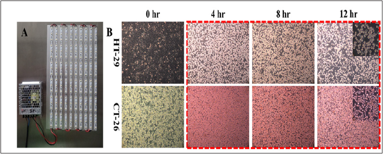
Human and murine colon carcinoma cell lines used were HT-29 (ATCC® number: HTB-38™) and CT-26 (ATCC® number: CRL-2638™). HT-29 and CT-26 were respectively cultured in McCoy’s 5a medium modified and RPMI-1640 medium (GIBCO®) and were maintained in its specific media supplemented with 10% fetal bovine serum (FBS), 100 U/ml penicillin and 100 mg/ml streptomycin (Invitrogen), in a humidified 5% CO2 incubator at 37°C. Effect of phototoxicity under LED lighting for human and murine colon cancer cell lines in vitro was evaluated. Cell morphology was observed between the in vitro experiments. Cells were treated with LED lighting 8 hr per day for 14 days in the 6 well culture plate. Observation of cell morphology with/without LED lighting (11,700 lm; Figure 1A) was by light microscope (Axiovert 200 M, Zeiss). Data were shown that LED light significantly induce cell death of HT-29 and CT-26 cells after 4 hours LED light exposure. Between 4 to 12 hours LED light exposure, all HT-29 and CT-26 cells were died according to their cell morphology (Figure 1B).
Figure 1: Phototoxic device and in vitro effects. (A) Appearance of LED light. (B) LED light is phototoxic for colon cancer cell lines, HT-29 and CT-26, by direct cytocidal effect in vitro. HT-29: Human colon cancer cell line; CT-26: Murine colon cancer cell line.

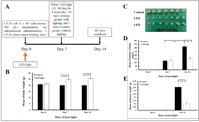
Figure 2: Design of this study and phototoxic device and in vivo effects. (A) Design of this study. (B) LED light is benefit for the increase of body weight of mice. ***p < 0.001. (C) Gross appearance of CT-26 colon tumor mass. (D) LED light is photo suppression for the tumor volume of CT-26 murine colon carcinoma in an ectopic allograft murine model in vivo. **p < 0.01. (E) LED light is photo suppression for the tumor weight of CT-26 murine colon carcinoma in an ectopic allograft murine model in vivo. **p < 0.01. No. of mice in LED group is 14; No. of mice in Control group is 7.

Animal care and all in vivo experiments were performed in compliance with the guidelines of the Agricultural Technology Research Institute (ATRI): Institutional Animal Care & Utilization Committee. BALB/c mice (6 to 8 weeks old) were purchased from National Laboratory Animal Center, Taipei, Taiwan. Mice were provided a standard laboratory diet and distilled water and kept on a 12 h light/dark cycle at 25 ± 2°C in the GLP Animal Test Room, Animal Technology Laboratories, ATRI, Miaoli, Taiwan. The experimental design in vivo was performed. To test the effects of LED light on CT-26 cells in vivo, CT-26 was established by subcutaneous implantation of CT-26 cells into BALB/c mice. BALB/c mice (n = 7 mice/control group; 14 mice/LED group) were randomly assigned to two groups: (1) a control group, vehicle with CT-26 cell implantation and without LED lighting; (2) a LED groups, CT-26 cell implantation with LED lighting (8 hr/day for 2 weeks). After 7 days CT-26 cell implantation, LED lighting was performed (Figure 2A). All animal experiments were approved by the Institutional Animal Care and Use Committee of ATRI, Hsinchu, Taiwan (approval No.: 107025). CT-26 cells (5 × 10⁶ cells per mouse) were suspended in 100 ml of serum free RPMI-1640 medium and injected subcutaneously into mice, aged 6 to 8 weeks. When tumors were detected, tumor volume was measured using the formula: 1/2 × the largest diameter × (the smallest diameter)2, as reported in previous cancer studies [4,5]. Measurement of tumor volume was periodically performed with calipers. At the end of the experiment, all mice in each group were sacrificed and tumor mass were collected. Herein, LED light with wavelength at 450 nm significantly elevated body weight of mice (Figure 2B). In vivo data were demonstrated that LED light with wavelength at 450 nm significantly suppressed CT-26 tumor weight and tumor volume in BALB/c mice with an ectopic allograft model (Figure 2C-2E).


