Impact Factor : 0.548
- NLM ID: 101723284
- OCoLC: 999826537
- LCCN: 2017202541
Monika Bekiesinska-Figatowska1* and Ewa Krupska
Received: July 30, 2018; Published: August 07, 2018
*Corresponding author: Monika Bekiesinska-Figatowska, Department of Diagnostic Imaging, Institute of Mother and Child, 01-211 Warsaw, Poland, Kasprzaka 17a
DOI: 10.26717/BJSTR.2018.07.001539
Introduction of MR-compatible incubators (INC) has revolutionized imaging preterm neonates and this fact has not been noted strongly enough in the medical literature so far. Built of non-magnetic materials, INC is equipped with gas cylinders, respirator, and pulse oximeter as well as head and body coils for imaging. Before era of INC premature newborns were deprived of possibility of MRI because they could not have been transported early in their lives to the MRI units and could not have been scanned in the unfriendly environment without the required temperature and humidity. INC has changed this situation and made MRI available to the youngest and sickest babies. In most cases it is brain that requires early assessment in preterm new borns, but body imaging is more and more demanded in INC in premature infants as well.
The coils from INC can also be used to study minor body parts in older children providing better signal-to-noise ratio than in “adult” coils. INC contributed significantly to scientific research on the development of prematurely born children and on the impact of the negative factors of prematurity on this development. The scant literature devoted to this subject increases thanks to these investigations.
Keywords: MR-compatible incubator, Neonate
Magnetic resonance imaging (MRI) is to-date the most informative non-invasive method of diagnostic imaging and as such is used – among others – in the diagnostics of newborn babies. Even though the MR-compatible incubators (INC) were introduced to the clinical practice many years ago (first publication in 2003 – [1]), the literature devoted to these examinations is very scant. In PubMed database, with a key word “MR-compatible incubator”, one can find 20 articles after 15 years. From this I derive the title of this mini review because introduction of this device has revolutionized imaging preterm neonates and this fact has not been noted strongly enough in the medical literature so far.
INC is built of non-magnetic materials and its equipment – apart from coils for imaging – consists of up to 4 gas cylinders (5L of oxygen, maximum pressure - 200 bar, gas cylinder pressure reducing valve with flow adjustment in the range of 0–5 L/min), respirator with air pressure reducing valve, and pulse oximeter (measurement of heart rate within the range of 20–300 bpm partial oxygen pressure within the range of 1–100%), and an uninterruptible power supply. INC is transported within the hospital on a non-magnetic trolley which enters safely MRI suite; another trolley is used for the transport in an ambulance.The first INC were equipped with head coils only and head coil is still the only one in the standard system. Nowadays one can buy also a whole-body 12-channel, phase-array coil consisting of an 8-channel part integrated with the incubator bed and a 4-channel surface coil and so we did in our center in which INC has been used since 2013 and 642 examinations have been performed so far with a constantly growing share of non-neuroradiological studies – up to 20% now. Of course, term babies have always been examined in MR scanners using “adult” coils first and later, with the advancement of technological development, with dedicated pediatric coils improving signal-to-noise ratio [2].
However, premature newborns were deprived of this possibility because they could not have been transported early in their lives to the MRI units and could not have been scanned in the unfriendly environment without the required temperature and humidity. INC has changed this situation thanks to protection inside the temperature- and humidity-controlled environment during the entire transport and MR examination process. Introduction of INC to clinical practice made MRI available to the youngest and sickest babies, even with a body weight of 600 g at the time of the examination [3,4]. In most cases it is brain that requires early assessment in preterm newborns. In our material at the moment we are looking for potential association of cerebellar injury and the risk of autism spectrum disorder in extremely premature infants [5]. In these cases, MRI scans are obtained at term equivalent age but in many cases, we search for the reason of neurological abnormalities (e.g. seizures) much earlier in the babies’ lives as some pathologies require prompt treatment – venous sinuses thrombosis, meningitis, brain abscesses etc. [6].
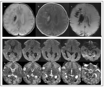
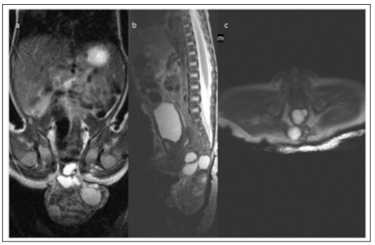
There is also a question of further management in cases of very injured brains in which pre-Wallerian degeneration that can be detected only on MRI worsens prognosis (Figure 1) [7]. Body imaging is more and more demanded in INC in premature infants as well. It is performed among others in the evaluation of congenital tumors. A good example is our patient born at the gestational age of 30 weeks and examined preoperatively on the second day of life with a body mass of 1800 g in order to rule out the suspected contact of sacrococcygeal teratoma with spinal canal (Figure 2). Another example is Wilms tumor of the left kidney confirmed on MRI on the first day of life in a 32-week-old baby with a body mass of 2570 g (Figure 3). As radiologists we do our best to convince clinicians that fetal MRI, especially when performed close to term, is sufficient in most cases to manage the newborn. However, even for the administration of a contrast agent and a better tumor assessment before treatment, it is not infrequently necessary to scan the baby postnatally which is nowadays feasible even in preterms. Moreover, MRI in INC is used as a follow-up study already in the neonatal period after surgery when sonography (US) is inconclusive. By the way, the coils from INC can also be used to study minor body parts in older children providing better signal-to-noise ratio than in “adult” coils.
Figure 1:A 33-week-old male neonate with a body mass of 1590 g, examined at day 5 after birth due to unclear sonographic picture. Bilateral hemorrhages (1A – a. FSE/T2WI, b. SE/T1WI, c. SWI), more massive in the right cerebral hemisphere, with pre-Wallerian degeneration along the cerebrospinal tracts (1B – a. DWI, b. corresponding ADC maps).

Figure 2:A 30-week-old female neonate with a body mass of 1800 g, examined at day 2. Sacrococcygeal teratoma type II according to the American Academy of Pediatric Surgery Section Survey without the contact with spinal canal which was suspected on US. T2WI in coronal (a), sagittal (b) and axial (c) projection.

Figure 3:A 32-week-old newborn with a body mass of 2570 g, examined at day 1. Wilms tumor of the left kidney with inhomogeneous signal intensity on T2WI (a – coronal, b – sagittal, c – axial plane), inhomogeneous contrast enhancement (d) and diffusion restriction (e – DWI, f-corresponding ADC map).

For example, we have recently examined the ankle of an 8-month-old girl in a head coil form INC obtaining conclusive images of good quality and revealing not only a nodule close to the lateral malleolus which was detected on US but (inflammatory?) involvement of the whole synovial membrane of the talo-crural joint. Dedicated neonatal MR scanners do also exist but with head coils only and with even smaller amount of publications (5 in PubMed) although the first publication dates earlier than those devoted to INC – to 1999 [8]. Such scanners placed in the neonatal intensive care units (NICU) allow to avoid transportation of the babies to the radiology departments which is a great advantage. However, these scanners are very limited and INC – that can be used everywhere with a standard, non-dedicated scanner – seems to remain the main tool for imaging neonates for a long time. An interesting review of the technical possibilities of neonatal MRI was published a few years ago and this is a valuable technical supplement to my clinical comments in this mini review [9].
Technical progress, which allowed the invention of INC, significantly contributed to the availability of diagnostic MR examinations to the smallest premature neonates. It also contributed significantly to scientific research on the development of prematurely born children and on the impact of the negative factors of prematurity on this development. The scant literature devoted to this subject increase thanks to these investigations.


