Impact Factor : 0.548
- NLM ID: 101723284
- OCoLC: 999826537
- LCCN: 2017202541
Uiju Cho1 and Dong Sup Lee*2
Received: July 25, 2018; Published: August 01, 2018
*Corresponding author: Dong Sup Lee, Department of Urology, St. Vincent’s Hospital, The Catholic University of Korea, 93-6 Ji-dong Paldal-gu, Suwon, South Korea
DOI: 10.26717/BJSTR.2018.07.001518
Keywords: Bladder Tumour, Recurrence, Cystoscopy, Computed Tomography
Abbreviations: TURB: Transurethral Resection of Bladder Tumour, CT: Computed Tomography
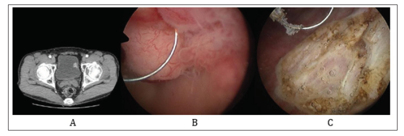
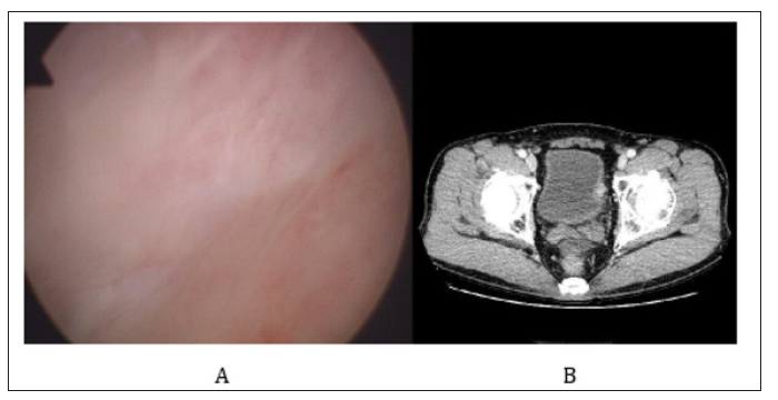
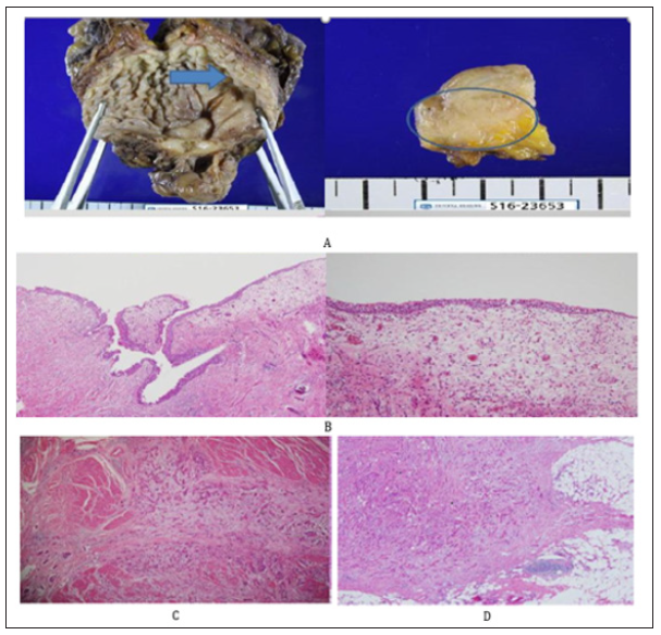
With respect to T1 high grade bladder tumour, 69% to 80% recurrence and 33% to 48% progression have been reported after transurethral resection of bladder tumour (TURB) alone [1,2]. Over half of recurrence after TURB for T1 bladder tumour occurred in the previous site that means incomplete TURB contributed to the recurrence [3]. Therefore, cystoscopy should be repeated every 3 months for 2 years in T1 tumour [4]. A 68-year old man complained of asymptomatic hematuria for 2 weeks. He had no underlying medical disease. He had never smoked. About a 2cm sized single papillary mass with wide base was located on the left lateral wall of urinary bladder (Figure 1). He underwent TURB, and T1 high-grade without carcinoma in situ was proven pathologically. A repeat TURB showed negative pathology. The patient did not want radical cystectomy at that time. 6 cycles of bacillus Calmette-Guérin intravesical instillation were done. There was no abnormal lesion in cystoscopy at 3 and 6 months after TURB. However, the round mass was detected in dynamic computed tomography (CT) 6 months after TURB (Figure 2). In the specimen from radical cystectomy, the tumour was not seen on the mucosa but it had invaded into perivesical fat tissue (Figure 3). 6 cycles of adjuvant chemotherapy with Gemcitabine and Cisplatin were applied immediately after radical cystectomy. The patient has been followed up for 3 years with semi-annually CT after radical cystectomy without evidence of recurrence. Because of any possibility of incomplete TURB for pathological T1 high grade bladder tumor, or pathologically underestimated T stage of T2 bladder tumor, any imaging study such as CT scan or bladder ultrasonography should be considered for T1 high bladder tumor, although a guideline recommends imaging study yearly [5].
Figure 1: Initial bladder tumour at computed tomography and cystoscopy.
Note:
A. Computed tomography showed a protruding enhancing mass on the left lateral wall of urinary bladder.
B. A papillary mass was on the left lateral wall of urinary bladder.
C. The tumour was resected by electro-loop.

Figure 2: Cystoscopy and computed tomography 6months after initial transurethral resection of bladder tumour.
Note:
A. No definite mass lesion on follow-up cystoscopy.
B. Computed tomography showed that the enhancing mass was outside the left lateral mucosal wall of the urinary bladder.

Figure 3:Pathological review
Note:
A. Gross anatomy showed that the tumour lesion was embedded in the left lateral wall of urinary bladder with intact mucosa (Arrow and Circle indicates the tumour)..
B. Intact mucosal surface (x40, H&E), mucosal surface (x100, H&E).
C. Tumour cells invading the muscle proper (x100, H&E).
D. Perivesical fat invasion (x40, H&E).


