info@biomedres.us
+1 (502) 904-2126
One Westbrook Corporate Center, Suite 300, Westchester, IL 60154, USA
Site Map
 ISSN: 2574 -1241
ISSN: 2574 -1241
ISSN: 2574 -1241
Received: July 16, 2018; Published: July 20, 2018
*Corresponding author: Juan Miguel Gil Jaurena, Pediatric Cardiac Surgery, Hospital Gregorio Marañón, C/O´Donnell 50. 28009. Madrid, Spain
DOI: 10.26717/BJSTR.2018.07.001455
Surgical closure of cardiac defects via a full mid-line sternotomy has been considered the gold standard for over 50 years. Due to the boom in interventional cardiology and the proliferation of techniques such as laparoscopy or thoracoscopy in other surgical specialties, several teams have adopted alternative approaches to median sternotomy [1-7]. The initial enthusiasm, fuelled by the technical advances of the past twenty-five years, stood against the reticence of those with a traditional approach. Doubts regarding the complexity and operative time of the technique, as well as its outcomes, prevented minimally invasive surgery from becoming widely used in congenital heart disease. Among the most frequent alternative approaches (Figure 1) we find: lower mini-sternotomy [8-11], right sub-mammary [1,12-16], postero-lateral thoracotomy [17,18] and right axillary incisions [19-23]. Main advantages are cosmesis and earlier recovery, as well as saving blood products and lower infection rates.
Figure 1: Clockwise from top left: drawing displaying several alternatives to midline sternotomy. Axillary approach, lower mini-sternotomy and sub-mammary incision.
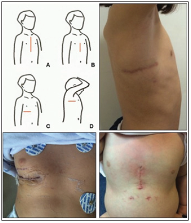
On the other hand, a steep learning curve and technical difficulties in handling some steps (myocardial protection, de-airing maneuvers, and so on) discourage many surgeons to include these minimally invasive procedures within their routine practice. Our group has been performing minimally-invasive congenital heart surgery for the last 20 years [24,25]. Sub-mammary incision in adolescent and young ladies was our first approach, which has evolved to a vast array of alternatives including several cardiac conditions since. Early in our experience, the rationale was to perform the whole procedure (full cannulation and cardiac repair) through an incision which could spare a complete median sternotomy. Then, lateral-posterior, axillary thoracotomy and lower partial sternotomies were sequentially added to our armamentarium (Figure 1). Interestingly, trends in adult surgery pushing forward less invasive techniques for valvular repair paved the way for its adoption in the congenital setting.
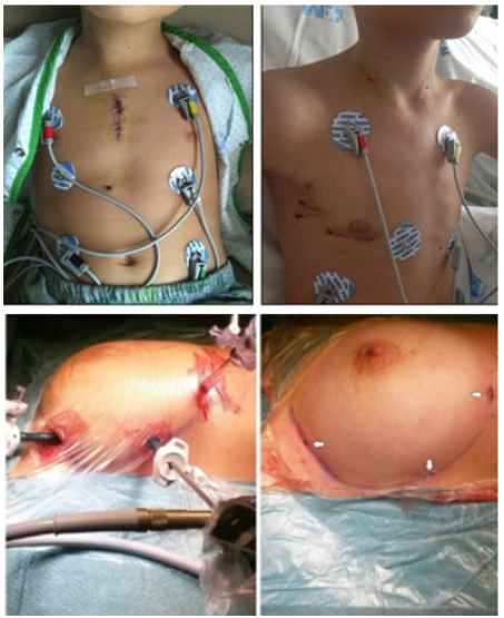
Thus, upper mini-sternotomy for aortic valve procedures and video-assisted anterior mini-thoracotomies for atrial septal defects (ASD), borrowed from mitral surgery, were implemented in our strategy (Figure 2). The latter means a shift in our early philosophy of single-different incision, since we have moved to a multi-small-access for peripheral cannulation plus additional ports. Needless to say that new tools such us shafted instruments, bendable sternal/rib spreaders, soft tissue retractors, etc. ease that change. Special attention merits video-assisted facilities learnt from our thoracic colleagues. Arrhythmia surgery can be carried out, even in pediatric patients, only by thoracic ports (Figure 2) as we recently reported [26,27]. Currently, surgical approach depends on age/weight and cardiac condition. Small babies (eg, ventricular septal defects) are best approached by lower sternotomy wheres an ASD can be closed through the right axilla in a 15 Kg. child or, alternatively, by the sub-mammary crease (with/without video-assistance) in an adolescent.
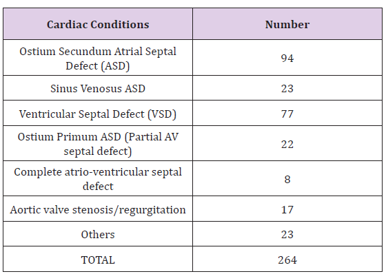
Tables 1 & 2 summarize our group data along the last five years. As experience is gathered, every patient is best suited according body-size and pathology as above mentioned [24,25]. Many groups have shifted towards the minimally invasive surgical approaches in pediatrics [1-7]. The rationale, beyond cosmesis, is offering the same results with new incisions, when catheter-based interventional procedures are also difficult or contra-indicated. Some teams are keen on a single particular approach, whereas others prefer to be familiar with many of them [4-6,24]. Whether this is a strategy or a matter of evolution is beyond the scope of this paper. Currently, the range of incisions different from a full mid-line sternotomy is rich enough to provide us many options. Interestingly, among the literature reviewed, some papers underline the steps to set up programs [25,28-30]. Particularly relevant is the publication by Bonaros et al. [31], in which the authors split every procedure in several parts and analyze them separately, so as to accurately depict anyone ́s learning curve.
Table 1: Surgical minimally-invasive approaches.
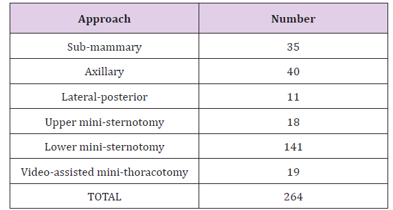
Table 2: Cardiac conditions approached.
The take-home message in a minimally invasive program is trying to convert any patient (when needed) to another minimally invasive approach in an expeditious way, for the sake of safety. Obviously, a partial sternotomy (either upper or lower one) can be easily enlarged to a full sternotomy. An axillary incision can be converted to a postero-lateral one (again, minimally invasive) by just prolonging posteriorly the already drawn surgical mark and severing the latissimus dorsi muscle [32]. The new program of video-assisted mini-thoracotomy is growing-up under the readiness to convert incisions to a full sub-mammary one, if needed. When approaching the chest thoracoscopically, the ports are fashioned in such a way that an axillary or sub-mammary incision can be produced, if requested (Figure 2, bottom pictures).
Figure 2: Top left: upper mini-sternotomy (aortic valve procedure). Top right: video-assisted 4 cm. minithoracotomy, with two additional 10mm. ports under the right arm (ASD closure). Bottom: Thoracoscopic ablation with three ports along the sub-mammary crease.
After gathering some experience, the question is how to move forward with the program? There is no clear answer, since not all surgeons in the same Institution are at the same level of proficiency or are still in their learning curve. Thinking in terms of contraindications rather than indications, as a last step of training, could be a reasonable marker. In other words, we are not expecting for the “perfect patient” to come and be an ideal candidate for a minimally invasive approach. We rather think about the contraindications, if any, for a minimally invasive procedure in every patient. Minimally invasive pediatric cardiac surgery is currently becoming a routine practice in many centers worldwide. The different approaches need their own learning curve, either straightforward or a steep one.
Our recent experience [25] demonstrates that a comprehensive schedule allows a safe and custom-made approach to train new surgeons in the field and enhances enthusiasm in developing further strategies on their own. A record of conversion-rate and complications should be used as marker of performance and quality standard. The new adopters can take their own training pace according to their level and skills. Interestingly, the wider the offer of approaches, the more ideas come up for new alternative minimally invasive methods. Maybe the future will rely on totally robotic or endoscopic [33-37] surgery but, for the time being, offering alternative approaches is worth trying.

International Association of Landscape Archaeology, Czech Glass Society, Czech Republic

Department of Chemistry, Semenov Institute of Chemical Physics, USSR Academy of Sciences, Moscow, Russia

Neurology, LA BioMed Research Institute, USA

Associate Professor at Department of Breast and Thyorid Surgey, Chongqing General Hospital, China

Professor of Nuclear Medicine, Faculty of Medicine and Surgery, University of Milan, Milan, Italy





 Abstract
 PDF
PDF



























