Impact Factor : 0.548
- NLM ID: 101723284
- OCoLC: 999826537
- LCCN: 2017202541
Arindam Sur*, Sayari Banerjee and Jayati Roy Choudhury
Received: June 24, 2018; Published: July 09, 2018
*Corresponding author: Arindam Sur, Demonstrator, College of Medicine & Sagore Dutta Hospital, Sodepur, West Bengal, Pin: 700113, India
DOI: 10.26717/BJSTR.2018.06.001366
Mucopolysaccharidoses (MPS) are hereditary diseases caused by mutations of genes coding for lysosomal enzymes needed to degrade glycosaminoglycans (GAG) leading to progressive accumulation of glycosaminoglycans in various organs. The patients often present with clinical features like coarse facies, organomegaly, dysostosis multiplex, developmental delay and abnormalities in vision or hearing. Incidence of mucopolysaccharoidosis is 1 in every 100,000 live births. Until date, very few such cases has been reported worldwide. We hereby report a rare case of mucopolysaccharoidosis 1admitted in hospital with atypical presentation (fever, convulsion and rash) and diagnosed by presence of coarse facial features, bullet shaped phalanges in x ray of hand, presence of GAGSin urine examination and confirmed by enzyme assayin a tertiary care hospital in West Bengal.
Keywords: Mucopolysaccharoidosis; Glycosaminoglycan; Coarse Facies
Mucopolysaccharidoses(MPS) are hereditary, progressive diseases caused by mutations of genes coding for lysosomal enzymes needed to degrade glycosaminoglycans.Glycosaminoglycan(GAG) is a long chain complex carbohydrate composed of uronic acids, amino sugars & neutral sugars. Major GAGs are chondriotin-4- sulphate, chondriotin-6- sulphate, heparan sulphate, keratan sulphate & hyaluronan. These substances are linked to proteins to form proteoglycans. Proteoglycans are major constituents of ground substance of connective tissue, as well as nuclear & cell membranes. Degradation of proteoglycans starts with the proteolytic removal of protein core, followed by stepwise degradation of GAG moiety. Failure of this degradation due to absent or grossly reduced activity of mutated lysosomal enzymes leads to accumulation of GAG fragments inside lysosomes.This leads to interference in cell function, leading to characteristic pattern of clinical, radiological & biochemical abnormalities(Figure 1). Patients often exhibit coarse facial features along with organomegaly (like hepatomegaly or splenomegaly).Patients also exhibit dysostosis multiplex (characterized by severe abnormalities in the development of cartilage & bone) and mental retardation.
Other signs include abnormalities of hearing, vision and cardiovascular system.The mucopolysaccharidoses are usually inherited in an autosomal recessive manner, with Hurler & Hunter syndrome perhaps being most widely studied. In some cases, a family history of mucopolysaccharidoses is obtained. Incidence of mucopolysaccharidoses in Poland is 1 in 20000 live births[1].MPS represents major part of lysosomal storage disorders & ultimately leads to severe mental disability & premature death[2].In the present instance a case of suspected mucopolysaccharidoses was admitted to indoor of Department of Pediatric Medicine in College of Medicine & Sagore Dutta Hospital, Kamarhati with history of high fever for 3 days and erythematous macular rash on back(Figure 2). He also had 2-3 episodes of generalized tonic clonic convulsion on third day of fever. On physical examination, he had coarse facies & hepatomegaly (3cm). Radiological findings includebullet shaped phalanges inX-ray of both hands. Urine examination for GAGs showed positive cetrimide test indicating presence of GAGS .Serum was tested for enzyme α L Iduronidase.
A one-year old boy was admitted to pediatric indoor of College of Medicine & Sagore Dutta Hospital, Kamarhati with history of fever with rash for 3 days and 2-3 episodes of convulsion. On examination the child was sick with dribbling of saliva from angle of mouth, had coarse facies & hepatomegaly. Detailed history was obtained from the mother who revealed, due to distress in utero the child had to be delivered via cesarean section. No developmental delay was found in this child. He had an elder brother, aged 17 years who was quite normal of his age. His family history was also found to be negative.All routine hematological & biochemical tests were performed which showed following findings[3].TC was 17200/ cmm, Platelet count was found to be within normal limit & CRP: Reactive.
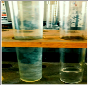
Urine routine examination revealed Gross proteinuria.Liver function test parameters was found to be within normal limit. On peripheral smear malaria parasites were not found & dengue serology was also Non-reactive. Xray of both hand was done which revealed ‘Bullet shaped phalanx’ & X ray of L-S Spine was within normal limit. Treatment was started with antipyretics & anticonvulsants.ENT review was sought & it was found be within normal limit. On Eye examination corneal clouding was detected. Urine examination for GAGs revealed positive cetrimide test showing presence of GAGS(Figure 3).
Figure 3: Positive cetrimide test in urine sample (left test tube) and control test (right side test tube).

Citric acid and trisodium citrate were used to prepare citrate buffer was adjusted at 6. Cetrimide solution (5%) was prepared with the help of citrate buffer. Urine sample was added to cetrimide solution and after 1-hour turbidity was observed. In a control test tube normal urine sample was added but no turbidity was observed. We also found positive Esbach test & Biuret test for protein in patient’s urine sample. The child was provisionally diagnosed to be suffering from mucopolysaccharidoses 1(hurler’s syndrome). Enzyme assay was ordered for confirming the diagnosis. Enzyme analysis by tandem mass spectrometry finally reveals deficiency of enzyme α-L-iduronidase.
Intraliposomal accumulation of undegraded products causes a group of lysosomal storage disorders known as mucopolysaccharidoses (MPSs)[3]. MPS 1 is caused by mutations of IUA gene on chromosome 4p16, 3 encoding α-L-iduronidase. Mutation analyses reveal 2 major alleles. Mutation analysis reveals 2 major alleles; W402X &Q70X.The mutations introduce stop codons with ensuing absence of functional enzyme.Homozygosity or heterozygosity gives rise to Hurler’s disease.Deficiency of α-L-iduronidase results in broad spectrum, varying from severe Hurler disease to mild Scheie disease. Hurler disease is a severe, progressive disorder with multiple organ & tissue involvement, resulting in premature death (usually by 10 years).Infant appears normal by birth, but inguinal hernias are often present. Diagnosis is usually made with the evidence of hepatospleenomegaly, coarse facies, corneal clouding, large tongue, short stature & skeletal dysplasia.
Cardiomyopathy is also found in some infants[4].In the present instance suspected case ofmucopolysaccharidoses was admitted to pediatric indoor of College of Medicine &Sagore Dutta Hospital, Kamarhati with history of fever with rash for 3 days and 2-3 episodes of convulsion, he also had coarse facies & hepatomegaly and hand Xray reveals bullet shaped phalanges. Urine examination for GAGs revealed positive cetrimide test showing presence of GAGS & Positive Biuret test for protein. Patients with MPS 1 may show dental abnormalities including enamel defects, carious teeth and dentigerous cysts. However, our patient did not show such features. Regular dental and radiological evaluation should be performed every 6 months and parents, or caregivers should be educated about dental home care.
Scheie disease is comparatively mild disorder characterized by joint stiffness, aortic valve disease, corneal clouding & mild dysostosis multiplex. Onset of symptoms is usually after 5 years, with diagnosis made between 10 to 20 yrs.They have normal intelligence & stature but have significant joint involvement.Carpal tunnel syndrome is very common in them.Ophthalmological features include glaucoma, corneal clouding & retinal degenerationwith the advent of hematopoietic stem cell transplantation and, more recently, enzyme replacement therapy, there is a need for early diagnosis, better disease recognition and management. Early detection of the disease and appropriate management through a multidisciplinary approach is recommended to improve the quality of life.


