Impact Factor : 0.548
- NLM ID: 101723284
- OCoLC: 999826537
- LCCN: 2017202541
D Borin, P Gallina, N Scuor and O Sbaizero*
Received: June 19, 2018; Published: June 26, 2018
*Corresponding author: Orfeo Sbaizero, Department of Engineering and Architecture, University of Trieste, Via A. Valerio 10, 34127 Trieste, Italy
DOI: 10.26717/BJSTR.2018.06.001304
This study addressesissues specifically related to the underwater dynamic actuation of Micro Electro Mechanical Systems (MEMS), such as micro-cantilevers or comb-drive microstructures. As a practical application, cantilevers made using MUMPs (Multi-User MEMS Processes) foundry are considered. Since the gap between the cantilevers and the underlying surface is very narrow (about 2 μm), water does not always flow easily underneath the cantilevers, especially when the surface is hydrophobic with respect to the liquid environment. As long as the MEMS surface is hydrophobic, the lower the gap, the higher the pressure required to flow into micro-channels or gaps .As a consequence, this issue significantly modifies cantilevers’ performances. To gain an insight of the phenomenon, a new measurement technique was developed, referred to as MFTEC (MEMS Freezing by Thermo Electric Cooling), which can verify the presence of water under cantilevers or other micro-devices.
Keywords: MEMS; Thermo Electric Cooling; Microdevices, Wettability
Abbreviations: MEMS: Micro Electro Mechanical Systems; AFM: Atomic Force Microscopy; XRT: X-Ray Micro-Tomography; FES: Focus Error Signal
There is a large and emerging demand for the use of Micro Electro Mechanical Systems (MEMS) working in aqueous environments, especially for biosensors and biotechnology applications. Applications span from general purpose devices, like Atomic Force Microscopy (AFM) probes used in liquid environment [1] to highly specialized devices exploiting horizontal cantilevers or vertical micropillars [2-3] used for the detection of the biomolecule of interest in the analyte solution [4]. In all these cases, the devices are obtained from silicon or silicon derivatives (e.g. silicon dioxide or silicon nitride) using standard or custom-designed MEMS processes. One possibility for the fabrication of cantilevers is the multi-user MEMS processes (MUMPs), relatively inexpensive and therefore suitable for low-cost prototyping. MUMPs provide three poly-silicon layers (0.5, 2 and 1.5 μm thick respectively) and a metal layer, typically used to provide electrodes or as a surface for a subsequent functionalization.
The minimum feature spacing using MUMPs technology is around 2 μm see Figure 1. MUMPs provide two structural poly-silicon layers for moving structures, respectively called: POLY1 and POLY2.If POLY1 is used, alone or in combination with POLY2, a nominal gap of 2 m can be achieved between these structures and the base plane, using a PSG (Phospho Silicate Glass) sacrificial layer. If only POLY2 is used, the designer has the choice of a 2m or 3.5 m gap, since one or two PSG layers can be added during manufacturing. Since the gap is, in any case, very narrow, the behaviour and the role of the liquid environment around the MEMS are not easily predictable. One of the relevant issues is whether the gap between the moving structure and the base is completely filled with liquid once the MEMS are submersed in the liquid environment. Obtaining the answer is unfortunately not always simple, since this phenomenon is affected by many parameters, such as liquid surface tension, MEMS shape, temperature, presence of micro-bubbles in the liquid, and the liquid filling procedure. To the best of our knowledge, in actual fact, a specific instrument for measuring the presence of water under MEMS structures with sufficient accuracy does not exist at present.
There are many reasons for this, mainly related to the very higher solution required for an inspecting technology and the not easily accessible working volume of interest. However, we believe that such an instrument would be very useful. The static and dynamic behaviour of actuated MEMS drastically changes in relation to the presence of water between the moving structure and the base. For example, a cantilever can be considered, operating underwater with electrostatic actuation, in which an amplitude modulation of a high frequency carrier technique is used to avoid electrolysis [5]. When water is trapped between the cantilever and the base, the electrostatic attractive force is 80 times higher than if no water was present between the cantilever and the base, since the relative dielectric constant of water is 80 times higher than that of air. Therefore, our aim in this paper is to design and test an accurate and inexpensive technique for investigating the presence of water under cantilevers or other MEMS structures. We named it MFTEC (MEMS Freezing by Thermo Electric Cooling) and it is presented in section 3. Section 2 compares and discusses other techniques, less accurate than MFTEC. Effectiveness of MFTEC is shown with experimental results in section 4.
One way to gain an insight of the phenomenon related to the actual presence of liquid under a submersed structure may be applying the concept of surface tension and Young-Laplace theory [6], which deals with the concepts of hydrophilicity and hydrophobicity. Hydrophilicity, which literally means water-loving, is the characteristic of materials exhibiting an affinity for water. By contrast, hydrophobicity refers to materials that have little or no tendency to absorb water. Water tends thus to gather on their surfaces forming discrete droplets. According to the Young-Laplace theory, if the micro device surface is hydrophilic with respect to the liquid, the liquid spontaneously flows under the structure due to capillarity, thus filling the space previously occupied by air and completely removing it.
By contrast, if the surface is hydrophobic with respect to the surrounding liquid, it can reasonably be assumed that the liquid doesn’t easily flow into the gaps, thus failing to fill them completely. The main reason is the high water surface tensionH2O, preventing the liquid from filling the gap under the poly-silicon structures. Let us consider the worst-casescenario of a mechanism made up of hydrophobic material with respect to the liquid (the opposite case is trivial, since the liquid tends to spontaneously fill the gap) - see Figure 2a. The figure shows the section of a structure with a gap underneath, which could represent the section of a cantilever. After a drop of liquid is placed over the device, the MEMS are usually covered by a microscope glass slide in order to flatten the spherical drop surface, so that the microscope image results undistorted. Due to the hydrophobicity of the material considered, only when high hydraulic pressure is applied can the water flow inside the gap.
Let us then calculate the shape of the meniscus between the structural layer and the base. First, we can calculate the shape of the meniscus between the covering glass slide and the MEMS base plane. Indeed, when the glass is pushed down until a desired, constant distance dg from the base plane, water tends to flow laterally until an equilibrium meniscus is reached. Such a meniscus is characterised by an advancing contact angle c [7] whose value depends mainly on the surface properties of the liquid/solid materials in contact with it. The meniscus radius rl is
According to the Young-Laplace equation and neglecting the hydrostatic pressure, the pressure in the liquid is
Where γ is the liquid surface tension. The same pressure acts on the meniscus between the POLY1 layer and the base plane. Therefore, exploiting the Young-Laplace equation again, it can be inferred that this meniscus has the same radius as the side meniscus: . By simple geometrical considerations, its contact angle γ is
It is noteworthy that the contact angle is lower than the advancing contact angle. Therefore the meniscus cannot proceed through the gap. In this case, the theoretical maximum penetration of the water inside the gap would be
Let us now consider the case in which the MEMS are placed in a chamber hermetically closed by the glass slide. Although the MEMS surface is assumed to be hydrophobic, if the liquid is pressurized, it can flow inside the gap (Figure 2). The minimum pressure value which can cause this behaviour is given by
Figure 2: Cantilever section: a) hydrophobic material with respect to the liquid in a partially open system; b) hydrophobic material with respect to the liquid in a pressurized system, where the liquid can flow inside the gap.

Even though these theoretical models are well known and broadly applicable to bi- and multi-phase systems, they show some drawbacks for the prediction of the effective presence of liquids inside small gaps:
a) The theory does not consider the presence of gas bubbles which can remain trapped under the structures - this phenomenon depends on the liquid properties, on the method chosen to fill-in the working environment, on the effectiveness of the liquid degassing procedure and on the shape of the MEMS;
b) The surface tension depends on surface morphological parameters such as roughness [8] and special patterns [9] and not only on the liquid/surface chemical affinity;
c) The surface tension also depends on temperature [10] and the presence of electric fields [11];
d) The concentration of a solute in the solution can vary from point to point, causing a non-homogenous wettability;
e) Unwanted fluid dynamic effects during filling cannot be investigated. For these reasons, predicting the presence of water under the MEMS structures or inside small gaps by simple theoretical considerations is not a reliable procedure.
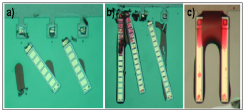
A simple way to confirm the effective presence or absence of liquid under MEMS structures is the modification of the liquid composition by addition of a low-volatility staining agent, which is traceable with a simple optical or fluorescence microscope. After the solution has almost completely evaporated, the residue, mainly consisting of the low vapour-pressure staining agent, reveals the areas originally occupied by liquid. An example is given in Figure 3. The MEMS was dipped into deionized water mixed with a staining solution (basic fuchsin). After the solution evaporated, cantilevers were mechanically removed using a micro-manipulator. Consider Figure 3a. Three cantilevers were removed: as can be seen, apparently not all the volume under them was occupied by the solution. This can be inferred by the presence of staining only under the distal portion of the cantilever, where the presence of a meniscus is furthermore clearly suggested by the round convex shape assumed by the staining. This could be explained considering the upward bending of the cantilevers due to the residual stresses introduced in their constituting layers by the manufacturing process [12].
Figure 3: Representative pictures of the stain method: a) stain solution occupies part of volume under the cantilevers; b) mechanical scratching not properly performed; c) enhanced wettability due to the increase in the stain concentration.

This method is inexpensive, but at the same time many drawbacks are evident:
a) modification of the liquid properties in relation to the presence of the staining agent, even if it is added at a low concentration level: the liquid surface tension is related to the staining agent’s concentration and, in particular, the higher the staining agent’s concentration, the lower the surface tension, causing the surface to become hydrophilic. During evaporation, the staining agent’s concentration increases, thus enhancing the wettability of the MEMS surfaces. This phenomenon is clear in Figure 3c since the meniscus shape is concave;
b) If the mechanical scratching is not properly performed, information about the stain print is lost see Figure 3b;
c) The ideal staining solution should produce an easily observable colour contrast, without affecting the liquid environment as concerns the surface tension. Unfortunately, in most staining solutions such properties are in conflict. In conclusion, evidence obtained with a staining solution is sometimes in disagreement with the theoretical considerations set out in the previous section. Even though the method is simple to implement and inexpensive, it is not sufficiently accurate.
Tomography is a non-invasive and non-destructive method for three-dimensional imaging, frequently used in materials technology and medicine. This technique consists of taking a set of images of the X-ray beam transmitted through a sample while the sample is rotated to different positions, from 0 to 360 degrees. Micro-tomography is the notation applied when the sample dimensions are below centimetre range, but the principles are otherwise identical to those just described. X-ray micro-tomography (XRT) requires an X-ray beam with a high degree of beam parallelism, preferentially with high brilliance and good penetration ability. For these reasons, X-rays of high photon energies from synchrotron sources are needed. This makes micro-tomography an expensive and non-feasible technique. The contrast in XRT is based on the differences in absorption of X-rays by the constituents of the sample. As a result, air can be distinguished from water [13] and an underwater 3D reconstruction of the MEMS can be provided. Unfortunately this technique, in addition to its high costs, in most cases does not reach adequate resolution. Only resolutions of a few microns can be achieved [14]. X-ray micro-tomography could be indeed an interesting tool for quantifying the presence of water under MEMS structures nevertheless, high experimental set-up costs suggest looking for alternative solutions.
With the definition indirect measurements we refer to all those methods that consist of measuring one or more MEMS properties affected by the presence of liquid under the structure. In MEMS, actuation comes from electro-thermal or electrostatic actuators. In the former (both standard thermal actuators and the Chevron thermal actuators), the presence of water under the moving structure increases dissipation. Therefore, a higher amount of electric-power is required to bend the arms [15]. In the latter (comb drivers [16], electro statically-actuated cantilevers [17] and electrostatic resonators), the presence of water increases the electrostatic force that is proportional to the dielectric constant of the medium. Moreover, the liquid introduces a mechanical damping effect related to the liquid’s viscosity. A well-known relevant phenomenon is squeeze film damping, which occurs for low gaps [18].
Therefore, one could operate the MEMS, measure values relevant to the aforesaid parameters and compare the experimental results with the theoretical models (with liquid or without liquid under the structures). For instance, in [19] an electro statically actuated cantilever beam was tested both in air and deionised water. The cantilever chosen for the test was 125 μm long, 10 μm wide and 2 μm thick (1.5 μm POLY2 plus 0.5 μm metal). The gap between the cantilever and the base was 3.5μm high. The cantilever was electrically insulated from the underlying silicon wafer, while it was connected to a contact pad by means of a conductive P-doped silicon path.The actual displacement depends on the intensity of the applied voltage, which can be made to vary in time, for example, as a sinusoid with an assigned frequency and amplitude.
To work in a liquid environment, thus effectively avoiding the formation of bubbles and corrosion caused by electrolytic phenomena which take place even when deionized water is being used, an amplitude modulation (AM) technique was used. In the proposed experimental set-up, a carrier frequency of 1 MHz was used in the experiments carried out in water, with 100% modulation (i.e. Voffset =Vmax ). The same set-up was used to easily compare the results, also for the experiments conducted in air. In both cases, a Vmax value of 2.5 V, that is 5 V peak to peak, was used. This driving voltage and the effective electrical load are well within the capabilities of a usual, low-cost signal generator, which can be thus directly wired to the MEMS device for this purpose. The frequency of the modulating signal was continuously varied from 10 up to 90 kHz and the vertical displacement measured using a DVD optical detector.
The heart of the system is a DVD optical head developed in our laboratory, which is briefly described below. The laser diodes used in DVD-ROM optical pickups typically operate at a wavelength around 630 nm. The absorption coefficient in liquid water is about 0.003 cm-1 (typically one-tenth of that for the CD-ROM units). The light source is a laser diode, which emits an astigmatic elliptical beam of light. Passing through diffraction grating, an airy pattern is formed. The light beam then encounters a polarizing beam splitter, a quarter wave plate, a collimating lens and, finally, the objective lens. When properly focused on a reflective surface, the beam reflects back along the original path, up to the polarizing beam splitter. Here, due to the changes in the polarizing direction, which occurred previously, it passes straight through this component and a cylindrical lens, finally striking the photo-detector. The combined effect of the effective position of the focus spot with respect to the reflective surface, and that of the cylindrical lens produce a characteristic light pattern on the surface of the quartered photodiode which, after being properly elaborated, results in a Focus Error Signal (FES).
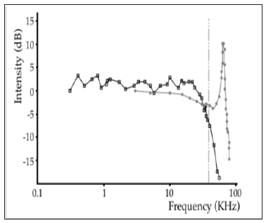
This is used to give a feedback signal to the moving-coil focusing actuator in the optical head. The RF signal arising from the reading of the pits embossed in the active layer of the storage medium is instead derived from the total intensity of the light that is striking the four-quadrant photodiode. As usual, the signal produced by each photodiode is in the form of a very small current, thus requiring further elaboration by means of a high-performance trans-impedance amplifier. The normalized cantilever displacements (expressed in dB) versus modulating frequency are shown in Figure 4. Squares mark experiment data in water, while dotsrefer to air.
Figure 4: Normalized cantilever displacements versus modulating frequency. Squared marks are relative to liquid, while dots refer to air.

It can be noted that in water the system is over-damped because of water viscosity. However what was relevant was that in some tests the ratio between the maximum displacement in water and in air fell in the 1530 ranges, when one expected a theoretical ratio of 80 because of the dielectric constant of water. This behaviour leads us to assume that the cantilever was not completely surrounded by water and/or that some air could have been trapped underneath the cantilever in the form of micro bubbles. It was this uncertainty that led us to develop a reliable method for mapping the presence of water under the cantilever. In conclusion, indirect measurement methods can reveal that the liquid is not properly distributed around the MEMS even when the surface is hydrophobic .On the other hand; they are not reliable, since mechanical displacements depend on many other parameters, which are not always easy to estimate. Moreover, indirect measurement methods cannot map how air present underneath MEMS structures is distributed.
A fast and low-cost technique was developed to verify whether or not water flows under cantilevers. This technique allows MEMS freezing to be performed in order to block water arrangement at a certain instant and then verify under the microscope the possible presence of frozen water under the beam structure. The prototype is shown in Figure 5. It is made up of a 30 by 30 mm working area Peltier cell (Marlow Industries), a water cooler apparatus, a power supply module and a microscope (Nikon Optiphot).The cell is fixed to the top of an aluminium cylindrical chamber, which fits under the microscope objective lenses. The cell, powered by a current of 1.5 ampere, requires an active cooler system in order to dissipate the excess heat produced on its bottom face. A continuous water flow (inlet temperature: 25 °C; flow: 12 cm3/s) passes through the chamber.
After several tests, a protocol to freeze the liquid was selected, which consists of the following steps:
a) The surface of the cell is cleaned with isopropyl alcohol in order to eliminate condensed water;
b) The MEMS to be analysed is glued using cyanacrylic adhesive on a glass slide;
c) A liquid drop is placed directly on the cell surface;
d) The glass with the glued MEMS is turned upside-down and placed over the liquid drop in such a way that the MEMS are completely surrounded by the liquid. To do so, a proper spacer is interposed between the glass slide and the cell surface. This way, the distance between the topside of the MEMS and the cell surface is about 0.5 mm;
e) The cell is turned on and after 20 s the drop is completely frozen (Figure 5). If some air is trapped under the cantilever, the possible scenario is the one shown in Figure 6a;
Figure 6: Scheme of the working principle of the MFTEC: a) before cantilever removal; b) after cantilever removal.

f) After 120 s the glass slide is manually removed by applying a vertical force. If the MEMS have been properly glued, it remains attached to the slide and a print can be observed on the frozen drop. Since poly-silicon layers (such as cantilevers, combs, folded springs) tend to adhere to the frozen liquid, the removal of the MEMS causes the poly-silicon layers to break in correspondence to their anchors to the substrate and their entrapment into the ice (Figure 6b).
g) Lastly, the frozen drop is analysed under the microscope. It is noteworthy that the poly-silicon layers on the drop show their bottom face (they are upside-down).Therefore, the presence of air or bubbles can be easily verified.
In our experiments, to remove air micro bubbles, some distilled water was degassed under vacuum at ambient temperature. Moreover, it was observed that the time required to freeze the drop affects the test. In particular, a long freezing time creates larger ice crystals, thus modifying the ice pattern with respect to the original water pattern. For this reason, before placing the MEMS on the drop, this was pre-cooled to 5-10°C.
An examination of the lower surfaces of the cantilevers after the removal of the MEMS yields some important data. This inspection has to be performed in just a few seconds, because of water condensation. When the MEMS are removed, some water could condense and freeze over the cantilevers over a very short time, in a way that could significantly modify the real configuration of the system. In the first set of experiments, non-degassed water was used. It was observed that many air bubbles remained entrapped on the MEMS-water interface see Figure 7a. Therefore, it was decided to degas water before MEMS immersion. When working with MEMS in a liquid environment, it is therefore important to take into account the likely and relevant presence of air bubbles in the water, which could considerably modify the dielectric characteristics of the medium interposed between the electrodes.
Figure 7: Different experimental observations with the MFTEC: a) air bubbles entrapped at the MEMS-water interface; b) presence of large crystals due to long freezing; c) continuous shape of ice after water degassing.

In Figure 7b, the presence of large crystals of ice can be noted. Large crystals are due to a relatively long freezing time and should be avoided, because they modify the initial pattern of water. In the subsequent experiments, degassing and pre-cooling of water were adopted before MEMS immersion. It is interesting to observe how the configuration of the total system changed: the micro-bubbles disappeared or became increasingly smaller (a few microns); the ice shows a continuous and irregular shape and single crystals cannot be distinguished as in the previous tests (Figure 7c). Degassing and pre-cooling procedures were adopted as an optimal protocol to study the problem. Many tests were performed and results were sufficiently variable depending on the shape of the cantilevers, on their bending, on the presence of small bubbles under and around them, and many other unpredictable factors. Therefore, every possible configuration of the lower surfaces of the cantilevers was observed: almost completely free from ice, partially covered and completely covered in ice.
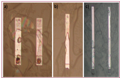
The second condition was the most recurrent result. When water partially flows under cantilevers, it can spread substantially in three different ways: In some cases, as shown in Figure 8a, it randomly flows, mainly depending on the distributions of small bubbles all around the structure. In other cases, it flows more under the free ends of the cantilevers than under the constrained ones. This phenomenon is preferred with long and curled-up cantilevers because of residual stresses caused by the different thermal expansion coefficient between poly-silicon and gold, as explained above see Figure 8b. It is possible to encounter the opposite case, too, as shown in Figure 8c. This is more reasonably the case of cantilevers in stiction - short for static friction - over the substrate. In the same figure, some water over the free end of the cantilever on the right can be noted: this is due to the presence of the dimple under the tip of the cantilevers that makes the free end of the cantilever stay 750 nm above substrate.
Figure 8: a) water randomly flowed under the cantilevers; b) water flowed under the cantilever free end; c) probable configuration of cantilevers in stiction

In some other cases, as shown in Figure 9a, cantilevers completely covered in ice were encountered. In the end, on a small number of occasions, cantilevers completely free from ice were observed (Figure 9b). However, it is difficult to ascertain whether these cantilevers were in stiction or rose above the substrate. The case of a comb drive was then studied. This system differs from that of cantilevers because of the substrate: it is made of silicon nitride in the case of cantilevers, while it is made of poly-silicon (POLY0) in the case of comb rotators. This is a necessary solution, since the substrate needs to have the same potential of the moving structures in order to prevent their adhesion. However, this difference is not appreciable, since poly-silicon and silicon nitride have approximately the same hydrophobicity. Besides, in the case of the comb drive, the distance between the moving structures and the substrate is lower than in the case of cantilevers: the comb rotator moves over a POLY0 base, touching the surface by means of dimples, so the gap is 750 nm.
In the end, in this second test it is more interesting to reveal the distribution of water between the comb fingers instead of that under the structure, since the active electrostatic force develops precisely between fingers. MFTEC is also capable of performing this type of investigation. The same protocol procedures were adopted and water distribution under the moving structures (comb rotors, shaft and springs) was studied. The comb stator, glued to the substrate, was obviously stripped off at the removal of the MEMS. Different situations were also encountered in the case of the comb drive. As shown in Figure 9a, water can completely flow under the moving structure; otherwise, as shown in Figure 9b, some parts of the structure are dry, implying that water did not flow in those zones. In both instances, the presence of water between the fingers can be revealed. These observations and each result described in the previous paragraph were obtained in a fast, simple and economic way using the MFTEC.
Micro-devices (MEMS) produced using silicon-based MUMS technology and working in a liquid environment were investigated. Direct and indirect measures were performed to detect the presence of water under the moving structures. In particular, a new, simple, economic and fast method for directly examining the configuration of the system was developed and tested. This technique, named MEMS Freezing by Thermo Electric Cooling (MFTEC), allows direct observation of the lower surface of the moving structures (cantilevers, comb rotors, etc.), revealing the distribution of water in the zone between the structures and the substrate. It showed that in most cases water partially flows under moving structures. In addition, the presence of many air micro-bubbles entrapped at the MEMS-water interface was revealed. Degassing of water, when possible, before MEMS immersion should therefore be considered a strictly necessary procedure. Since uncertainty on the value of surface tension, variability in the protocol to flow water (or any other fluid) and geometrical errors make it difficult to control for the presence of bubbles within complicated MEMS structures, the proposed MFTEC technique can be used for also improving the MEMS structure. In this case, particular MEMS should be made. It must replicate, on a different scale and with different parametric ratios, the geometries to be achieved with the final MEMS structure. In this way it will be possible to extract useful information for improving and optimizing MEMS structures.


