Impact Factor : 0.548
- NLM ID: 101723284
- OCoLC: 999826537
- LCCN: 2017202541
Hussam M Abdel-kader* and Ahmed SM Ammar
Received: June 01, 2018; Published: June 14, 2018
*Corresponding author: Hussam M Abdel-kader, Department of Orthodontics Faculty of Dental Medicine (Boys) Al-Azhar University, Cairo, Egypt
DOI: 10.26717/BJSTR.2018.05.001225
Introduction: The objective of this retrospective study was to investigate maxillary canine internal root resorption (IRR) concomitant to orthodontic retraction evaluated by cone beam computed tomography (CBCT).
Materials and Methods: The current retrospective study was conducted on a total sample of 10 CBCT of right and left maxillary canines of 5 maxillary first premolars extraction orthodontic patients with age range between 14 to 18 years old with mean age of 15.82 1.74 years. The sample was selected at random from a total sample of 32 CBCT which had been previously used to study external maxillary canine root resorption (ERR) of 16 orthodontic patients. Cone beam computed tomography (CBCT) image of maxillary teeth had been taken before treatment and after canine retraction using optimal orthodontic force (150g). Internal resorption of maxillary canines had been evaluated using much more sensitive scores previously used to study canine external root resorption.
Results: The results of the current study were calculated from CBCT of 20 right and left maxillary canine teeth. The average rate of orthodontic canine retraction was 1.34 + 0.31mm/month and the average time of canine retraction was 188.75 + 45.00 days.
Data interpretation: pulp and/or root canal chamber; increase or decrease in any direction, will be considered as internal resorption or build-up respectively. The results showed statistically insignificant difference in IRR between; before and after right and left maxillary canine retraction. There was also statistically insignificant correlation between internal root resorption (IRR) and rate of canine retraction.
Conclusion: With the use of the appropriate mechanics for orthodontic maxillary canine retraction into the first premolar extraction space, internal root resorption (IRR) will not be expected. On the other hand the use of appropriate mechanics in orthodontics is the keystone to achieve orthodontic treatment outcome without any hazard.
The literature is very rich with different studies covering external root resorption (ERR) subsequent to orthodontic treatment of dental malocclusion, which is investigating by different radiographs ending by the use of cone-beam computed tomography (CPCT) [1- 19]. In a recent study on ERR using CBCT, it was concluded that with the use of optimum orthodontic force ERR is of negligible [20]. On the other hand, data base lacking studies covering internal root resorption (IRR) associated with orthodontic tooth movement. In a case report the authors reported extensive internal resorption affecting the crown of orthodontically treated teeth which could extend into the root canal and ‘pink tooth’ [21]. In view of this shortage, it will be worth to investigate internal root resorption concomitant to orthodontic retraction of maxillary canine in first premolars extraction orthodontics, using CBCT.
The current retrospective study was conducted on a total sample of 10 CBCT of right and left maxillary canines of 5 maxillary first premolars extraction orthodontic patients with age range between 14 to 18 years old with mean age of 15.82 + 1.74 years. The sample was selected at random from a total sample of 32 CBCT which had been previously used to study external maxillary canine root resorption (ERR) of 16 orthodontic patients [20]. Cone beam computed tomography (CBCT) image of maxillary canine teeth was taken before-and after orthodontic canine retraction into the first premolars extraction space with optimal orthodontic force of (150g).
Measurements of internal root resorption (IRR): To measure the changes in pulp chamber:
a) When coronal view was studied, each root canal chamber was divided into three thirds; cervical, middle and apical third. As the canine was retracted orthodontically distally into the first premolar extraction space, the mesiodistal dimension of root canal chamber of each root canal third was measured.
b) when the axial view was studied, the vertical dimension from the tip of the pulp chamber to the apex of the root canal chamber. Internal root pulp chamber resorption (IRR): In order to measure the difference in internal root canal chamber length and dimension, maxillary canine pulp tip and root apex tip were identified on sagittal and the three coronal views obtained from CBCT 3D image respectively, and the software directly measure the actual changes in root canal measurements. These measurements were performed on both pre- and post- canine retraction CBCT images. Data interpretation of CBCT: pulp and/or root canal chamber; increase or decrease in any direction, will be considered as internal resorption or build-up respectively. All measurements were taken to the nearest 0.1mm.
The collected data was tabulated and statistically analyzed using SPSS statistical package. The result of the current study was considered of significant value at the level of p ≤ 0.05.
The current retrospective study was conducted on a total sample of 10 CBCT of right and left maxillary canines of 5 maxillary first premolars extraction orthodontic patients with age range between 14 to 18 years old with mean age of 15.82 + 1.74 years. The sample was selected at random from a total sample of 32 CBCT which had been previously used to study external maxillary canine root resorption (ERR) of 16 orthodontic patients [20].
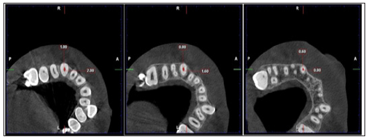
a) The following (Figures 1-7) from 1 to 7 illustrates CBCT analysis of the maxillary right and left canines teeth internal resorption concomitant to orthodontic retraction, using the optimal force, if any, of one of selected samples in the current study.
b) The following table from 1 to 3 illustrates the statistical analysis of the results.
Figure 1: Pre-treatment (left) and post canine retraction (right) CBCT image (coronal view) showing root canal length from the tip of pulp chamber to the apical end of right maxillary canine root canal.
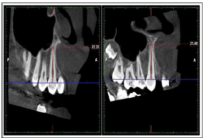
Figure 2: Pre-treatment (left) and post canine retraction (right) CBCT image (sagital view) showing root canal length from the tip of pulp chamber to the apical end of right maxillary canine root canal.
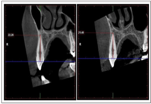
Figure 3-A: Pre-treatment (left) and post canine retraction (right) CBCT image (sagital view) showing root canal thirds (cervical, middle, and apical) and root canal length from the cement- enamel junction to the apical end of right maxillary canine root canal. (Axial slice (blue line) passing in the cervical third of maxillary canine root).
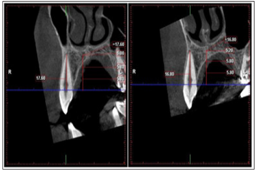
Figure 3-B: Pre-treatment (left) and post canine retraction (right) CBCT image (axial view) showing root canal dimensions in the cervical third of right maxillary canine root.
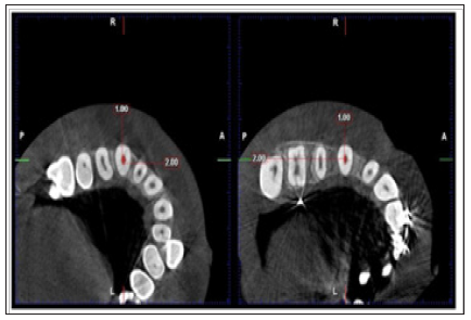
Figure 4-A: Pre-treatment (left) and post canine retraction (right) CBCT image (sagital view) showing root canal thirds (cervical, middle, and apical) and root canal length from the cement- enamel junction to the apical end of right maxillary canine root canal. (Axial slice (blue line) passing in the middle third of maxillary canine root).
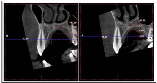
Figure 4-B: Pre-treatment (left) and post canine retraction (right) CBCT image (axial view) showing root canal dimensions in the cervical third of right maxillary canine root.
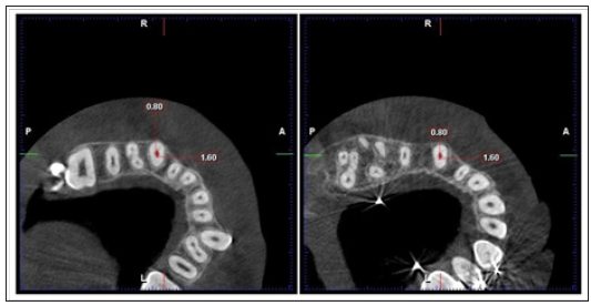
Figure 5-A: Pre-treatment (left) and post canine retraction (right) CBCT image (sagital view) showing root canal thirds (cervical, middle, and apical) and root canal length from the cement- enamel junction to the apical end of right maxillary canine root canal. (Axial slice (blue line) passing in the apical third of maxillary canine root).
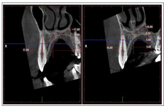
Figure 5-B: Pre-treatment (left) and post canine retraction (right) CBCT image (axial view) showing root canal dimensions in the middle third of right maxillary canine root.
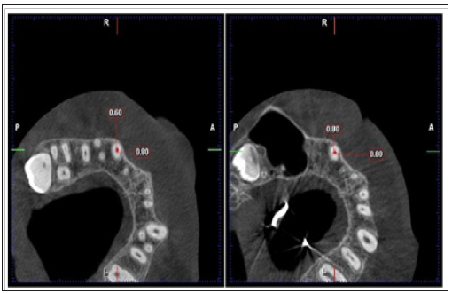
Figure 6: Pre-treatment CBCT image (upper row showing sagital view with axial line(blue color) passing in cervical, middle, and apical respectively from left to right) and ( lower row showing root canal dimensions in cervical, middle, and apical respectively from left to right) of right maxillary canine root canal.
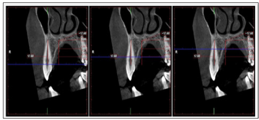
Figure 7: Post canine retraction CBCT image (upper row showing sagital view with axial line(blue color) passing in cervical, middle, and apical respectively from left to right) and ( lower row showing root canal dimensions in cervical, middle, and apical respectively from left to right) of right maxillary canine root canal.
Data interpretation of CBCT: pulp and/or root canal chamber; increase or decrease in any direction, will be considered as internal resorption or build-up respectively. The results of the current study are summarized in the following tables from 1 to 3. Table 1 showing the mean value and standard deviation of different measurements of the study including patient’s age, time, amount, and rate of canine retraction in different groups in the study. Table 2 shows the descriptive statistics of maxillary canine pulp chamber length before and after canine retraction and the difference between them and showing statistically insignificant difference, in the total sample group and the right and left side subgroup. In evaluation of the differences between right and left side pulp chamber length (resorption) and rate of canine retraction, Table 3 shows statistically insignificant difference in pulp chamber length between right and left side from numerical results obtained from CBCT.
S.D.= Standard deviation. n= numbers of maxillary canines R= Right canine. L= Left canine S.E. Diff.= Standard error of difference
* = Significant at (p ≤ 0.05)
Table 2: Descriptive statistics and paired-sample t test of maxillary canine pulp chamber length before and after canine retraction in different groups of the study.

S.D. = Standard deviation.
S.E. = Standard error.
Diff =difference
P = Probability level (paired t test).
* = Significant at (p ≤ 0.05)
n = numbers of maxillary canines
Table 3: aired t tests for side difference rate of canine retraction and pulp chamber length between, before and after canine retraction (mm).

S.D. = Standard deviation.
S.E. = Standard error.
Diff =difference
P = Probability level (paired t test).
* = Significant at (p ≤ 0.05)
n = numbers of maxillary canines
The literature is very rich regarding the use of CBCT to study external root resorption (ERR) of the maxillary canine in premolar extraction orthodontics [12,15,18,19]. On the other hand, searching the data base revealed shorting in studies covering internal maxillary canine resorption concomitant to orthodontic retraction in first premolar extraction orthodontics using CBCT. This encouraging the proposal of the current study. In accordance the result of the current study was discussed in view of thorough interpretation of the results. The current retrospective study was based on CBCT data of previous study by the authors on external root resorption. Internal root resorption (IRR) of the maxillary canines (right and left) had been investigated by using CBCT before and after complete maxillary canine retraction into the extraction. The results of CMPT sagittal view interpretation in the current study showed insignificant canine internal vertical length changes from the tip of the pulp chamber to the apical tip of the pulp. Which mean that there is insignificant internal vertical resorption of the canine. On the other hand the result of CBCT three coronal views interpretation showed insignificant changes. This means that there is insignificant coronal root canal internal resorption as well. The current study showed negligible maxillary cane internal root resorption after complete orthodontic retraction into the first premolars extraction space. This result is explained in view of using the appropriate mechanics for maxillary canine retraction (force range of 150gm). The result of the current study is in agreement and supported by the result of previous study of the same sample group which aimed to determine external root resorption (ERR) due to orthodontic canine retraction using cone beam computed tomography (CBCT) [20]. In evaluation of differences between right and left side regarding internal root resorption the current study did not detect any significant difference in internal root resorption between right and left side from numerical results obtained from CBCT at the level of p ≤ 0.05. This finding could be explained by the similarity of both right and left canines in terms of treatment mechanics.
With the use of the appropriate mechanics for orthodontic maxillary canine retraction into the first premolar extraction space, internal root resorption (IRR) is not expected. On the other hand the use of appropriate mechanics in orthodontics is the keystone to achieve orthodontic treatment outcome without any hazard.


