info@biomedres.us
+1 (502) 904-2126
One Westbrook Corporate Center, Suite 300, Westchester, IL 60154, USA
Site Map
Received: September 02, 2017; Published: September 15, 2017
Corresponding author: Murugesan S, 1Division of Algal Biotechnology and Bio-Nanotechnology, PG and Research Department of Botany, Pachaiyappa’s College, Chennai-600030, India
DOI: 10.26717/BJSTR.2017.01.000359
Silver nano particles have known to possess anticancer properties in the early stages of virus proliferation. In the present study biosynthesis of silver nano particles from marine red alga Halymenia porphyroides was synthesized and characterized. Studies on protein disulphide bonds were done using Metal Detector Predicts V2.0 software. All Cys and His residues in amino acid sequences were identified. The study revealed that silver nano particles couple with the sulfhydryl groups led to decrease the disulphide bonds and denatures the E7 protein thus eliminating the possibility of the occurrence of the disease.
Keywords: Bioinformatics; E7 protein; Silver nano particles; Metal binding sites.
Nano particles exhibit unique biological, physical and chemical properties based on specific characteristics such as shape, size, distribution and surface morphology. Synthesis of nano particles through biochemical routes, using plant and algal extracts as reducing and capping agents, has received special attention among others, due to maintaining an aseptic environment during the synthetic process [1-7]. Therefore, medicinal plants and algae having well established therapeutic importance are being widely used for the size and shape controlled synthesis of silver nano particles [8-11].
Silver nano particles are one of the most marketable nano materials, because they are extensively applied as biocides due to their strong antimicrobial activity. The silver nano particles possess high surface area enhancing the antimicrobial activity of nano particles [12] and their conductive [13], optic [14], as well as catalytic properties [15] widen their scope in many applications. Silver nano particles are also potential antiviral agents against HIV-1 [16], hepatitis B virus [17], respiratory syncytial virus [18], herpes simplex virus type 1 [19] and monkey pox virus [20]. Human papilloma viruses (HPVs) have been found in over 90% of cervical cancer, as well as in other carcinomas [21]. Certain HPV types have been classified as “high risk” types and others as “low risk”, based on the clinical prognosis of lesions which they cause. The major transforming proteins of the high risk HPVs have been identified as the early proteins E6 and E7; expression of these proteins is maintained in carcinoma cells lines [22], and expression of these two proteins induces immortalization and transformation in a variety of rodent and human cell types [23]. The aim of the study was to investigate the bioinformatics prediction of interaction of the obtained silver nano particles from H. porphyroides corresponding to the silver with Ag formula (number 94565) from ChemSpider website on the disulphide bonds of the E7 protein (www.chemispider.com).
Silver nano particles were biosynthesized from the red seaweed Halymenia porphyroides. 500 mg of the dry seaweed was taken in 10-3 M aqueous (AgNO3) solution and the incubation was done at 35-37 0C for 24-48 hrs. The pH was checked and found to be 5.09 during the synthesis. The silver nanoparticles were characterized using UV visible spectroscopy, X-Ray diffraction studies, FT-IR spectroscopy, Scanning Electron Microscopy and HR- Transmission Electron Microscopy
The specific disease selection and all the proteins involved in the pathway are surveyed from KEGG database. The amino acid sequence of the selected protein for cancer is E7 protein which was obtained from Swissprot. The existing template modelled designed structures were searched in PDB. Since there is no model available in PDB, Swiss model and Ramachandran plot were created. For this purpose swisspd bv was used. A login ID was created in SWISS MODEL. AUTOMATED mode was selected for designing. The raw amino acid sequence of E7 was submitted. BLASTp was run to determine the most similar template for E7. The programmer was run to predict the most effective structure. The Ramachandran plot was constructed to find out the stability, as well as the alignment of all the amino acids. The overall quality of the modelled protein was checked by logging on to structure verification server site (SAVS). The target molecule file was uploaded to the SAVS. The SAVS process the molecule and interpret the results. The problematic amino acids, overall quality factor and other results were obtained. The modelled protein was obtained and the structure variation is viewed through SPDV.
The web server Metal Detector is freely available at (http:// metaldetector.dsi.unifi.it/v2.0) which classifies histidine residues in proteins into two states (free or metal bound) and cysteine’s into three states (free, metal bound or disulphide bridged). This web server takes the protein sequence as input and provides predictions of transition-metal binding sites for cysteine and histidine residues as output; for cysteine it also predicts disulphide bonding bridges. The residues were predicted to coordinate the same ion and will also share the same identifier. Every identifier is an integer ranging from 1 to 4. Its value has no special biochemical semantics but lower value corresponds to a higher level of confidence for the predictor.
The studies on disulphide bonds were performed using Metal Detector Predicts v2.0 software. All Cys and His residues in amino acid sequences were identified. (http://metaldetector.dsi.unifi.it).
Biosynthesis of silver nanoparticles from the red seaweed Halymenia porphyroides extract was formed by the reduction of Ag+ at 35-370C. The change in pale yellow to brown colour change indicated the formation of silver nano particles in the solution and confirmed the excitation of surface plasmon vibrations during the formation of silver nano particles. The biosynthesized silver nano particles was thoroughly characterized and was presented in our previous investigation.
The absorption peak at 420 nm in the UV-visible spectroscopy showed AgNPs formation and confirmation of surface plasmon resonance [24]. The vibrations peaks from 2354.71 to 3884.4 cm-1 from the FT-IR spectrum analysis indicated the presence of several functional moieties of organic compounds associated with silver nano particles. The various organic compounds present were O-H groups, free alcohol groups, free amines and intra molecular hydrogen bonds. The secondary metabolites such as flavanoids, alkaloids, terpenoids and steroids could possibly act as capping ligand for stabilization of the silver nano particles [25]. The SEM analysis of the biosynthesized silver nano particles showed the average size of the particles and was in spherical shape. The formation of the stable spherical and isotropic nano particles maybe due to the biomolecules associated with the silver nano particles [26].
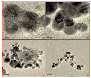
The HR-TEM images of mediated silver nanoparticle synthesis showed the formation of spherical shaped silver nano particles (Figure 1). The size of the biosynthesized silver nano particles ranged between 5 to 50 nm and the average mean size of silver nano particles was 32 nm.
Figure 1: HR-TEM images of silver nano particles of biosynthesized H. porphyroides. (a) 5 nm (b) 10 nm (c) 20 nm (d) 50 nm.
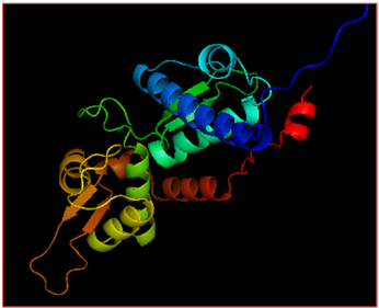
The Swissprot was used to identify the amino acid sequence of the selected protein for cancer E7 protein (Figure 2). The stability and the alignment of the amino acids were confirmed by constructing the Ramachandran plot (Figure 3). SPDV was used to see the structure variation and the constructed modelled protein (Figure 4). E7 protein structure contains chain A and chain B and its length is 371 amino acids, and its molecular weight is 17 kD. Over 90% of malignant carcinomas of the genital tract have HPV DNA sequences; the majority of which contain the high risk HPV-16 and 18 types which are linked to the progression of cervical lesions. The most abundant viral transcripts in tumor and tumor cell lines come from the E6 and E7 open reading frames that are known to be oncogenic. These two genes from HPV are necessary and sufficient to induce HPV-mediated transformation of murine cells. HPV16 E7 binds to the unphosphorylated form of the retinoblastoma protein. E7 proteins of other types (HPVs 6, 11, 18) have also been shown to bind to pRB [27]. Silver nano particles act at the beginning stages of viral propagation (penetration or spread) and at pre-penetration stages [28]. The binding of silver ions to sulfhydryl groups causes protein denaturation through reducing disulphide bonds [29]. The results of the present study showed that the first and most important part of this protein binds E7 and pRB protein. In Molegro software the basis of the three dimensional structure of this protein is positioned between 20 to 29 amino acids where sulphide bonds are located. Breaking this binding can change the spatial structure of this protein, therefore prevents this part from binding with pRB.
Figure 2: Protein sequence from Swissprot.

Figure 3: Ramachandran plot showing the protein clearance in SAVS.
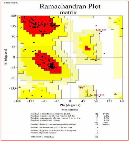
Figure 4: Modelled E7 protein structure.
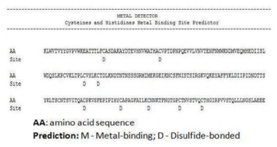
According to the results taken from Metal Detector Predicts v2.0 online server, the disulphide bonds in E7 protein amino acid sequence are sited between cysteine amino acids in various locations (Figure 5). The 27th sequence is in the first and most important part of this protein to bind E7 and pRB proteins. In Molegro software on the basis of the three dimensional structure of this protein is positioned between 20 to 29 amino acids where sulphide bonds are located. Breaking this binding can change the spatial structure of this protein and therefore, prevent this part from binding to pRB.
Figure 5: Disulphide bonds and metal binding site in E7 protein.
Silver nano particles can act as antivirus agent for deactivation of the virus in a short period of time. This process is done through interaction of nano silver with 2 disulphide bonds located in carboxyl (E7) so that silver ions, through interaction with thiol group, decrease disulphide bonds leading to protein denaturation. Considering other similar scientific investigations, one can conclude that the mentioned process is fulfilled through substitution of single valence Ag+ with H+ existed in thiol group. Bioinformatics results using software and servers show that silver ions make interaction with thiol group through which the disulphide bonds decrease and the virus and cancer proliferation were inhibited.


