 Mini Review
ABSTRACT
Proteins or other compounds that cancer cells produce in greater quantities than healthy cells have historically been used as tumour markers Tumour markers can reveal details about a cancer, including its aggressiveness, its ability to be treated with targeted therapies, and its response to therapy. Surface plasmon resonance (SPR) has received a lot of interest and offers a wide range of uses in biosensors and chemical sensors. The significant aspects of this innovation, such as its sensitivity, real-time identification, and label-free assay, set it apart from prior sensing platform breakthroughs. SPR technique has been proved a significant used for the detection of tumour markers. This review concludes various applications of Surface Plasmon Resonance in detection of tumour markers.
Keywords: Surface Plasmon Resonance (SPR); Tumour Markers, microRNA (miRNA); Biomarkers; Exosomes
Abbreviations: SPR: Surface Plasmon Resonance; miRNA: Micro Ribonucleic Acid; ELISA: Enzyme-Linked Immunosorbent Assay; WHO: World Health Organization; AuNPs: Gold Nanoparticles; LOD: Limit of Detection; PDAC: Pancreatic Ductal Adenocarcinoma; CSCs: Cancer Stem Cells; AML: Myeloid Leukaemia
Introduction
The second most prevalent globally, cancer is a serious issue for human wellbeing [1]. As per World Health Organization (WHO) statistics from 2019, cancer is the third or fourth major cause of mortality before the age of 70 in 23 nations, while it is the top or second main cause in 112 of 183 countries [2]. Cancer is the uncontrollable proliferation of leukemic cells that causes significant modifications in physiological mechanisms [3]. Normal cells, chemicals, and blood arteries that surround and nourish a tumour are affected by cancer cells, which have the ability to elude the immune system [4]. The digestive, neurological, and circulatory systems may be affected by the tumours, which have the ability to spread and expand to other parts of the body. They may also emit hormones that affect how the body works. For anticancer drug development, designing molecules that can selectively inhibit the proliferation of abnormal cells with minimal or no effect on normal cells is critical [5]. Therefore, developing anticancer drugs is of utmost importance worldwide. Developing compounds that may selectively limit the growth of aberrant cells with little to no impact on healthy cells is crucial for the development of anticancer drugs [6]. Therefore, creating anticancer medications is of highest significance everywhere. In 2020, it is anticipated that were 19.3 million cases of cancer globally (18.1 million with the exception of nonmelanoma skin cancer) and over 10 million cancer deaths (9.9 million excluding nonmelanoma skin cancer).
With an expected 2.3 million new cases (11.7%), female breast carcinoma has overtaken lung cancer as the most often detected malignancy. Lung (11.4%), colorectal (10%), prostate (7.3%), and stomach (5.6%) cancers are the next most commonly detected malignancies. With an expected 1.8 million fatalities (18%), lung cancer continued to be the most common kind of cancer. It was then followed by colorectal (9.4%), liver (8.3%), stomach (7.7%), and female breast (6.9%) tumours [7]. Surface plasmon resonance (SPR) has received a lot of interest and offers a wide range of uses in biosensors and chemical sensors [8]. Over than 80 years ago, Wood first identified a phenomenon, and it wasn’t until Liedberg et al. that the first gas sensing plus biosensing principles were developed [9]. The significant aspects of this innovation, such as its sensitivity, real-time identification, and label-free assay, set it apart from prior sensing platform breakthroughs [10]. The core idea of SPR sensor design is the resonance of a strong electromagnetic field oscillation at the interface of a nanometal sheet and a dielectric medium with p-polarized light as the incoming light, which results in a dark band pattern in the light reflectivity at a certain wavelength [11].
Applications of Surface Plasmon Resonance
Various applications of SPR have been outlined in Figure 1.
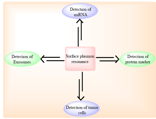
Figure 1: Applications of Surface plasmon resonance.
SPR-Detection of Tumour Cells
Tumour is a mass of tissue that develops abnormally when cells grow and multiply more often than normal or do not die when they should. Both benign and malignant tumours are possible (cancer). Although benign tumours have the potential to become enormous, they do not penetrate or spread to neighbouring tissues or other body regions [12]. Fathi et al., designed and reported the establishment of a real-time, label-free surface plasmon resonance (SPR)-based biosensor for the identification of cancer stem cells (CSCs) employing the cellular biomarker CD133. A few patients with severe myeloid leukaemia (AML) had this marker detected using the constructed biosensor, and the outcomes were evaluated to those of the flow cytometry (FC) approach. After separating mononuclear cells from the individuals’ bone marrow, CD133 antibody was immobilised on the gold chip surface using the EDC/NHS coupling approach, and binding of the candidate cells to the altered gold sensor surface was observed. The technique was verified using a number of criteria, including cell density and CD133-antibody content. Seven AML patients’ CD133-marked cells were examined.
The findings of the FC technique and all SPR outcomes were compared. The SPR sensing chip’s gold sheets with 25 μg/ml of CD133 antibody put on them exhibited the greatest angle shift, and at a flow rate of 20 ×μl/min, the best grab potential was achieved with 1×10⁵ cells/ml. The association among SPR and FC responses in regard to the densities of CD133-marked cells was extremely strong (R2 = 0.96). In summary, a label-free and real-time SPR cytometry approach was created in this work and effectively used to identify CD133 and track this tumour stem cell biomarker in AML patients [13]. Chiu and co-workers designed a brand-new class of extremely effective biosensing technology is functionalized graphene oxide. Using the cytolerayin 19 (CK19) protein biomarker in spiking human plasma, authors describe a carboxyl-functionalized graphene oxide (GO-COOH)-based surface plasmon resonance (SPR) device for the quick and accurate detection of non-small cell lung cancer (NSCLC). The authors showed that kinetic study of connections between GO-COOH and anti-CK19 and CK19 protein was binding selective. They also determined the relationship of SPR angle and the refractive index of GO-COOH with one another and showed that -COOH modified GO sheets on Au film can increase the field energy transmission intensity of an SPR sensor, leading to a higher sensitivity for the detection of CK19 protein than a traditional Au-based SPR chip. By immobilising CK19 antibody at a trace amount (10 μg/mL) on an SPR chip, the immunosensor was planned and operated.
One fg/mL was the minimal observable concentration. It was possible to generate a boosted 10 percent human plasma CK19 detection limit of 0.05 pg/mL, which is much lower than the physiologically appropriate amount of serum protein (3.3 ng/ mL). For the identification of clinical total plasma biomarkers and potential use in disease diagnosis, a carboxyl-GO based SPR biosensor consequently seems to offer good sensitivity and specificity. In comparison to a traditional SPR chip, this GO-COOH based SPR chip had a quicker reaction time, a better detection limit, and a good linear range (0.001 to 100 pg/mL). We also showed that our carboxyl-GO-based SPR biosensor could detect CK19 at levels as low as 0.05 pg/mL in 10% blood plasma and at levels as low as 0.001 pg/mL in PBS solution. When compared to outcomes in PBS solution, the identification of protein in spiked plasma was demonstrated with an accuracy of R2=0.965 [14].
Surface Plasmon Resonance -Detection of Exosomes
Extracellular vesicles called exosomes comprise the protein, DNA, and RNA of the cells that produce them. They are absorbed by far-off cells, in which they can alter cellular activities and functioning. Exosomes are carrier atoms that cells spontaneously discharge [15]. Cell communication is made possible by these particles. Exosomes provide channels for cell communication and deliver genetic material and proteins to every cell in your body. Currently available techniques for isolation and quantification of exosomes are either requires higher quantities of sample, less specificity and less purity results. To overcome these limitations, researchers have designed some other technologies such as microfluidic device connected with surface plasmon sensors. These sensors exhibited accurate results and isolate exosomes with perfectly sized under the limits [16]. Chen and co-workers designed and reported a label-free, real-time surface plasmon resonance imaging biosensor based on the hydrogel-AuNP supramolecular sphere (H-Au) for the very precise and targeted detection of exosomes produced by prostate carcinoma cells.
The SPR biosensor for exosome identification displayed a vast linear range from 1.00×10⁵ to 1.00×10⁷ particles/mL with a limit of identification of 1.00×10⁵ particles/mL after embedding the signal amplification influence of the mass accumulated hydrogel and the LSPR impact of AuNPs with relatively high aptamer. Most notably, this biosensor demonstrated excellent practical applicability for human serum evaluation, which demonstrates promising applications in diagnosis of the disease and bioanalysis, as demonstrated by a strong correlation between the SPRi signal and the t-PSA value assessed by the clinical chemiluminescence immunoassay. The mass cumulative multilayer hydrogels and the LSPR effect of the AuNPs are primarily responsible for the exceptional sensitivity of this approach. Figure 2 displayed the whole procedure for detection of exosomes. Additionally, the devised test has been successfully used to analyse exosomes in the clinical serum of patients with prostate cancer. Most importantly, there is a substantial association between the t-PSA levels determined by therapeutic chemiluminescence immunoassay and the SPRi signals. Consequently, our technique is capable of differentiating between the sick group and the apparently healthy group, holding tremendous promise for exosome detection-based early cancer diagnosis and therapy monitoring. As a result, we believe that this study may open up a fresh, alternative route for the early detection of prostate cancer [17].
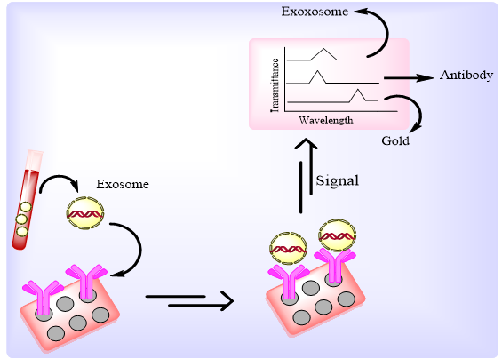
Figure 2: Detection of exosomes – Transmission spectra.
Jabin et al., proposed a biosensor for the prompt identification of various cancer-affected cells, a novel surface plasmon resonance (SPR)-based cancer sensor with an optimal bowl shape. Some significant variations in optical characteristics are seen when the refractive index (RI) of each cancer-contaminated cell is compared to its normal cell counterpart. Additionally, the concentration of malignant cells in liquid is estimated to be 80%, and 2100390 mesh components are used in the finite element model (FEM) for screening. The plasmonic band gap in between silica and cancer cell parts, which are isolated by a thin (35 nm) titanium film coating, is responsible for variation in spectrum shift. The suggested sensor has a maximal coupling length of 66 μM and a high birefringence of 0.04. The suggested structure, however, offers an optimal wavelength sensitivity level in between about 10000 nm/RIU and 17500 nm/ RIU, with a sensor precision between 1.5×10⁻² RIU and 9.3×310-3 RIU. Additionally, for carcinoma cells in major polarisation phase with a maximal detection limit of 0.025, the transmittance variance of the malignant cell ranges from around 3300 dB/RIU to 6100 dB/ RIU and the amplitude sensitivity is virtually between -340 RIU-1 and -420 RIU-1. Additionally, the total sensitivity performance is evaluated in relation to their normal cells, which may be superior to any previously presented structures [18].
SPR-Detection of miRNA
Non-coding RNAs known as microRNAs (miRNAs) have significant functions in controlling the expression of genes. Most miRNAs are produced via transcription of DNA sequences into primary miRNAs, precursor miRNAs, and then mature miRNAs [19]. By base-pairing with the target mRNA to inhibit its production, the miRNA acts as a guide. The silencing method is selected by how well the guide and mRNA target complement one other breakage of the target messenger RNA (mRNA), followed by its destruction or suppression of translation [20]. In the presence of single-stranded (ss)DNA/miRNA duplexes, Liyanage et al., (2019) proposed a novel transduction mechanism that incorporates the dispersion of photoexcited conduction electron wave functions of gold triangular nanoprisms (AuTNPs). The plasmoelectronic impact affects the LSPR characteristics of AuTNPs, improving the sensing capabilities. This sensor allowed for the highly specific identification of miR- 10b, miR-182, miR143, and miR145 in serum sample from bladder carcinoma patients with a limit of detection (LOD) as low as 140 zeptomolar (zM). In addition to having the potential to develop into a revolutionary liquid biopsy technology, this ultrasensitive assay may also be used to analyse single malignant cells and identify circulatory miRNA in patient serum for initial-stage, low-volume diagnostic tests for a range of disorders [21].
Recent research on the first LSPR-based sensing method in physiological medium was published by Joshi and group. They created a highly focused plasmonic biosensor to identify miRNA in bloodstream from pancreatic cancer patients with extreme sensitivity. Gold nanoprisms mounted to a glass surface and functionalized with ssDNA (HS-C6-ssDNA) corresponding to the target miRNA are used in this experiment. By observing the LSPR dipole peak (LSPR), the direct hybridization of the target miRNA was discovered. As possible circulatory diagnostic and prognostic indicators for pancreatic ductal adenocarcinoma (PDAC), miR-21 and miR10 levels may be precisely detected with great precision in the sub-femtomolar (fM) range using this biosensor. The LSPRbased observations indicate that, in contrast to conventional qRT-PCR, where a few RNA is lost during sample collection, the concentration of this miRNA is at least 2-fold greater. Any sample collection for the RNA target sequence (such as alteration, amplification, or tagging) has been eliminated in this case, and all issues with the existing sensing methods have been resolved.
Additionally, DNA RNA duplex splitting enzymes might be used to replenish the sensor without reducing its sensing effectiveness. This method transforms the sensor into a straightforward, affordable tool for the early screening of cancer using any miRNA [22]. Research conducted by Ding et al., discloses an SPR biosensor for very rapid screening of miRNA relying on streptavidin signal amplification and DNA super-sandwich complexes, which is intended to solve the drawbacks described earlier. In this test, a corresponding thiolate capturing hairpin probe is mounted on the sensor surface. The structural change is caused by the target miRNA’s hybridization with the thiolate probe. After miRNA hybridization, the capture probe’s hairpin loop reveals interaction sites for the supplementary probe AP1, which has been altered with a biotin tag. On the sensor surface, AP1 partly binds AP2, additional auxiliary probe, to create a super-sandwich. By introducing streptavidin to the surface of sensor, a signal augmentation cascade is further set off. Streptavidin binds to the biotin label of AP1 in the supersandwich to adhere to the chip surface. With higher overexpression in several tumour tissues, miR-21, a possible cancer biomarker was found using this sensing technology. A LOD of 470 pM was obtained with rapid identification, whereas a cascade of signal augmentation reduced the LOD to 9 pM. MiR21 was effectively identified in human breast adenocarcinoma MCF-7 cells following calibration of the sensing platform, exhibiting a non-compromised analysis in complicated components. The test also shown a high level of specificity in detecting complementary target miRNA, single-base mismatch target, double-base mismatch target, and non - specific miRNA sequences. The detection of miRNA with a LOD of 9 p mL-1 is straightforward, quick (30 min), and enzyme-free using this sensing technology. It was also possible to renew the assay, enabling the use of the same chip for at least 20 times [23].
Surface Plasmon Resonance- Protein Marker
Proteins or other compounds that cancer cells produce in greater quantities than healthy cells have historically been used as tumour markers [24]. Some cancer patients may have them in their tumours, blood, urine, faeces, or other body fluids or tissues [25]. Anything appear in or formed by tumour cells, other cells of the body, or certain benign (noncancerous) circumstances is referred to as a tumour marker [26]. Tumour markers can reveal details about a cancer, including its aggressiveness, its ability to be treated with targeted therapies, and its response to therapy [27]. Additional interesting biomarkers towards early detection of cancer are proteins, whose expression, quantity, location, and biochemical changes significantly causes impact on biological process. Only a small subset of those proteins are actually tested as biomarkers in clinical practice for cancer diagnosis and monitoring, despite extensive data showing that hundreds of proteins are differently expressed in human malignancies. Mainly, immunoassays and affinity techniques were used from past times for detection of protein and its quantification in which antibody and targeted protein bound together with another antibody. For this, we have compiled some protein biomarkers generally used clinically for detection of various cancer and their quantification at lower concentration. The whole procedure for detection of protein markers have been depicted in Figure 3. Eletxigerra and coworkers have developed a concise and accurate SPR method for the detection of tyrosine kinase specially (ErbB2) in human serum. the LOD of the sample was found to be 180 pg mL-1, which is under the limits [28].
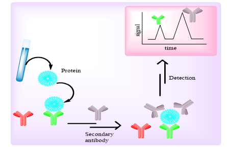
Figure 3: SPR method for determination of tyrosine kinases.
Conclusion
In this article, we have studied about new applications of surface plasmon resonance for isolation, detection and quantification of various types of tumour markers used in diagnosis of cancer. Most of the SPR methods exhibited better results and minimize the time duration of results than other assays like, ELISA. These methods also detect the markers at low concentration that’s why, the techniques are more reliable and required less amount of sample. These all considerations help SPR techniques for providing cost-effective, robust, accurate and sensitive methods for isolation, detection and quantification of various tumour markers.
References
- Turner MC, Andersen ZJ, Baccarelli A, Diver WR, Gapstur SM, et al. (2020) Outdoor air pollution and cancer: An overview of the current evidence and public health recommendations. CA: a cancer journal for clinicians 70(6): 460-79.
- Cao W, Chen H-D, Yu Y-W, Li N, Chen W-Q (2021) Changing profiles of cancer burden worldwide and in China: a secondary analysis of the global cancer statistics 2020. Chinese Medical Journal 134(07): 783-791.
- Hsieh C-Y, Ko P-W, Chang Y-J, Kapoor M, Liang Y-C, et al. (2019) Design and synthesis of benzimidazole-chalcone derivatives as potential anticancer agents. Molecules 24(18): 3259.
- Fadaka A, Ajiboye B, Ojo O, Adewale O, Olayide I, et al. (2017) Biology of glucose metabolization in cancer cells. Journal of Oncological Sciences 3(2): 45-51.
- Wang J, Li J, Xiao Y, Fu B, Qin Z (2020) TPP-based mitocans: A potent strategy for anticancer drug design. RSC Medicinal Chemistry 11(8): 858-875.
- Küpeli Akkol E, Genç Y, Karpuz B, Sobarzo-Sánchez E, Capasso R (2020) Coumarins and coumarin-related compounds in pharmacotherapy of cancer. Cancers 12(7): 1959.
- Sung H, Ferlay J, Siegel RL, Laversanne M, Soerjomataram I, et al. (2020) Global cancer statistics: GLOBOCAN estimates of incidence and mortality worldwide for 36 cancers in 185 countries. CA: a cancer journal for clinicians 71(3): 209-249.
- Wang Q, Ren Z-H, Zhao W-M, Wang L, Yan X, et al. (2022) Research advances on surface plasmon resonance biosensors. Nanoscale 14(3): 564-591.
- Liedberg B, Nylander C, Lunström I (1983) Surface plasmon resonance for gas detection and biosensing. Sensors and actuators 4: 299-304.
- Šípová H, Homola J (2013) Surface plasmon resonance sensing of nucleic acids: A review. Analytica chimica acta 773: 9-23.
- Li L, Hong M, Schmidt M, Zhong M, Malshe A, et al. (2011) Laser nano-manufacturing–state of the art and challenges. CIRP annals 60(2): 735-755.
- Yadav AR, Mohite SK (2020) Cancer-A silent killer: An overview. Asian Journal of Pharmaceutical Research 10(3): 213-216.
- Fathi F, Rahbarghazi R, Movassaghpour AA, Rashidi M-R (2019) Detection of CD133-marked cancer stem cells by surface plasmon resonance: Its application in leukemia patients. Biochimica et Biophysica Acta (BBA)-General Subjects 1863(10): 1575-1582.
- Chiu N-F, Lin T-L, Kuo C-T (2018) Highly sensitive carboxyl-graphene oxide-based surface plasmon resonance immunosensor for the detection of lung cancer for cytokeratin 19 biomarker in human plasma. Sensors and Actuators B: Chemical 265: 264-272.
- Niazi SK (2022) The Future of Pharmaceuticals: A Nonlinear Analysis: CRC Press.
- Boriachek K, Islam MN, Möller A, Salomon C, Nguyen NT, et al. (2018) Biological functions and current advances in isolation and detection strategies for exosome nanovesicles. Small 14(6): 1702153.
- Chen W, Li J, Wei X, Fan Y, Qian H, et al. (2020) Surface plasmon resonance biosensor using hydrogel-AuNP supramolecular spheres for determination of prostate cancer-derived exosomes. Microchimica Acta 187(11): 1-10.
- Jabin MA, Ahmed K, Rana MJ, Paul BK, Islam M, et al. (2019) Surface plasmon resonance-based titanium coated biosensor for cancer cell detection. IEEE Photonics Journal 11(4): 1-10.
- O'Brien J, Hayder H, Zayed Y, Peng C (2018) Overview of microRNA biogenesis, mechanisms of actions, and circulation. Frontiers in endocrinology 9: 402.
- Kakumani PK (2022) AGO-RBP crosstalk on target mRNAs: Implications in miRNA-guided gene silencing and cancer. Translational Oncology 21: 101434.
- Liyanage T, Masterson AN, Oyem HH, Kaimakliotis H, Hang Nguyen, et al. (2019) Plasmoelectronic-based ultrasensitive assay of tumour suppressor micrornas directly in patient plasma: design of highly specific early cancer diagnostic technology. Analytical chemistry 91(3): 1894-1903.
- Joshi GK, Deitz-McElyea S, Johnson M, Mali S, Korc M (2014) Highly specific plasmonic biosensors for ultrasensitive microRNA detection in plasma from pancreatic cancer patients. Nano letters 14(12): 6955-63.
- Ding X, Yan Y, Li S, Zhang Y, Cheng Q, et al. (2015) Surface plasmon resonance biosensor for highly sensitive detection of microRNA based on DNA super-sandwich assemblies and streptavidin signal amplification. Analytica chimica acta 874: 59-65.
- Sadighbayan D, Sadighbayan K, Tohid-Kia MR, Khosroushahi AY, Hasanzadeh M (2019) Development of electrochemical biosensors for tumour marker determination towards cancer diagnosis: Recent progress. TrAC Trends in Analytical Chemistry 118: 73-88.
- Alorda-Clara M, Torrens-Mas M, Morla-Barcelo PM, Martinez-Bernabe T, Sastre-Serra J, et al. (2022) Use of Omics Technologies for the Detection of Colorectal Cancer Biomarkers. Cancers 14(3): 817.
- Vidavsky N, Kunitake JA, Estroff LA (2021) Multiple pathways for pathological calcification in the human body. Advanced healthcare materials 10(4): 2001271.
- Regel I, Mayerle J, Ujjwal Mukund M (2020) Current strategies and future perspectives for precision medicine in pancreatic cancer. Cancers 12(4): 1024.
- Eletxigerra U, Martinez-Perdiguero J, Barderas R, Pingarrón JM, Campuzano S (2016) Surface plasmon resonance immunosensor for ErbB2 breast cancer biomarker determination in human serum and raw cancer cell lysates. Analytica Chimica Acta 905: 156-162.
