
 Research Article
Abstract
To improve of care of patients with chronic inflammatory skin condition - psoriasis, diagnostic methods that can facilitate personalized medicine are needed. This study aimed to determine whether non-invasive measurements of inflammation related proteins from psoriasis lesional skin can be sampled using Transdermal Analysis Patch (TAP) to assess disease severity and to monitor treatment efficacy. For skin surface biomarker assessment psoriasis vulgaris patients and healthy volunteers were enrolled to exploratory study where biomarker measurements from skin were performed using FibroTx TAP and scores for psoriasis activity and severity (PASI) were documented. Differences in thickness of skin layers were determined using sonography. To evaluate the non-invasive biomarkers pharmacological response to therapeutics measurements from the skin surface of psoriasis patients undergoing whole-body treatment with narrow-band UVB was assessed. Using TAP technology clear differences in levels of IL-1, IL-1RA and CXCL-1/2 were found between psoriasis lesional and non-lesional skin. In addition, a weak correlation between CXCL-1/2 and desquamation and between CXCL-1/2 and SLEB thickness where observed. Monitoring IL-1RA and CXCL- 1/2 on skin lesions undergoing narrow-band UVB phototherapy clearly reflected normalisation of skin.
Abbreviations: BSA: Body Surface Area; hBD-1: Human Antimicrobial Beta Defensin 1; ELISA: Enzyme-Linked Immunosorbent Assay; IHC: Immuno-Histochemistry; IL-: Interleukin One Alpha; IL-1RA: Interleukin One Receptor Antagonist;IL-6: Interleukin Six; IL-12: Interleukin Twelve; IL-23: Interleukin Twenty-Three; hBD-1: Human Beta Defensin 1; CXCL-1: Chemokine (C-X-C motif) Ligand 1; CXCL-2: Chemokine (C-X-C motif) Ligand 1;CXCL-1/2: Chemokine (C-X-C motif) Ligand ½; K16: Keratin Sixteen; PASI: Psoriasis Area Severity Index; SCORAD: Scoring Atopic Dermatitis Severity Index; SLEB: Sub-Epidermal Low Echogenic Band; TAP: Transdermal Analysis Patch; TNF-: Tumor Necrosis Factor Alpha; UVB: Ultraviolet B; qPCR: Quantitative Polymerase Change Reaction
Introduction
Psoriasis is a chronic relapsing immune inflammatory dermatosis with different clinical manifestations that affects 1-3% of the world population [1]. Psoriasis vulgaris (PV) is the most common variant of psoriasis, characterized by erythematous scaly plaques of the skin caused by the interplay between immune cells, keratinocytes, and other skin-resident cells, mediated by adaptive and innate immune system components cause a hyperproliferation of keratinocytes and chronic inflammation in affected skin [2-4]. Psoriasis patients often develop additional systemic comorbidities such as arthritis, metabolic syndrome along diabetes, cardiovascular risk, and depression [5]. Despite this knowledge, it remains difficult to predict both onset and progression of psoriasis, and there is an unmet medical need for methods that can be used to measure if people are at risk for psoriasis, and/or that can predict how disease progresses, both with respect to severity, and with respect to comorbidities. Clinical evaluation of psoriasis is primarily performed visually. The Psoriasis Area Severity Index (PASI) is a clinical score based on assessment of the percentage of skin affected (on head, trunk, arms, and legs) and severity of the skin erythema, induration, and desquamation in these areas [6,7]. The PASI score allows monitoring of changes in affected skin areas over time, which may either reflect progression, relapse, or improvement of the disease. However, PASI also has its limitations: gross difference may occur between examiners, and it can present poor sensitivity in small areas of involvement, being not sensitive enough for patients with mild disease.
Another method for non-invasive evaluation of psoriatic skin is sonography, a “real-time” imaging technique based on ultrasound measurements that allow assessment of the morphological and structural appearance of psoriatic skin lesions at the moment of diagnosis, but also allows monitoring of changes of the underlying tissue during therapy [8-10]. Treatment of PV depends on the severity and areas affected in patients - treatment options range from local ointments for mild psoriasis, to more harsh therapies for moderate and severe psoriasis, such as phototherapy (e.g. UVB), photochemotherapy (e.g. PUVA), systemic treatment with conventional agents (e.g. methotrexate, cyclosporine, acitretin) or biological treatment (e.g. anti-tumor necrosis factor (anti–TNF-α) and interleukin inhibitors (anti-IL17, anti–IL-12/23 and anti- IL23)) [10-12]. Therapeutic efficacy, defined as a diminishment in psoriasis clinical scores, does not occur instantly, and patients may not respond to therapy at all. At the moment, there are no methods in the clinic that can objectively predict response to psoriasis treatment and/or methods that can objectively measure therapeutic efficacy. Nonetheless, patients are treated with harsh immune-suppressive treatments where there a risk for developing side effects, such as infections (biologic therapies), or an increased risk for skin cancer (PUVA, phototherapy) [13,14]. Hence, there is an unmet clinical need for methods that can objectively predict and/or measure response to therapy.
A method comply such needs is a biomarker assessment. Biomarkers are attractive especially due to their predictive value. It is shown that the development of rheumatoid arthritis can be predicted months before clinical signs by the presence of cycliccitrullinated peptide reactive antibodies in blood [15]. Portugal- Cohen has published a non-invasive method in which a limited number of soluble biomarkers (IL-1α, TNF-α and IL-6) could be assessed in skin-lavage from lesional compared to non-lesional skin of a limited number of psoriasis or from skin of healthy individuals [16,17]. Malaviya with colleagues has shown that the amount of cleaved Caspase-3-positive cells predicts accurately response to anti- TNF-α therapy months before response to therapy can be assessed based on clinical symptoms [18]. Biomarkers that similarly predict onset and / or progression of psoriasis may be identified, as well. Dand et al. reported HLA-C*06:02 genotype as a predictive biomarker of biologic treatment response in psoriasis for Adalimumab (anti–TNF-α) and Ustekinumab (anti–IL-12/23) [19]. Several studies have identified biomarkers which correlate longitudinally with the history of the disease and skin conditions for psoriasis and atopic dermatitis, including biomarkers for keratinocyte activity (e.g. presence of K16) and inflammatory response (e.g. up-regulation of IL-1α and TNF-α) [18, 20-27]. Majority of currently applied biomarker sampling methods have invasive nature with the exception semi-invasive tape stripping or non-invasive skin lavage.
We have previously introduced a non-invasive method to measure biomarkers directly from skin [28]. In addition, we have proven TAP to be able capturing biomarkers on skin surface of pediatric patients receiving topical- and systemic treatment [29]. To test the potential of non-invasive skin-surface proteins measurement for monitoring psoriasis severity and/or progression, we have studied IL-1 α, IL-1RA, CXCL-1/2 and hBD-1 on the skin surface of moderate-to-severe psoriasis patients using non-invasive FibroTx TAP technology. In the study, we have compared presence of these proteins on lesional and non-lesional skin with ultrasound measurements and with scores for psoriasis activity and severity. In addition, we assessed whether non-invasive TAP can be used effectively for skin surface protein measurements to monitor treatment effectiveness during narrow band ultraviolet B therapy.
Materials and Methods
Study Participants
The study was an explorative observational non-invasive study performed at the Dermatology Clinic of Tartu University Hospital in Estonia, under the approval of Tallinn Medical Research Ethical. Patients with moderate to severe PV visiting dermatologist at Tartu University Hospital Dermatology clinic were included to the study. Prior the study, the detailed aim of the study was explained to each of the volunteer and an informed consent to participate was signed voluntarily by each of patient. Local skin status and the severity of the psoriasis were assessed according to the degree of erythema, induration desquamation and PASI by the same dermatologist during the visit.
At first thirty adult patients with PV and 10 adult healthy volunteers were enrolled in the study in order to determine the differences of skin surface biomarkers, clinical scores and skin sonography of healthy and diseased skin. In addition, later fourteen adult PV patients were included for monitoring narrow-band UVB treatment in combination with calcipotriol/betamethasone dipropionate ointment (Dovobet®) daily.Patients included in the study had not received any systemic form of medical treatment and all kind of phototherapies for at least 4 weeks prior to study and have not received any topical form of medical treatment for at least 2 weeks prior to study. Pregnant or breastfeeding women and volunteers with a history of other skin diseases were excluded from participation.
TAP Biomarker Measurements from Skin
TAP capture antibody micro-arrays coated with anti-IL-1α, anti-IL-1RA, -anti CXCL-1/2 and anti-hBD-1 were applied to the non-lesional and lesional skin of psoriasis patients and onto the skin of healthy volunteers. TAP capture antibody micro-arrays were incubated on skin for 20 minutes. Following incubation, TAP capture antibody micro-arrays were removed from the skin and stored at 4°C until further analysis. Captured IL-1α and IL- 1RA, CXCL-1/2 and hBD-1 were visualised using spot-ELISA, as previously described 28.
Ultrasound Measurements
Determination of differences in thickness of skin layers (epidermis, sub-epidermal low-echogenic band (SLEB) and dermis) between non-lesional and lesional skin on psoriasis was carried out using DermaLab Combo from Cortex Technology according to manufacturer’s instructions. Ultrasound imaging was conducted from the exact same skin area as FibroTx TAP measurements, after TAP removal from non-lesional and lesional skin.
Narrow-Band UVB Treatment
In total 14 patients with psoriasis were enrolled for narrowband UVB treatment 3 times a week, 30 treatments all together during the 10 weeks, were performed. Treatment schedule, possible benefits and side effects of treatment was explained to all patients. Biomarker measurements with FibroTx TAP where performed before the first treatment (serves as base line) and before start of treatment week three and before start of treatment in week five.
Statistical Analyses
All statistical test was performed using statistics program JASP (version 0.9.2 for macOS). For statistical analysis the normality of the data was tested with the Shapiro-Wilk test. Statistical significance for related groups analysis was determined using matched paired Wilcoxon signed-rank test, for two unrelated groups Mann-Whitney non parametrical test was applied. For correlation analysis non-parametrical Spearman’s Rank correlation analysis was performed, statistical significances were verified with probability value (p - value). The level of statistical significance was set at 5% (p < 0.05).
Ethical Considerations
Ethical approval for the studies is covered by Decision No. 2551 from the Tallinn Medical Research Ethical Committee. The Declaration of Helsinki protocols were followed, all participants gave their informed and written consent. Participants data has been collected such that it cannot be traced back directly to patients by FibroTx employees.
Results
FibroTx TAP Protein Measurements on Lesional Skin Compared to Non-Lesional Skin and Healthy Volunteers
The skin-surface expression of IL-1 α, IL-1RA, CXCL-1/2 and hBD-1 was measured on lesional skin and non-lesional skin of psoriasis patients (N = 30), and on skin of healthy individuals (N =10), using FibroTx TAP tests. Significant differences were observed between measurements of IL-1α, IL-1RA, CXCL-1/2 and hBD-1 on lesional skin and non-lesional skin of psoriasis patients, depicted on paired (Figure 1A, panel A-D) and unpaired (Figure 1A, panel E-H) data analyses. The patterns of IL-1 α, IL-1RA, CXCL-1/2 and hBD-1 measurements were largely consistent for all patients, but substantial variations were found in expression levels of individual proteins on lesional and non-lesional skin amongst single patients (Figure1A, panel A-D).
The levels of IL-1 α found on lesional skin were remarkably lower than levels found on non-lesional skin; a pattern that was observed in 24 out of 30 psoriasis patients (p < 0.01, Figure 1A, panel E). In contrast, the levels of IL-1RA and CXCL-1/2 detected on lesional skin were notable higher compared to levels found on non-lesional skin of patients (p < 0.001 and p < 0.001, respectively) - a pattern that was observed in 26 and 17 of 30 psoriasis patients for IL1-RA and CXCL-1/2, respectively (Figure 1A, panels F&G). The expression levels of hBD-1 found on lesional skin of psoriasis patients were somewhat higher compared to the levels of hBD-1 captured on non-lesional skin of psoriasis patient (p < 0.05, Figure 1A, panel H). This pattern was noted in 17 psoriasis patients. Biomarker levels detected on non-lesional skin of psoriasis patients appeared similar to the levels captured on the skin of healthy individuals (Figure 1B, panel A-D). Thus, there is a clear correlation between expression levels of IL-1 α, IL-1RA, CXCL-1/2 and hBD-1 and the condition of skin in psoriasis patients.
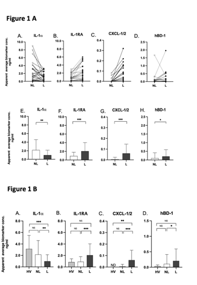
Figure 1A: Measurements of IL-1, IL-1RA, CXCL-1/2 and hBD-1 on non-lesional and lesional skin of psoriasis patients using FibroTx TAP. In panel A - D single measurements of IL-1 α , IL-1RA, CXCL-1/2 and hBD-1detected from non-lesional (NL) skin and lesional skin (L) of 30 psoriasis patients have been depicted, each line represents a single patient. In panel E - H the apparent average biomarker concentrations of IL-1 α ,IL-1RA, CXCL-1/2 and hBD-1detected from non-lesional skin (white bars) and lesional skin (black bars) have been plotted. Y-axis: Apparent concentration of analysed biomarker on skin in ng/ ml. X-axis: sampling site. Error bars on graphs present the standard deviations from average of combined measurements of the participants (N = 30). Statistical significance is indicated on panel E - H: *p < 0.05, ** p < 0.01, *** p < 0.001; NS- not significant.
Figure 1B: Measurements of IL-1, IL-1RA, CXCL-1/2 and hBD-1 on lesional and non-lesional skin of psoriasis patients and normal skin of healthy volunteers using FibroTx TAP. The apparent average biomarker concentrations of IL-1IL-1RA, CXCL-1/2 and hBD-1 detected from the skin surface of 10 healthy volunteers (N = 10; grey bars), on non-lesional skin (NL; white bars) and lesional skin (L; black bars) of 30 psoriasis patients have been plotted. Y-axis: Apparent concentration of analysed biomarker on skin in ng/ml. X-axis: sampling site. Error bars on graphs present the standard deviations from average of combined measurements of the participants. Statistical significance is indicated on panel A - D: *p < 0.05, ** p < 0.01, *** p < 0.001; NS- not significant.
The inverse expression patterns of IL-1α and IL-1IL-1RA on lesional and non-lesional skin of psoriasis patients, as well as the biological link between IL-1α and IL-1RA, prompted us to analyse the molar ratio between IL-1 α and IL-1RA on lesional and nonlesional skin of psoriasis patients. IL-1α and IL-1RA bind to the same receptor, the IL-1 receptor (IL-1R), as a pro-inflammatory agonist and an anti-inflammatory antagonist, respectively. Two forms of IL-1α exist, the immature form with a MW of 31 kDa, and the mature form of 18 kDa, that are both biologically active [30]. Both isoforms are recognised by antibodies used for FibroTx TAP. IL-1RA is predominantly expressed as a 17.1 kDa protein [31]. The analyses revealed that there is clear molecular excess of IL-1α over IL-1RA on non-lesional skin of psoriasis patients and skin of healthy volunteers, regardless whether IL-1α is present in immature or in mature form, or a combination there-of. Similarly, there is a clear excess of IL-1RA over IL-1a on lesional skin of psoriasis patients regardless of the form of IL-1α Table 1).

Table 1: Ratio of IL-1RA over IL-1on the skin of healthy volunteers and on the skin of psoriasis patients.
Note: The average concentration of IL-1 and IL-1RA on normal skin of healthy volunteers (N = 10), non-lesional and lesional skin of psoriasis patients (N = 30) is presented in Table 1 in ng/ml. The standard deviation (SD) presented in table present the standard deviation from average of combined measurements of the 10 healthy volunteers and 30 psoriasis patients, respectively. Additionally, molar ratio of IL-1RA over precursor and mature IL-1 (Ratio of IL-1RA/IL-1) is presented.
Correlations Between FibroTx TAP Protein Measurements and Psoriasis Clinical Scores
IL-1α, IL-1RA, CXCL-1/2, are cytokines directly involved in psoriasis skin-inflammation [32,33]. A possible explanation for the substantial differences in skin-surface levels of IL-1α, IL-1RA, CXCL-1/2 and hBD-1 on lesional skin of individual patients may be due to differences in disease severity between patients. To assess correlations between FibroTx TAP measurements of psoriatic skin and elements of the PASI, we analysed the correlation between the values of IL-1α, IL-1RA, CXCL-1/2 and hBD-1 measurements from psoriatic skin against the values (0 - 4 scale) of erythema, induration and desquamation, assessed by a dermatologist, at the area of FibroTx TAP measurements. The only correlation nearing to statistical significance was a weak positive correlation between the clinical score for skin thickness and levels of CXCL-1/2 on lesional skin (Table 2 and Supplementary).
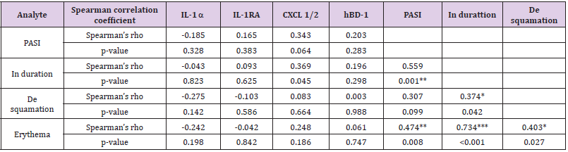
Table 2: Analysed correlations of FibroTx TAP measurements of IL-1α, IL-1RA, CXCL-1/2 and hBD-1 on lesional skin between PASI and local score of thickness, scaling and redness in psoriasis patients.
Note: Correlation between biomarker measurements and clinical scores of psoriasis patients (N = 30) was assessed using Spearman’s rank correlation analysis. Statistical significances were verified with probability value (p- value). Relevant correlations are flagged with asterisk (*p < 0.05, ** p < 0.01, *** p < 0.001).
No statistically significant correlations between measurements of either IL-1α or IL-1RA and severity scores for erythema, induration and desquamation were detected. Nevertheless, a tendency towards a negative correlation between TAP measurements of IL-1α and scaling was observed. The higher the levels of IL-1α on psoriatic lesions, the lower the scaling of lesions (Supplementary Figures). No apparent correlations were found for FibroTx TAP measurements of hBD-1 from psoriatic skin and clinical assessments of erythema, induration and desquamation.
Correlations Between FibroTx TAP Protein Measurements and Ultrasound Analysis on Skin
PV manifests itself in physical changes of the skin layers, such as thickening of the epidermis and presence of a characteristic low-density layer between epidermis and dermis, the so-called sub-epidermal low-echogenic band (SLEB), that can be measured via ultrasound [8,9]. To determine whether differences in the molecular expression patterns of IL-1α, IL-1RA, CXCL-1/2 and hBD-1, between non-lesional and lesional skin site of psoriasis patients correlate with alterations in physical properties of skin layers, FibroTx TAP measurements of these four proteins were correlated with ultrasound measurements from exactly the same skin of psoriasis patients. Using ultrasound, a clear and statistically significant thickening of epidermis (p < 0.001), SLEB (p < 0.001) and dermis (p < 0.001) was measured in lesional skin of psoriasis patients in comparison with non-lesional skin from the same patients (Figure 2A, panel A - C).
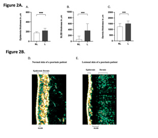
Figure 2A: Thickness of epidermis, SLEB and dermis in non-lesional and lesional skin of psoriasis patients measured by ultrasound. The apparent average epidermal thickness (panel A), SLEB thickness (panel B) and dermis thickness (panel C) analysed from non-lesional skin (NL; white bars) and lesional skin (L; black bars) have been blotted. The ultrasound measurements are performed at the exact lesion and healthy apparent skin of psoriasis patient where the FibroTx TAP measurements were performed and local clinical scores by physician where stated. Y-axis: Average thickness of epidermis, SLEB and dermis, respectively, in m. X-axis: sampling site. Error bars on graphs present the standard deviations from average of combined measurements of the patients (N = 30). Statistical significance was determined with paired sample Wilcoxon signed-rank test (* p 0.05, ** p 0.01, *** p 0.001).
Figure 2B: A representative image of the thickness of epidermis, SLEB and dermis in non-lesional (panel D) and lesional skin (panel E) of psoriasis patient measured by ultrasound. 20-MHz ultrasound image of non-lesional (panel A) and lesional skin area (panel B) of the same psoriasis patient is presented. The positions of epidermis, SLEB and dermis are indicated on the image.
Combining FibroTx TAP measurements of IL-1α, IL-1RA, CXCL- 1/2 and hBD-1 and ultrasound measurements of non-lesional skin from the same patients did not reveal any strong significant correlations between expression of IL-1α CXCL-1/2 or hBD-1 and thickness of the epidermis, dermis or SLEB (Table 3A). A weak positive correlation between IL-1RA and SLEB thickness was observed on non-lesional skin, but not between IL-1RA and epidermis or dermis thickness of lesional skin sites. Combining FibroTx TAP measurements of IL-1α, IL-1RA, CXCL-1/2 and hBD-1 and ultrasound measurements of lesional skin from the same patients also did not reveal any significant correlations between expression of IL-1α IL-1RA or hBD-1 and thickness of the epidermis, dermis or SLEB (Table 3B). The only positive correlation was observed between CXCL-1/2 and SLEB thickness on lesional skin. No such correlation was noted between the expression of the CXCL-1/2 and epidermis nor dermis thickness.
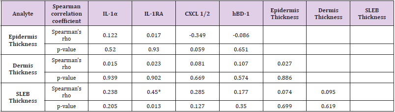
Table 3A: Correlation analysis between FibroTx TAP measurements of IL-1α, IL-1RA, CXCL-1/2 and hBD-1 on non-lesional skin of psoriasis patients combined with ultrasound measurements of epidermis-, dermis- and SLEB thickness at the same analysis site.
Note: Correlation between biomarker measurements and skin layer thickness of psoriasis patients (N = 30) was assessed using Spearman’s rank correlation analysis. Statistical significances were verified with probability value (p-value). Relevant correlations are flagged with asterisk (* p 0.05, ** p 0.01, *** p 0.001).
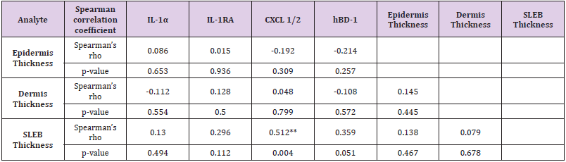
Table 3B: Correlation analysis between FibroTx TAP measurements of IL-1α, IL-1RA, CXCL-1/2 and hBD-1 on lesional skin of psoriasis patients and between ultrasound measurements of epidermis-, dermis- and SLEB thickness at the same analysis site.
Note: Correlation between biomarker measurements and skin layer thickness of psoriasis patients (N = 30) was assessed using Spearman’s rank correlation analysis. Statistical significances were verified with probability value (p-value). Relevant correlations are flagged with asterisk (* p 0.05, ** p 0.01, *** p 0.001).
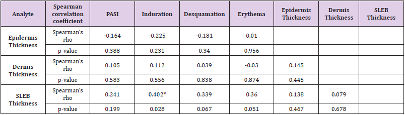
Table 3C: Correlation analysis between local clinical scores and epidermis-, dermis- and SLEB thickness measured from lesional skin by ultrasound.
Note: Correlation between local clinical scores and epidermal-, dermal- and SLEB thickness measured from lesional skin of psoriasis patients (N = 30) was assessed using Spearman’s rank correlation analysis. Statistical significances were verified with probability value (p- value). Relevant correlations are flagged with asterisk. The FibroTx TAP measurements, clinical scores and ultrasound measurements were performed all at the exact same skin lesion (* p 0.05, ** p 0.01, *** p 0.001).
Response to Narrow-band UVB Treatment Measured with TAP
To assess whether TAP has potential for biomarker assessment in response to therapy and whether IL-1α, IL-1RA, CXCL-1/2 and hBD-1 are merely qualitative markers of disease, rather than quantitative, we measured expression levels of IL-1α, IL-1RA, CXCL-1/2 and hBD-1 from the skin surface of 14 psoriasis patients undergoing whole-body treatment with narrow band ultraviolet B. FibroTx TAP was used to measure expression of IL-1α, IL-1RA, CXCL- 1/2 and hBD-1 on lesional and non-lesional skin of patients before treatment initiation (baseline), after two weeks and four weeks of treatment. Measurements were performed on exactly the same position on skin on each time-points. In parallel, visual assessment for erythema, induration, and desquamation were performed at the exact location of TAP measurements. In addition, the PASI score was determined before and after four weeks of treatment.
As a result of the narrow-band UVB treatment, the PASI score dropped on average 57.71 percent during treatment, a difference that was highly significant (p < 0.001) (Figure 3A). Scores for erythema, induration and desquamation showed highly significant improvements of the lesions measured by FibroTx TAP (p < 0.01, p < 0.01, p < 0.01, respectively, Figure 3A). During the four weeks course of UVB treatment, levels of IL-1α did not change on lesional skin of psoriasis patients, but there was a modest decline in IL-1α on non-lesional skin. In contrast, levels of IL-1RA (p < 0.01) and CXCL-1/2 (p < 0.05) showed a significant reduction on lesional skin in response to narrow-band UVB treatment. Whereas four weeks of treatment reduced IL-1RA on lesional skin to the level of IL-1RA on non-lesional skin before treatment, CXCL-1/2 showed a 75 percent reduction of the level of CXCL-1/2 observed on lesional skin before treatment.
No alterations were measured for IL-1RA on non-lesional skin during course of treatment and CXCL-1/2 remained undetectable. Analyses of the IL-1RA over IL-1α ratio also confirm the clinically observed pattern of normalisation of skin in lesions measured. The ratio between IL-1RA and IL-1α measured on lesions (ng/ml) declined from 4.89 to 2.25. In contrast, the ratio between IL-1RA and IL-1α measured on non-lesional skin remained stable, changing from 0.57 to 0.65 during treatment (Table 4).
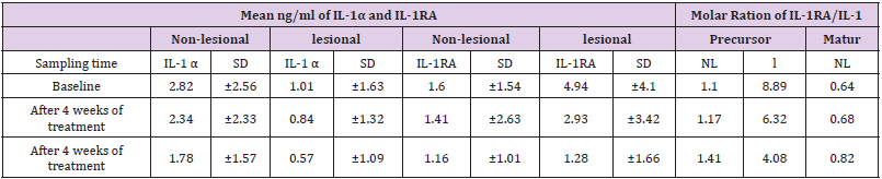
Table 4: Ratios between IL-1RA and IL-1α on non-lesional and lesional skin of psoriasis patients.
Note: The mean concentration (ng/ml) of IL-1α and IL-1RA sampled on lesional (L) and non-lesional (NL) skin of psoriasis patients (N=14) before treatment initiation (baseline), after two weeks and after four weeks of treatment is presented in Table 4. The Standard Deviation (SD) in table presents the standard deviation from average of combined measurements of psoriasis patient NL and L skin site, respectively. Additionally, molar ratio of IL-1RA over precursor and mature IL-1α (ratio of IL-1RA/IL-1α is presented.
The levels of antimicrobial peptide hBD-1 detected at base line on non-lesional skin are nearly 2 - fold lower compared to the levels captured on lesional skin, however after 4 weeks of narrowband UVB treatment the levels of hBD-1 detected on non-lesional skin are increased compared to the baseline approximately 2-fold contrary to the levels of hBD1 captured on lesional skin where nearly 4 - fold decrease compared to amounts of base line hBD-1 is detected (Figure 3C). Due to the UVB treatment the ratio of hBD- 1 detected on healthy apparent and lesional skin at base line has changed opposite after 4 weeks.No adverse events were reported in FibroTx TAP measurements, neither on non-lesional skin nor on lesional skin, neither in patients nor in healthy individuals were reported, neither by visual assessment (e.g. signs of redness) or upon inquiry (e.g. irritation, itching, pain).
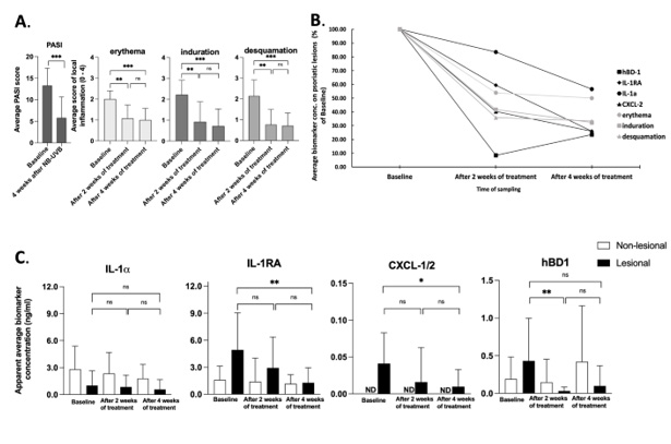
Figure 3A: Changes in the psoriasis area severity index (PASI) and local inflammation scores induced by narrow-band UVB treatment. The PASI score was documented before the treatment initiation and after four weeks of treatment. Local inflammation scores erythema, induration and desquamation were documented before the treatment initiation, after two weeks and four weeks of treatment at the exact same lesions. Each bar plotted on Figure 3 panel A represents an average measurement of analysed clinical score of psoriasis patients (N=14). Error bars on graphs present the standard deviations from average of combined measurements of the patients (N = 14). Statistical significance was determined with paired sample Wilcoxon signedrank test (* p 0.05, ** p 0.01, *** p 0.001).
Figure 3B: Measurements of IL-1⍺, IL-1RA, CXCL-1/2 and hBD-1 on lesional skin of psoriasis patients during narrow band UVB treatment. Combine average levels of biomarkers IL-1α, IL-1RA, CXCL-1/2 and hBD-1 sampled with TAP on lesional skin of psoriasis patient (N = 14) and scores of local erythema, induration, and desquamation of the same lesional site before and during treatment with narrow band UVB combined with Cyclosporine A. Data is presented as % of baseline values.
Figure 3C: Measurements of IL-1⍺, IL-1RA, CXCL-1/2 and hBD-1 on non-lesional and lesional skin of psoriasis patients during narrow band UVB treatment. Biomarker measurements were performed on healthy apparent and lesional skin before, after two and four weeks of treatment at the exact same skin site using FibroTx TAP. The apparent average biomarker concentration of non-lesional skin is plotted with white bars (panel A - D), average biomarker measurements of lesional skin site are presented with back bars (panel A - D). Y-axis: apparent concentration of IL-1α, IL-1RA, CXCL-1/2 and hBD-1 on skin in ng/ml. X-axis: time point of biomarker sampling. Error bars in graph A – D represent the standard deviations from average of combined measurements of the patients (N = 14); ND - not detected. Statistical significance was determined with paired sample Wilcoxon signed-rank test (* p 0.05, ** p 0.01, *** p 0.001).
Discussion and Conclusion
To improve psoriasis care, diagnostic methods are needed that can facilitate personalized medicine. Such diagnostic method should be objective, accurate, cost-effective and easy-to-use for both patients and health-care professionals. Proteins, such as interleukins, chemokines, cell surface receptors, growth factors and anti-microbial peptides drive the biological processes underlying both the physical and visual hallmarks of psoriatic skin. As such, this psoriasis ‘molecular footprint’ may be very suitable for the development of diagnostic methods that can monitor disease progression, as well as measure response to treatment. Particularly suitable may be proteins that can be assessed non-invasively from the skin surface (i.e. without disrupting the skin). A prerequisite is that skin surface molecules follow the state of disease like. Therefore, the first objective of this exploratory pilot study was to assess whether expression patterns of inflammatory proteins known to be involved in psoriasis could be measured non-invasively from the skin surface of adult psoriasis patients and whether the detected levels of these proteins correspond with physical and visual hallmarks of psoriatic skin, and secondly to explore the feasibility of TAP as a tool for monitoring skin surface proteins levels in response to therapy.
The panel of IL-1α, IL-1RA, CXCL-1/2 and hBD-1 was chosen for the pilot study because these proteins haw been shown reliable quantitative and qualitative measurements from the skin surface using TAP [28,29], and based on their role in inflammation of psoriasis reported in the literature. IL-1α and IL-1RA are examples of a pro- and anti-inflammatory interleukins, respectively, that are known to play important role in skin homeostasis and skin inflammation, including psoriasis [30-34,36]. The combination of chemokines CXCL-1 and -2 was chosen because of their role in attracting neutrophils to psoriatic skin lesions which leads to T-cell activation [32,35]. Hence CXCL-1/2 is a valuable biomarker for monitoring inflammatory related processes in skin. Epithelial produced antimicrobial peptide hBD-1 is detected also in immune cells like macrophages and monocytes [37] linking these signalling molecules with immune system regulation. Further, Uzuncakmak with colleagues has monitored invasively alteration of tissue expression of human beta defensin-1 in PV following phototherapy [38] therefore presenting hBD-1 as an interesting candidate for monitoring its pattern in response to therapy non-invasively.
The choice for measuring skin surface proteins using FibroTx TAP was based on the fact that TAP is a non-invasive sampling technology that does not affect skin, i.e. protein measurements are not biased by skin responding to the measurement method, and do not interfere with biological processes in and on the skin [34]. To underline this, no adverse events were reported during TAP measurements, neither on non-lesional skin nor on lesional skin, neither in patients nor in healthy individuals were reported, neither by visual assessment (e.g. signs of redness) or upon inquiry (e.g. irritation, itching, pain).Expression patterns of IL-1α, IL-1RA, CXCL-1/2 and hBD-1 on the skin surface, as measured by FibroTx TAP, reflect reported protein expression patterns as assessed by more invasive technologies, such as mRNA analyses and immunohistochemistry (IHC) using skin biopsies and protein-analyses after tape-stripping of the stratum corneum [32,34,36,38-40]. It appears thus that protein expression in the skin is reflected both qualitatively and quantitatively on the skin surface.
Importantly, the fact that we find some proteins, like IL-1RA, CXCL-1/2 and hBD-1, are present in higher amounts on lesional skin, whereas others, like ILα are found in reduced amounts on lesional skin in comparison with non-lesional and healthy skin, indicates that differences in proteins measured cannot simply be attributed to e.g. differences in skin texture, skin barrier function or amounts of dead cells on lesional skin. Instead, these differences rather indicate that amounts of proteins found on skin reflect regulation in the skin. This is supported by reports in the literature, describing an increase in IL-1RA, CXCL-1/2 and hBD-1, and a decrease in IL-1α, in psoriasis lesional skin in comparison with non-lesional skin, or skin or healthy individuals have been reported in the literature. Thus, it appears that non-invasive measurements of soluble proteins found on the skin, e.g. as measured by TAP, both qualitatively and quantitatively correlate with proteins found in the skin, as measured by invasive methods such as immunohistochemistry and qPCR from skin biopsies [32,34,36,38-40].
In the literature, there is ample evidence that IL-1α is found in decreased levels, and IL-1RA in increased levels in psoriatic lesional skin in comparison with non-lesional skin [33,36,39]. TAP measurements of IL-1α and IL-1RA on skin of adult psoriasis patients thus fit the bulk of evidence in the literature. One of possible reason for these levels of assessed IL-1a and IL-1RA on lesional versus non-lesional could be trough skin barrier disruption which is a distinguishing parameter for lesions. Preformed IL-1α is described to be stored in epidermis [41] is released and depleted due to the skin barrier damage compared to the healthy tissue where the skin barrier is intact. Despite the very clear association between disease and expression patterns of IL-1α, IL-1RA, CXCL-1/2 and hBD-1, as evidenced by the statistically significant differences in expression of these proteins on non-lesional and lesional skin, no firm correlations could be established between IL-1α, IL-1RA, hBD- 1 or CXCL-1/2 and PASI scores of the patients in the present study. A simple conclusion is that measurements of analysed biomarkers on a single lesion, or the ratio between IL-1RA and IL-1α may not be representative for ‘whole body’ diagnostic purposes. Rather, the lack of correlation between measurements of IL-1α, IL-1RA, CXCL- 1/2 and hBD-1 and PASI may be explained by the lack of significant correlations with clinical assessment for erythema, induration, and desquamation of the same lesions, which are among elements that comprise the PASI in addition to scoring other lesions and bodysurface area affected by disease. Nevertheless, the patient cohort of current study was limited, and a study with larger cohort of patients is needed for firm conclusions.
Ultrasound measurements clearly showed a statistically significant thickening of epidermis, SLEB and dermis in lesional skin in comparison with non-lesional skin. Despite a similar trend for TAP measurements of IL-1α, IL-1RA and hBD-1, no clear correlations could be found between protein measurements and ultrasound measurements of epidermis, SLEB or dermis, neither with respect to thickness nor to quality of individual skin layers. At least not with the small number of patients used. Mild positive correlation between CXCL-1/2 and SLEB thickness of lesional skin was observed. Subepidermal low echogenic band (SLEB) presents dermis reduced echogenity which is caused by inflammatory cell infiltration and edema at the inflamed lesions which in turn can be explained by elevated levels of CXCL-1/2 produced by T-cells and keratinocytes in inflamed lesions. Interestingly, neither ultrasound measurements and visual assessments of lesional skin correlated in a highly statistically significant sense; only a mild correlation between skin thickness and SLEB thickness was observed, and thus it appears that visual -, ultrasound – and protein-measurements quantify disease intensity in their own sense.
One of the objectives of current study was to explore the feasibility of non-invasive TAP measurements for soluble biomarkers assessment on the skin surface of psoriasis patients in response to therapy. Whether measurements of skin-surface IL- 1α, IL-1RA, CXCL-1/2 and hBD-1 reflect disease intensity merely in a qualitatively state, i.e. inflamed or not inflamed, or whether skin surface measurements of these biomarkers can reflect disease intensity quantitatively, we followed patients during the course of short-wave UVB treatment. Four weeks of narrow band UVB treatment resulted in notable decrease in PASI scores and improvement in local erythema, induration, and desquamation (Figure 3A). Further, changes in skin surface protein levels were detected. Interestingly, analysed biomarkers displayed different treatment response kinetics- whereas IL-1RA, CXCL-1/2 and hBD- 1 present close to 80% of reduction compared to baseline levels assessed on lesional skin, levels of IL-1a displayed slower and low response reaching just to 40% of reduction (Figure 3B) when compared to baseline. These distinction of L-1α lower response kinetics could be explained with the importance of these cytokine in the regulation of innate immunity defence mechanism and by constant production of local keratinocytes. Therefore, preproduced IL-1α could be emerge from the depths of epidermal layer whereas anti-inflammatory IL-1RA, synthesised previously in counter to inflammation, is reducing in response to therapy. This hypothesis seems to be supported by measurement of IL-1α and IL-1RA levels of the same psoriasis patents healthy apparent skin – response kinetics and alteration in levels of IL-1α detected on lesional skin resemblances with kinetics and reduction rate of skin surface IL-1α detected on healthy apparent skin whereas kinetics and decrease levels of IL-RA is more robust on psoriatic lesion when compared with measurements of non-lesional skin.
Interestingly, although skin surface hBD-1 stated higher levels on psoriatic lesions compared with healthy apparent skin and moreover followed the local inflammation scores during UVB therapy, reverse levels of hBD-1 were detected on psoriasis lesions compared to patients non-lesional skin after four weeks of treatment. We hypothesise it could be related on one hand with integrity of the skin barrier host defence mechanism of healthy skin and on the other hand due to the apoptosis caused by narrow band UVB of local keratinocytes producing hBD-1 in psoriasis lesions. Analysis of skin surface biomarker levels assessed with FibroTx TAP revealed skin surface measurements of IL-1RA, CXCL-1/2 displayed a different pattern than achieved by visual evaluation of local inflammation. Visual assessment for erythema, induration, and desquamation decreased after 2 weeks of treatment and displayed similar levels after 4 weeks of treatment whereas IL- 1RA and CXCL-1/2 normalized more gradually thorough therapy (Figure 3C). This confirms that measuring the ‘molecular root’ of inflammation appears to have value as an objective, non-invasive biomarker measurement for scoring disease intensity on its own right. The difference in kinetics of analysed biomarkers suggest that changes in skin-surface proteins are not a uniform reflection of skin healing, but rather reflect individual changes of expression in the skin.
Conclusion
In conclusion, using FibroTx TAP we could measure clear differences in amounts of IL-1α, IL-1RA, CXCL-1/2 and hBD-1 on skin from psoriasis patients, with clear differences between lesional and non-lesional skin. Monitoring skin surface protein levels with TAP in response to therapy revealed different treatment response kinetics of analysed proteins: clear patterns of normalisation for skin surface IL-1RA and CXCL-1/2 assessed with TAP. This pattern emerged gradually, thus confirming that TAP as a non-invasive skin surface measurement can be used to assess psoriasis intensity in a qualitative way in response to therapy.
Statements
Statement of Ethics
This research was conducted in accordance with the World Medical Association Declaration of Helsinki. Ethical approval for the studies is covered by Decision No. 2551 from the Tallinn Medical Research Ethical Committee. The Declaration of Helsinki protocols were followed, all participants gave their informed and written consent. Participant´s data has been collected such that it cannot be traced back directly to patients by FibroTx employees.
Conflict of Interest Statement
KO, KS, JA, AM and TN are all employed by FibroTx. TN is a founder and shareholder of FibroTx. PS is a consultant at FibroTx.
Funding Sources
Current research was funded by FibroTx LLC and by the Estonian Research Council as personal research Grant PUT1465 and Estonian Research Council Team grant PRG1189 to KK.
Author Contributions
KO analysed the data, KO and PS wrote the manuscript. KK, KA, MK, KO and KS conducted TAP biomarker measurements from the skin of psoriasis patients. KO and KS designed, and KO, KS and JA performed experiments related to TAP biomarker measurement performed on of skin of healthy volunteers. KA and AM performed ultrasound measurements. TN and PS were responsible for the overall study design. All authors have read and approved the final manuscript.
Data Availability Statement
The data is available upon reasonable request.
References
- Ayala F (2007) Clinical presentation of Psoriasis. Reumatismo 59(suppl 1): 40-45.
- Schön MP, Boehncke WH, Brocker EB (2005) Psoriasis: Clinical manifestations, pathogenesis and therapeutic perspectives. Discov Med 5(27): 253-258.
- Langley R, Kreuger G, Griffiths C (2005) Psoriasis: epidemiology, clinical features, and quality of life. Ann Rheum Dis 64(Suppl 2): 18-23.
- Bata Csörgö Z Szell M (2012) The Psoriatic Keratinocytes. Expert Rev Dermatol 7(5): 473-481.
- S Prey, C Paul, V Bronsard, E Puzenat, PA Gourraud, et al. (2010) Assessment of risk of psoriatic arthritis in patients with plaque psoriasis: a systematic review of the literature. J Eur Acad Dermatol Venereol 24: 31-35.
- Schmitt J, Wozel G (2005) The psoriasis area and severity index is the adequate criterion to define severity in chronic plaque-type psoriasis. Dermatology 210(3): 194-199.
- Carlin CS, Feldman SR, Krueger JG, Menter A, Krueger GG (2004) A 50% reduction in the Psoriasis Area and Severity Index (PASI 50) is a clinically significant endpoint in the assessment of psoriasis. Journal of the American Academy of Dermatology 50(6): 859-866.
- Cucoş M, Crişan M, Lenghel M, Dudea M, Croitoru R, et al. (2014) Conventional ultrasonography and sonoelastography in the assessment of plaque psoriasis under topical corticosteroid treatment - work in progress. Med Ultrason 16(2): 107-113.
- Nguyen T, Gattu S, Pugashetti R, Koo J (2009) Practice of phototherapy in the treatment of moderate-to-severe psoriasis. Curr Probl Dermatol 38: 59-78.
- Mihaela Cristina Șomlea , Andreea Nicoleta Boca, Alexandra Dana Pop, Roxana Flavia Ilieș, Stefan Cristian Vesa, et al. (2019) High-frequency ultrasonography of psoriatic skin: A non-invasive technique in the evaluation of the entire skin of patients with psoriasis: A pilot study. Experimental and Therapeutic Medicine 18(6): 4981-4986.
- Kim IH, West CE, Kwatra SG, Feldman SR, O Neill JL (2012) Comparative efficacy of biologics in psoriasis: a review. Am J Clin Dermatol 13(6): 365-374.
- Laws PM, Young HS (2012) Current and emerging systemic treatment strategies for psoriasis. Drugs 72(14): 1867-1880.
- Rustin MH (2012) Long-term safety of biologics in the treatment of moderate-to-severe plaque psoriasis: review of current data. Br J Dermatol 167: 3-11.
- Patel RV, Clark LN, Lebwohl M, Weinberg JM (2009) Treatments for psoriasis and the risk of malignancy. J Am Acad Dermatol 60(6): 1001-1017.
- Alice B Gottlieb, Francesca Chamian, Salman Masud, Irma Cardinale, Maria Veronica Abello, et al. (2005) TNF inhibition rapidly down-regulates multiple proinflammatory pathways in psoriasis plaques. J Immunol 175: 2721-2729.
- Portugal Cohen M, Kohen R (2013) Non-invasive evaluation of skin cytokines secretion: an innovative complementary method for monitoring skin disorders. Methods 61(1): 63-68.
- Portugal Cohen M, L Horev, Claas Ruffer, G Schlippe, W Voss, et al. (2012) Non-invasive skin biomarkers quantification of psoriasis and atopic dermatitis: cytokines, antioxidants, and psoriatic skin auto-fluorescence. Biomed Pharmacother 66: 293-299.
- Rama Malaviya, Yvonne Sun, Jennifer K Tan, Melissa Magliocco, Alice B Gottlieb (2006) Induction of lesional and circulating leukocyte apoptosis by infliximab in a patient with moderate to severe psoriasis. J Drugs Dermatol 5(9): 890-893.
- Nick Dand, Michael Duckworth, David Baudry, Alice Russell, Charles J Curtis, et al. (2019) HLA-C* 06: 02 genotype is a predictive biomarker of biologic treatment response in psoriasis. Journal of Allergy and Clinical Immunology 143(6): 2120-2130.
- Gottlieb AB (2003) Infliximab in psoriasis. J Am Acad Dermatol 49: 112-117.
- JG Krueger, JT Wolfe, RT Nabeya, VP Vallat, P Gilleaudeau, et al. (1995) Successful ultraviolet B treatment of psoriasis is accompanied by a reversal of keratinocyte pathology and by selective depletion of intraepidermal T cells. J Exp Med 182: 2057-1068.
- Paul F Lizzul, Abhishek Aphale, Rama Malaviya, Yvonne Sun, Salman Masud, et al. (2005) Differential expression of phosphorylated NF-kappaB/RelA in normal and psoriatic epidermis and downregulation of NF-kappaB in response to treatment with etanercept. J Invest Dermatol 124: 1275-1283.
- Michelle A Lowes, Francesca Chamian, Maria Veronica Abello, Judilyn Fuentes Duculan, Shao Lee Lin, et al. (2005) Increase in TNF-alpha and inducible nitric oxide synthase-expressing dendritic cells in psoriasis and reduction with efalizumab (anti-CD11a). Proc Natl Acad Sci USA 102: 19057-19062.
- Cabrijan L, Lipozencic J, batinac T, Lenkovic M Zgombic ZS (2008) Influence of PUVA and UVB radiation on expression of ICAM-1 and VCAM-1 molecules in psoriasis vulgaris. Coll Antropol 32: 53-56.
- Usmani N, Murphy A, Veale D, Goulden V, Goodfield M (2008) Photochemotherapy for localized morphoea: effect on clinical and molecular markers. Clin Exp Dermatol 33: 698-704.
- Chodorowska G, Wojnowska D, Juszkiewicz Borowiec M (2004) C-reactive protein and alpha2-macroglobulin plasma activity in medium-severe and severe psoriasis. J Eur Acad Dermatol Venereol 18: 180-183.
- Bukulmez G, Akan T, Ciliv G (2000) Serum adenosine deaminase levels in patients with psoriasis: a prospective case-control study. Eur J Dermatol 10: 274-276.
- Kadri Orro, Olga Smirnova, Jelena Arshavskaja, Kristiina Salk, Anne Meikas, et al. (2014) Development of TAP, a non-invasive test for qualitative and quantitative measurements of biomarkers from the skin surface. Biomark. Res 2: 20.
- Mirjam J Schaap, Finola M Bruins, Xuehui He, Kadri Orro, Malou Peppelman, et al. (2021) Skin Surface Protein Detection by Transdermal Analysis Patches in Pediatric Psoriasis. Skin pharmacology and physiology 34(5): 271-280.
- Busun Kim, Youngmin Lee, Eunsom Kim, Areum Kwak, Soyoon Ryoo, et al. (2014) The interleukin-1α precursor is biologically active and is likely a key alarmin in the IL-1 family of cytokines. Frontiers in immunology 4: 391.
- Malyak M, Guthridge JM, Hance KR, Dower SK, Freed JH, et al. (1998) Characterization of a low molecular weight isoform of IL-1 receptor antagonist. J Immunol 161: 1997-2003.
- Liisi Šahmatova, Elena Sügis, Marina Šunina, Helen Hermann, Ele Prans, et al. (2017) Signs of innate immune activation and premature immunosenescence in psoriasis patients. Scientific reports 7(1):7553.
- T Terui, T Hirao, Y Sato, T Uesugi, M Honda, et al. (1998) An increased ratio of interleukin‐1 receptor antagonist to interleukin‐1α in inflammatory skin diseases. Experimental dermatology 7: 327-334.
- Falcone D, Spee P, Salk K, Peppelman M, van de Kerkhof PCM, et al. (2017) Measurement of skin surface biomakers by transdermal analyses patch following different in vivo models of irritation: a pilot study. Skin Res Technol 23: 336-345.
- Andrea Chiricozzi, Emma Guttman Yassky, Mayte Suárez Fariñas, Kristine E Nograles, Suyan Tian, et al. (2011) Integrative responses to IL-17 and TNF-α in human keratinocytes account for key inflammatory pathogenic circuits in psoriasis. Journal of Investigative Dermatology 131: 677-687.
- Janssens AS, Pavel S, Tensenn CP, Teunissen MBM, Out Luiting JJ, et al. (2009) Reduced IL‐1Ra/IL‐1 ratio in ultraviolet B‐exposed skin of patients with polymorphic light eruption. Experimental dermatology 18: 212-217.
- Lisa K Ryan, Jihong Dai, Zhiwei Yin, Nicholas Megjugorac, Victoria Uhlhorn, et al. (2011) Modulation of human β‐defensin‐1 (hBD‐1) in plasmacytoid dendritic cells (PDC), monocytes, and epithelial cells by influenza virus, Herpes simplex virus, and Sendai virus and its possible role in innate immunity. Journal of leukocyte biology 90(2): 343-356.
- Tugba Kevser Uzuncakmak, Ayse Serap Karadag, Seyma Ozkanli, Ozge Akbulak, Emin Ozlu, et al. (2019) Alteration of tissue expression of human beta defensin-1 and human beta defensin-2 in psoriasis vulgaris following phototherapy. Biotechnic & Histochemistry 95: 243-248.
- Tamilselvi E, Haripriya D, Hemamalini M, Pushpa G, Swapna S (2013) Association of Disease Severity with IL‐1 levels in Methotrexate‐treated Psoriasis Patients. Scandinavian journal of immunology 78: 545-553.
- Emin Ozlu, Ayse Serap Karadag, Seyma Ozkanli, Serpil Oguztuzun, Ozge Akbulak. et al. (2017) The investigation of antimicrobial peptides expression and its related interaction with methotrexate treatment in patients with psoriasis vulgaris. Cutaneous and ocular toxicology 36: 321-326.
- Hulshof L, Hack DP, Hasnoe Q, B Dontje, I Jakasa, et al. (2019) A minimally invasive tool to study immune response and skin barrier in children with atopic dermatitis. Br J Dermatol 180(3): 621-630.
