 Mini Review
ABSTRACT
Fluorescence bioimaging is highly sensitive and noninvasive technique, which is desirable for the visualization of the structure–function property in biological systems. Owing to the easy operation, high selectivity and sensitivity, excellent brightness, photostability, and real time monitoring of the complicated biological processes, organic fluorescent molecules with aggregation induced emission (AIEgens) ability have attracted worldwide attention in biomedical science. Inspired by the widespread use of AIEgens in biomedical science, herein this review, we have summarized some of the recently published literature regarding the AIEgens applications as fluorescent biosensors.
Introduction
Bioimaging is sensitive and noninvasive technique, which is highly desirable for visualization of the structure–function property in biological systems [1]. Owing to the increasing trend of bioimaging in the biomedical fields, numbers of imaging techniques, such as magnetic resonance imaging, computed tomography, positron emission tomography, have been developed and successfully applied for the practical biomedical applications [2]. However, some drawbacks associated with these techniques, such as high cost, long time operation, complicated procedures, low specificity and low biocompatibility are encouraging the scientific community to discover novel ones to overcome such drawbacks. Fluorescence imaging is a superior alternative in contrast to the above imaging methods, which has been proven to be a powerful tool for sensing and visualizing of bioanalytes, biological structures and processes in real time with high spatial resolution [3-5].
Numbers of fluorescent materials, such as organic dyes, organic nanoparticles, inorganic nanoparticles, metallic nanoparticles, and fluorescent proteins have been recently developed and successfully utilized as bio-probes. Among them, organic luminescent materials are of particular interest owing to their better biocompatibility and easy preparation processes [6]. Conventional fluorescent probes, such as rhodamine, fluorescein, BODIPY, and cyanine suffer from the aggregation-caused quenching effect and seriously undergo photobleaching during the long-term imaging, which greatly limit their practical clinical applications [7]. Inorganic nanoparticles, such as quantum dots usually have high brightness and are photostable, but their potential toxicity caused by their heavy metal contents is always a main concern when used for biomedical purposes [8-10]. An alternative bioprobe family, AIE luminogens, usually possessing high photostability and low cytotoxicity, are considered as ideal candidates to overcome these drawbacks.
AIEgens generally have no or weak fluorescence in their solution states, owing to the availability of the free rotors in their molecular structures, which undergo dynamic rotation and active vibrations during the excitation of the AIE-molecules that largely consume the exited state energy and result in the nonradiative relaxation. On the other hand, such molecular motions and vibrations are restricted in the aggregated states, which leads toward high fluorescence in the aggregated/solid states of AIEgens [11]. The concept of AIEgens was first coined in 2001 [12], since then AIEgens have found multiple applications in the imaging of pathogens and mammalian cells.
AIEgens and Mammalian Cells Imaging
Cell is the basic structural and functional unit in all living organisms and for the basic biomedical research, the exploration of cell is necessary. In this regard, fluorescence imaging, which is highly sensitive and non-invasive technique is attracting the global attention. For example, we have recently reported a photostable naphthalimide based probe (TPE-NIM+) for staining Plasma Membrane (PM) of mammalian cells [13] (Figure 1). It can be seen from Figure 1 that our designed TPE-NIM+ has better PM staining performance and much longer retention time at the PM compared to the commercially available PM dyes. Long retention time at PM is one of the important parameters in studying the important biological process occurring at cellular level, such as phagocytosis, necrosis, apoptosis, etc., where the deformation of PM occurs, hence provides useful information about a particular process.
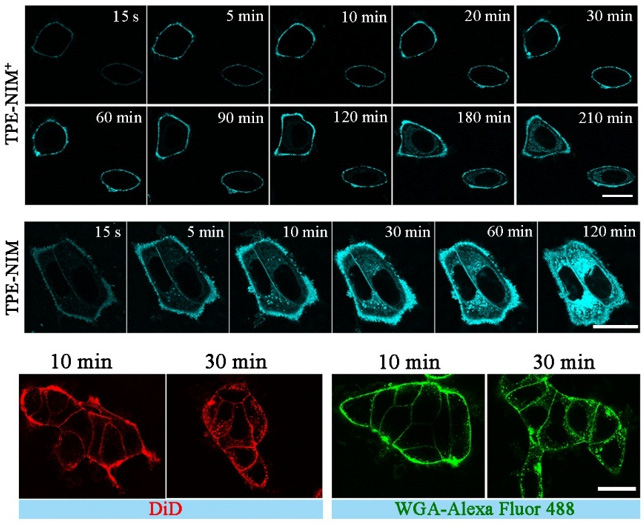
Figure 1: Real-time confocal fluorescence images of MCF-7 live cells after treatment with TPE-NIM+ and TPE-NIM), DiD, and WGA-Alexa Fluor 488.
TPE-NIM+ with high photostability, biocompatibility, ultra-fast and wash-free staining ability, and long retention time is superior to the commercially available PM probes, and it will find a number of applications in the biological/biomedical fields. Similarly, Yu et al. also reported a purine-based water soluble organic fluorophore, pent-TMP, for wash-free and ultrafast imaging of plasma membrane in different complex biosystems both in vitro and in vivo [14]. Imaging of Lipid Droplets (LDs) [15,16] is also important, as it plays crucial roles in many cellular functions, such as the storage and metabolism of lipid, protein degradation, energy storage, membrane formation, signal transduction, etc. [17] In addition, LDs is an important biomarker for various diseases and the abnormality in the size and amount of LDs in the cell is closely related to the dysfunction of LDs, which is associated with many diseases, such as coronary artery diseases, type 2 diabetes, cardiovascular and fatty lever diseases, and obesity [18-20].
We have recently reported a dibenzothiophene based probe for the imaging of lipid droplets in diverse biological systems [21]. The designed probe has the ability of dynamic tracking of lipid droplets and recording the process of lipophagy. Imaging of other cell organelles, such as lysosomes [22], mitochondria [23], and endoplasmic reticulum [24] etc. have also been reported in the literature.
AIEgens and Bacterial Sensing
Pathogenic infections caused by bacteria pose a serious threat to foods, air, and water, resulting in public health issues, climate change, and environmental pollution [25]. To treat the pathogenic infections, the first step is the detection and identification of pathogens. For this purpose, cheap and fast analytical tools are of particular interest. In this regard, fluorescence imaging provides an ideal platform for the detection and identification of pathogenic bacteria. For example, we have recently reported naphthalimide based probes for the selective sensing of Gram-positive bacteria via a fast and wash-free protocol [26]. The designed probes have the ability to selectively target Gram positive bacteria even in the mixtures of Gram positive (S. aureus) and Gram-negative bacteria (E. coli), as shown in Figure 2 (A-F). The designed probes can also target biofilms of Gram-positive bacteria and are bacterial friendly that have extremely low hemolysis rates, hence provide a reliable, cheap, and fast platform for the detection and identification of Gram-positive bacteria that will find practical applications in the clinical analysis of pathogens. Similarly, Tang et al. have also reported numbers of AIEgens with long emission and absorption wavelength to selectively stain G+ bacteria [27]. The designed probes were applied to different kinds of bacteria, both G+ and G– bacteria, where the probes exhibited high degree of selectivity towards G+ bacteria. The designed AIEgens with quaternary amine terminal units were then applied as photosensitizers to kill G+ bacteria both in vivo and in vitro analysis.
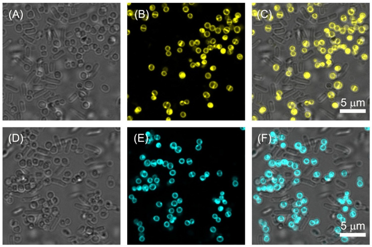
Figure 2: Treatment of Gram positive and Gram negative bacteria with TPE-NIM and TPA-NIM.
Conclusion
In conclusion, owing to the high sensitivity and noninvasive nature, fluorescence imaging is an ideal sensing platform for imaging of cell organelles, tracing of biological processes, disease diagnosis and treatment, and for the detection and differentiation of pathogenic bacteria. Thus, it provides a quick and sensitive approach for the pathogens detection and treatment of the diseases, and tracing of biological processes at cellular level. Therefore, it is necessary to develop novel fluorescent organic molecules with aggregation induced emission for the early medical diagnosis, drug development, and basic biomedical research.
Conflicts of Interest
The author declares no conflict of interest.
References
- Weissleder R, Pittet MJ (2008) Imaging in the era of molecular oncology. Nature 452(7187): 580-589.
- Zheng Z, Zhang T, Liu H, Chen Y, Kwok RTK, et al. (2018) Bright near-infrared aggregation-induced emission luminogens with strong two-photon absorption, excellent organelle specificity, and efficient photodynamic therapy potential. ACS Nano 12(8): 8145-8159.
- Michalet X, Pinaud FF, Bentolila LA, Tsay JM, Doose S, et al. (2005) Quantum dots for live cells, in vivo imaging, and diagnostics. Science 307(5709): 538-544.
- Chan J, Dodani SC, Chang CJ (2012) Reaction-based small-molecule fluorescent probes for chemoselective bioimaging. Nature Chemistry 4(12): 973-984.
- Smith AM, Mancini MC, Nie S (2009) Second window for in vivo Nature Nanotechnology 4: 710-711.
- Mei J, Huang Y, Tian H (2018) Progress and trends in AIE-based bioprobes: A brief overview. ACS Applied Materials & Interfaces 10(15): 12217-12261.
- Kobayashi H, Ogawa M, Alford R, Choyke PL, Urano Y (2010) New strategies for fluorescent probe design in medical diagnostic imaging. Chemical Reviews 110(5): 2620-2640.
- Cai Y, Wei Z, Song C, Tang C, Han W, et al. (2019) Optical nano-agents in the second near-infrared window for biomedical applications. Chemical Society Reviews 48: 22-37.
- Soenen SJ, Parak WJ, Rejman J, Manshian B (2015) (Intra) cellular stability of inorganic nanoparticles: Effects on cytotoxicity, particle functionality, and biomedical applications. Chemical Reviews 115(5): 2109-2135.
- Richards D, Ivanisevic A (2012) Inorganic material coatings and their effect on cytotoxicity. Chemical Society Reviews 41: 2052-2060.
- Gu X, Kwok RTK, Lam JWY, Tang BZ (2017) AIEgens for biological process monitoring and disease theranostics. Biomaterials 146: 115-135.
- Luo J, Xie Z, Lam JWY, Cheng L, Chen H, et al. (2001) Aggregation-induced emission of 1-methyl-1,2,3,4,5-pentaphenylsilole. Chemical Communications 18: 1740-1741.
- Sayed SM, Haoran J, Yaowen J, Yaxuan Z, Liang M, et al. (2021) Photostable AIE probes for wash-free, ultrafast, and high-quality plasma membrane staining. Journal of Materials Chemistry B 9(21): 4303-4308.
- Shi L, Liu YH, Li K, Sharma A, Yu KK, et al. (2020) An AIE‐Based probe for rapid and ultrasensitive imaging of plasma membranes in biosystems. Angewandte Chemie International Edition 59(25): 9962-9966.
- Hu R, Chen B, Wang Z, Qin A, Zhao Z, et al. (2019) Intriguing “chameleon” fluorescent bioprobes for the visualization of lipid droplet-lysosome interplay. Biomaterials 203: 43-51.
- Liu Z, Zou H, Zhao Z, Zhang P, Shan GG, et al. (2019) Tuning organelle specificity and photodynamic therapy efficiency by molecular function design. ACS Nano 13(10): 11283-11293.
- Walther TC, Farese RV (2012) Lipid droplets and cellular lipid metabolism, Annual Review of Biochemistry 81: 687-714.
- Cohen JC, Horton JD, Hobbs HH (2011) Human fatty liver disease: Old questions and new insights, Science 332(6037): 1519-1523.
- Greenberg AS, Coleman RA, Kraemer FB, McManaman JL, Obin MS, et al. (2011) The role of lipid droplets in metabolic disease in rodents and humans. Journal of Clinical Investigation 121(6): 2102-2110.
- Collot M, Fam TK, Ashokkumar P, Faklaris O, Galli T, et al. (2018) Ultrabright and fluorogenic probes for multicolor imaging and tracking of lipid droplets in cells and tissues. Journal of the American Chemical Society 140(16): 5401-5411.
- Sayed SM, Xiangfe L, Smaran D, Haoran J, Fugen W, et al. (2021) A dibenzothiophene core-based small-molecule AIE probe for wash-free and selective staining of lipid droplets in live mammalian and fungal cells. Sensors and Actuators B: Chemical 343: 130128
- Zheng X, Zhu W, Ni F, Ai H, Gong S, et al. (2019) Simultaneous dual-colour tracking lipid droplets and lysosomes dynamics using a fluorescent probe. Chemical Science 10: 2342-2348.
- Wang YF, Zhang T, Liang XJ (2016) Aggregation‐induced emission: Lighting up cells, revealing life. Small 12(47): 6451-6477.
- Alam P, He W, Leung NLC, Ma C, Kwok RTK, et al. (2020) Red AIE-active fluorescent probes with tunable organelle-specific targeting, Advanced Functional Materials 30(10): 1909268.
- Hu R, Zhou F, Zhou T, Shen J, Wang Z, et al. (2018) Specific discrimination of Gram-positive bacteria and direct visualization of its infection towards mammalian cells by a DPAN-based AIEgen. Biomaterials 187: 47-54.
- Sayed SM, Kefei X, Haoran J, Feifei Y, Liang M, et al. (2021) Naphthalimide-based multifunctional AIEgens: Selective, fast, and wash-free fluorescence tracking and identification of Gram-positive bacteria. Analytica Chimica Acta 1146: 41-52.
- Kang M, Zhou C, Wu S, Yu B, Zhang Z, et al. (2019) Evaluation of structure–function relationships of aggregation-induced emission luminogens for simultaneous dual applications of specific discrimination and efficient photodynamic killing of Gram-positive bacteria. Journal of the American Chemical Society 141(42): 16781-16789.
