 Research Article
SUMMARY
Early detection of lesions and maintenance of the normal functioning of the retinal tissue at an early stage of non-proliferative diabetic retinopathy (NPDR) is considered an extremely important step in its secondary prevention, which makes it advisable to include angioprotective drugs in the complex of conservative therapy. One of the drugs of interest is “Doxy-Chem” - (calcium dobesilate) a drug that improves retinal microcirculation, is able to prevent and correct biochemical changes in nerve tissues and has an endothelioprotective effect. The aim of the study was to evaluate the effectiveness of the use of the drug “Doxy-chem” in patients with an early stage of NPDR. Analysis of the results showed that in the group of patients who received “Doxychem” there was an improvement in visual acuity by an average of 0.20±0.02 (p<0.05), a decrease in the thickness of the retina (CTS) by an average of 1.60 micron (p<0.05), as well as a decrease in the thickness of the retina in other parts of the central zone in 75% of cases (44 eyes). While in the control group, the studied functional and Doppler indicators did not change significantly (p>0.05). The present study showed that therapy with Doxychem improves ocular blood flow in the retrobulbar vessels, especially in the central retinal artery and the short posterior ciliary artery.
Keywords: Diabetic Retinopathy; Microcirculation; Hemodynamics; Conservative Treatment of Diabetic Retinopathy
Abbreviations: IDF: Diabetes federation; DM2: Diabetes Mellitus Type 2; DR: Diabetic Retinopathy; FDV: Final Diastolic Velocity of Blood Flow; CAR: Central Artery Retinal; CVR: Central Vienna Retina
Background
According to international Diabetes federation (IDF), more than 400 million people in the world suffer, and half of the cases of the diabetes mellitus type 2 (DM 2) is not diagnosed. Changes in the body developing in patients with DM 2 lead to a violation of all types of metabolism, angiopathy, polyneuropathy, as well as to violation of the function of almost all organs and tissues [1-3]. One of the vascular complications of DM 2 is diabetic retinopathy (DR), which is the main cause of weakness and blindness [4,5]. The prevalence of the DM 2 and the severity of its complications, in particular, DR determine the enormous medical and social significance of this disease. Early detection of foci of lesion and maintaining the normal functioning of the retinal tissue and the optic nerve at the initial stages of DR is considered an extremely important step of its secondary prophylaxis [4,6]. In this case, conservative treatment of DR using a number of angioprotective and antioxidant drugs is published [5,7]. At the same time, the progression of DR leads to hypoxic and morphological damage to neuroepithelial cells, which makes it appropriate for the inclusion of neuroprotective drugs into a complex of conservative therapy [8,9].
One of the pieces of interest is the doxy-hem - (calcium docking) angioprotector, the drug that improves the retinal microcirculation capable of preventing and corrected biochemical changes in the nerve tissues that has an endotheloprotective effect. It is also proven that the therapeutic dosage of the drug leads to a significant decrease in the volume of edema arising from a pronounced lymphatic drainage effect. The drug shows a variety of pharmacological effects in relation to the main pathophysiological processes at DR, as well as other vascular changes in patients with diabetes. The medicine Dox-Hem® reduces the increased permeability of the vessels, increases the resistance of the capillar stakes, moderately reduces the aggregation of platelets and blood viscosity, increases the elasticity of the erythrocyte membrane. The action is associated to a certain extent with an increase in the activity of plasma kinines, as well as with its chemical structure, which allows you to interact with free radicals, suppressing peroxidation oxidation of lipids. In clinical and experimental studies, the angioprotective effect of the Dox-Hem® as a result of the suppression of apoptosis, which occurs due to the prevention of changes in the permeability of the membrane and DNA fragmentation. The use of Dox-Hem® orally in the experiment made it possible to protect the retina from damage to free radicals, the Dox-Hem® stabilizes the GRS, reduces the output of the albumin, thereby contributing to the preservation of the normal retinal thickness. Dox-Hem® affects NO-dependent vasodilation, inhibiting endothelin-1 Thus, the use of Dox-Hem® contributes not only to optimizing endothelialdependent vasodilation, but also to reduce the intensity of retinal neurodegeneration. Another most important effect of the Dox- Hem® is its effect on angiogenesis, which is a key point in the development of the proliferative stage of other experimental studies, proved the powerful dose-dependent anti-angiogenic effect of Dox-Hem® associated both inhibition of fibroblast growth factor and the VEGF factor that promotes the proliferation of endothelial cells and an increase in vascular permeability.
The Purpose of the Study
Is to evaluate the effectiveness of the application of “doxy-chem” in patients with preclinical and early stages of diabetic retinopathy.
Materials and Methods
Surveyed 60 patients (120 eyes), the average age of which amounted to 59.4 ± 6.2 years. The study included patients with preclinical and early stages of non-proliferative diabetic retinopathy without any other eye pathology. All patients were divided into 2 homogeneous groups depending on the treatment carried out: patients in the main group (n = 60), in addition to the standard treatment on the main disease, was appointed Dox-Hem® according to the scheme (in the first 3 weeks - 1 capsule in time or after meals, further use the drug per day for 10 months); In the control group (n = 60), only a standard treatment for the main disease was carried out.
Ophthalmic examination of patients, in addition to basic research methods, such as: visual acuity wit optimal optical correction, biomicroscopy, ophthalmoscopy and tonometry [10], also included static perimetry with the definition of medium sensitivity of the retina and fowolar photosensitivity, optical coherent tomography (OCT) with an estimate of the thickness of the central fox and Makula in 4 meridians and color Doppler mapping of vessels [11]. All patients were used by the Color Doppler Ultrasound method for estimating the peak systolic velocity of blood flow (PSV), the final diastolic velocity of blood flow (FDV) and the resistance index (RI) in the following arteries: eye artery (EA), Central Artery Retinal (CAR), Central Vienna Retina (CVR), Short Back Cylinder Artery (SBCA) The survey included an integrated ultrasound study of the eye and orbits in EA CAR, CVR and SBCA on the VOLUSONE 8 device (GE, Healt Ere) to using a linear sensor with a frequency of 10 to 16 MHz. The spectrum of the doppler shift of the frequencies was recorded and the main quantitative indicators of blood flow were determined: VSYST, VDiaSti Ri.
Results and Discussion
In the primary inspection of patients, a decrease in visual acuity was revealed on average to 0.61 ± 0.03 in 72.5% of cases (44 eyes). At an ophthalmoscopy in 84.3% of cases (50 eyes), the microaneurisms of the rear area of the eye, localized mainly in the macular region, in 80.1% (48 eyes) are point hemorrhages in 20.1% (12 eyes), small solid (30 %, 18 eyes) Exudates. At OCT of the Fovea Center, a non-uniform thickening of the neuroepitelium of the retina was revealed in 23% of cases (14 eyes), which, apparently, is associated with hypoxia phenomena and microcirculation disorders in patients with diabetic retinopathy. The results of analyzing the thickness of the Macula in patients of the main and control groups remained within the age norm. As a result of the treatment, a significant increase in visual acuity was noted in the main group - on average by 0.20 ± 0.02 (p <0.05), with 84.5% of cases (50 eyes) there was a positive trend. In the control group, the visual acuity has not changed significantly, and its increase was not statistically significant (p> 0.05).
According to the results of the optical coherent tomography of patients of the main group, a decrease in the thickness of the retina in the Fovea is found by an average of 1.60 μm (p <0.05), as well as a decrease in thickness in other parts of the central zone. In general, the positive dynamics was marked in 75% of cases (44 eyes). In the control group, when comparing the results of the thickness of the retina before and after the treatment of statistically significant changes, (P> 0.05) is noted (Table 1). Analysis of changes in the eye picture pattern (microaneurism, the number and dynamics of hemorrhage, solid and soft exudates, retinal edema) indicated statistically significant changes in the main group, starting with 3 months of observations. In the control group, the statistical authenticity of the positive dynamics of the process in the specified period was absent An analysis of the remote results of the study showed that 3 months after treatment, the patients with main group showed a slight decrease in visual acuity compared with the results obtained immediately after treatment, but this figure remained reliably higher than the initial results by an average of 20.78% (p <0 05).
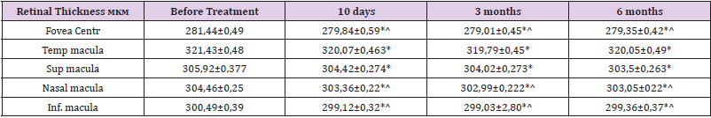
Table 1: The retina before and after the treatment of statistically significant changes.
In the control group, in 3 months, visual acuity was similar to the initial values. After 6 months after treatment, a decrease in visual acuity was noted in all studied groups, while in the main group this indicator remained above the initial results on average by 18.67% (p <0.05), and in the control group decreased compared to the initial Indicators on average by 4.9% (p> 0.05). OCT-scanning of the central retinal zone in patients of the main group after 3 months revealed a further minor decrease in the thickness of the central fox and the makeup with a tendency to increase by 6 months after treatment, though the values were preserved significantly below the source data (p <0.05). Statistically significant changes in patients of the control group were not (P> 0.05) (Table 2) shows a progressive reduction of blood flow at the second stage of IB and the third IC NDR in the central artery of the retina (CAК) in the initial state [4,5]. The progressive phased decrease in blood flow in the short rear ciliary artery (SRCA) was also revealed.
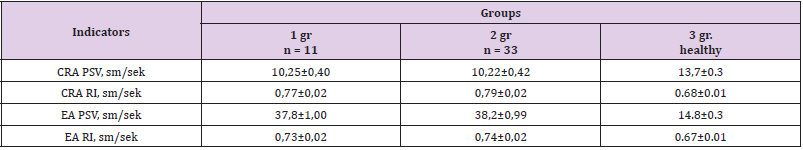
Table 2: Results of ultrasonic doppler vascular vessels by groups. (abs. Numbers and%).
Note: * NPDR = non-proliferative diabetic retinopathy, PSV = peak systolic rate of blood flow, CRA = central artery retina, CVR = central vein retina, SPCA = short rear cylinder artery, EA = eye artery, ri = resistant index; * p <0.05; ** p <0.01 - from healthy, # p <0.05 - from the NDR IA, ^ P <0.05- from the NDRR IB; SD = Standard deviation.
Peak systolic speed (PSS) in the central vein of the retina (CVR) changed from 6.48 cm / s at stage Ia to 3.97 cm / s at stage IC, which indicates dilatation of veins increasing as retinopathy progression. Ultrasound studies of the condition of blood flow in the vessels of the eye were performed in 12 patients (24 eyes) with various stages of the Central Tshold Group II and in 14 patients (28 eyes) of the III group, of which 10 eyes with the non-acudative stage of TsDD in the group II group and 12 eyes in the group III, 8 Eye with an exudative stage of TsDD in group II and 8 eyes in the third group, with a scar stage of TsDD in the group II 6 eyes, in the third eye group. When analyzing hemodynamic indicators in persons with non-assessive stage of TsDD in the II and III group of patients, TCDDs revealed increased microcirculation in the CAR and the WCCC system, which manifest itself with an increase in the systolic velocity of blood flow: in patients with group II 1.2 times and III groups 1.5 times and decrease Resistance index, respectively.
These parameters in patients of the Group III significantly correlated with indicators of visual acuity (Table 3). Thus, the analysis of the results, showed that in the group of patients who received “doxy-hem” after treatment, there was a reliable positive dynamic of a number of studied functional and doppler indicators, which is associated with the effect of the drug on the microcirculation of the retina and its protection against the effects of metabolic and hypoxic defeats in patients dr. These clinical and functional studies have shown the effectiveness of the drug “Doxy - Hem” in the treatment of non-proliferative DR. The use of this drug contributes to an increase in visual acuity, a decrease in the thickness of the retina, improving the hemodynamics indicators. Positive changes in visual functions obtained as a result of treatment are preserved for up to 6 months. All this makes it possible to recommend the specified method for the secondary prevention of the development of diabetic retinopathy and the rehabilitation treatment of patients with early stages of non-proliferative DR.
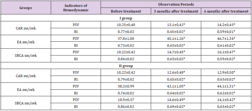
Table 3: Comparative assessment of the results of treatment on the dynamics of CDC indicators in patients with NDPR.
Note *P <0.05 The accuracy of differences in relation to the data before treatment. ** - p <0.05 accuracy differences between groups.
Conclusion
1. This study showed that the treatment with the drug “Doxy- Hem” helps to improve eye blood flow in retrobulbar vessels, especially in the central artery of the retina and the short rear ciliary artery.
2. psV and RI in CRA and SPCA can be potentially useful for early diagnosis and subsequent observation of others.
3. The places of increased resistance or reduced blood flow velocity can be used to predict a higher risk of developing heavy DR, which is important to determine the further patient’s tactics.
References
- Lorenzi M, Gerhardinger C (2001) Early cellular and molecular changes induced by diabetes in the retina. Diabetologia 44: 791-804.
- Lieth E, Gardner TW, Barber AJ, Antonetti DA (2000) Penn State Retina Research Group. Retinal neurodegeneration: early pathology in diabetes. Clin Exp Ophthalmol 28: 3-8.
- Mohamed Q, Gillies MC, Wong TY (2007) Management of diabetic retinopathy: a systematic review. Prog Retin Eye Res 298: 902–916.
- Astakhov YuS, AB Lebina, FE Shadrichev (2003) Modern directions of drug treatment of non-proliferative diabetic retinopathy. Wedge Ophthalmology RMW 4(3): 96-101.
- Tetrov KS, Kolchiketal OV (2017) Pategenetic approaches to the prevention and treatment of diabetic retinopathy. Consilium Medicum 19(4): 43-49.
- Cheung N, Mitchell P, Wong TY (2010) Diabetic retinopathy. Lancet 376: 124-36.
- Аmbiase A, Aloe L, Centofanti M, Parisi V, Báo SN, et al. (2008) Experimental and clinical evidence of neuroprotection by nerve growth factor eye drops. Proc Natl AcadSci USA 106: 13469-13474.
- Bearsejr MA, Adams AJ, Han Y, Schneck ME, Ng J, et al. (2006) A multifocal electroretinogram model predicting the development of diabetic retinopathy. ProgRetin Eye Res 25: 425-448
- Castillo M, Bellot JL, Garcia-Cabanes С, Miquel J, Orts A, et al. (2002) Effects of hypoxia on retinal pigmented epithelium cells: protection by antioxidants. Opthal Res 6: 338-342.
- Shirao Y, Kawasaki K (1998) Electrical responses from diabetic retina. ProgRetin Eye Res 17: 59-76.
- Villarroel M, Ciudin A, Hernбndez C, Simу R (2010) Neurodegeneration: an early event of diabetic retinopathy. World J Diabetes 1: 57–64.
