 Case Report
Abstract
The inflammatory phase, which begins in the healing process right after the hemostasis phase, is basically a prelude to the actual healing process. However, an excessive or too long inflammatory phase can lead to impaired healing and the formation of non-healing wounds. On the other hand, the most important from the point of view of biomedical engineering is the fact that each immune reaction, and inflammatory in particular, is manifested by a change in the temperature of the tissues in which it occurs, and this change in this parameter can be measured and visualized. For this reason properly developed visual data can be of strategic importance for the accuracy of a doctor’s decision-making.
Keywords: Fusion of Images; Multimodal Imaging; Medical Imaging, Thermography; Wounds
Introduction
Medical infrared imaging is a non-invasive diagnostic method that allows the examiner to evaluate and quantify changes in skin surface temperature [1]. However, the use of infrared for medical imaging has its limitations related to the range of measurements, which results from the imperfection of image processing software. This article presents a new approach to the problem and also a new perspective that allows to obtain a more complete range of data of interest to the doctor in one coherent picture [2,3]. This novel approach is strategic for making the right decision about how to manage the wound [4-7]. The multimodal imaging presented in this paper is an excellent method for the initial assessment of difficultto- heal wounds prior to the determination of infectious agents (microorganisms) using electromigration and MALDI MS (matrix assisted laser desorption and ionization mass spectrometry) techniques. It should be taken into account that the use of a MALDI MS coupled with electromigration techniques is a very selective method of detection and also constitutes a kind of imaging [8-10]. The use of thermographic image in conjunction with separation techniques would be a huge tool in the diagnosis of difficult-to-heal wounds. The same as in the case of studies on the effectiveness of signaling proteins, especially immunomodulatory proteins, in supporting the healing of difficult-to-heal wounds [11-13].
Case Report
A 73-year-old female patient, diagnosed with long-term diabetes type II, advanced diabetic angiopathy and neuropathy, trophic changes in the lower limbs, edema, discoloration characteristic of circulatory disorders. The patient is under the care of a medical doctor, not very mobile. She came to the surgeon for consultation of the open wound of the right lower limb in the tibia area, closer to the ankle. Diabetic gangrene with ischemic and necrotic areas associated with severe inflammation was diagnosed.
Methods
The imaging was performed using the FLIR E95 (FLIR E96, S/N 90200296) infrared camera and then modelled in FLIR ResearchIR (FLIR ResearchIR, version 4.40.11) and FLIR Tools (FLIR Tools, version 6.3.17227.1001) software. Infrared imaging was performed with the use of filters allowing the assessment of the hottest and coldest places - ischemic and inflamed areas, respectively, to obtain an accurate temperature topography of the damaged tissue surface and adjacent areas. The last processing was done manually in the basic MS Paint (Microsoft Windows 10 Paint, version 21H1) image editing program.
Results
Two primary thermal images were obtained and processed.
The first image shows the temperature regions that overexpress
inflammation (Figure 1, Phase 1A). In this case, the location of the
area indicates an obstruction of blood flow towards the wound.
The second area with the coldest points (Phase 1B.) indicates
tissue necrosis caused by damage to the vascular network. Both
assumptions were surgically confirmed by a doctor. In the next
phase, the fragment of the thermographic image was superimposed
on the real image of the limb (Phase 2A & 2B). The images were
processed using the FLIR Tool and FLIR ResearchIR software. The
aim for this measurement was connected with obtaining a different
perspective of the problem, and more precisely, to superimpose
isotherms on the real image. However, such a solution still did not
reflect the full picture of tissue pathology and was not satisfactory
for the surgeon making the clinical evaluation.
In the phase 3 (Phase 3A & 3B) the conversion was performed,
still in the firmware, by “dissolving” the background of the
thermographic shot, which is not an isotherm of tissue changes.
However, such an imposition requires the adoption of a correction
as it is done with a shift downwards and to the left from the
perspective of the viewer. Otherwise, the actual image placed on
the underside, and more precisely the tissue lesion, would become
unreadable. In the last phase (Phase 4), an image satisfying for the
doctor was obtained, which meets all the requirements. Therefore,
it was decided to create a complete thermographic image, showing
all interesting aspects. For this purpose, the image was manually
edited and the isotherms below the assumed product were
superimposed on the isotherms above the assumed threshold.
Thanks to this approach, the result of necrosis and inflammation
was obtained on one consistent image, which allowed the surgeon
to introduce appropriate therapy.
Diabetic foot problems, such as ulcerations, infections, and
gangrene, are the most common cause of hospitalization among
diabetic patients. At least half of all amputations occur in people
with diabetes, most commonly because of an infected diabetic foot
ulcer. For this reason the main objectives of the study should be
connected with the identification of the spectrum of multidrugresistant
bacteria associated with these infections, their antibiotic
sensitivity pattern, and to detect the biofilm formation. The
previously described research by Buszewski and coworkers [8-10]
concerned the identification of microorganisms in postoperative
wounds and in diseases related to the diabetic foot. This technique
has become a very good diagnostic test for identifying pathogens.
In the future, the combination of imaging using multimodal
thermography and separation techniques, such as capillary
electrophoresis, microscopic imaging, MALDI MS technique will
allow to obtain a complete diagnostic picture and facilitate the
introduction of targeted therapy.
Conclusion
The new approach to image recording presented in this paper
combines two completely different modalities of infrared medical
images: infrared images of the warmest isolated places and the
coldest places - both with the determination of the temperature
threshold. As a result of the research, a thermal visualization with
the features of a multimodal image was obtained, representing
two extreme / marginal aspects of a wound difficult to heal -
overexpression of inflammation and necrosis. Infrared is essentially
a 2D technique and its image does not provide useful anatomical
and topographic information related to it, such as slow-healing
wounds. Summarizing the conducted research, it should be stated
that there is a need for further development of this approach in
two main directions: automation of associating various modalities
with the choice of temperature assumptions; the use of associated,
multimodal images as a texture to be superimposed on a three-axis
rotational spatial image, made with the use of 3D scanners [14].
This would be a huge towards obtaining a rendered 3D image
that could be used as a virtual phantom to plot temperature
changes, as a disorder due to ischemia (temperature below normal)
or inflammation (temperature above normal) and be used as a
tool to improve the medical diagnosis of pathologies associated
with hard to heal wounds. An ambitious direction would be to
create software supporting making medical decisions based on the
performed measurements and hybridization of imaging [15-17]. It
would also be a big challenge to apply a millimeter grid to the model
- both flat and 3D - in order to obtain the best diagnostic precision.
This imaging together with the use of separation techniques
(electromigration techniques, MALDI MS) will give a full diagnostic
picture and facilitate the understanding of the infection mechanism.
Ultimately, coupling the spatial model with 3D bioprinters could
result in a bespoke dressing suited to the condition and pathology
of the wound [18-21].
Conflict of Interest
The Author(s) declared no potential conflicts of interest with respect to the research, authorship, and/or publication of this article.
Author Contributions
Conceptualization: Wojciech Karwowski
Formal Analysis: Wojciech Karwowski, Jarosław Skórski
Investigation: Wojciech Karwowski, Jarosław Skórski, Ewa
Kłodzińska
Methodology: Wojciech Karwowski, Ryszard Sroka
Software: Wojciech Karwowski
Supervision: Wojciech Karwowski, Ewa Kłodzińska, Bogusław
Buszewski
Visualization: Wojciech Karwowski
Writing - Original Draft: Wojciech Karwowski
Writing - Review & Editing: Wojciech Karwowski, Jarosław
Skórski, Ewa Kłodzińska, Ryszard Sroka, Bogusław Buszewski.
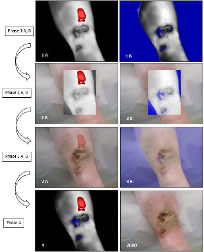
Figure 1: Successive transformations of thermographic images from the original form, through modified ones, to the final form
acceptable to the diagnostician.
Phase 1: Standard images - the warmest (A) and coldest areas (B).
Phase 2A & 2B: Crops of thermographic images superimposed and matched with real images of the limb.
Phase 3A & 3B: Blurring of thermographic images for penetration of real images - the effect achieved by software in the FLIR
software.
Phase 4: Manual overlay of two modalities of thermographic images - the hottest and coldest areas and merging them into one
coherent image. Image ZERO as reference showing the actual appearance of the wounds.
References
- Jain R, Kasturi R, Schunck BG (1995) Machine Vision, McGraw-Hill.
- Podbielska H, Skrzek A (2014) Biomedical applications of thermal imaging. Publishing House of Wrocław University of Technology.
- Eddie YK, NG, Etehadtavakol, Mahnaz (2017) Application of Infrared to Biomedical Sciences. Springer Singapore.
- Karwowski W (2018) Wound diagnosis of various etiologies by thermographic method., Conference materials: 1st Wound Treatment Forum "Management of Difficult-to-heal Wounds - Facts and Myths", Zakopane, Poland.
- Karwowski W, Kucharzewski M (2018) Thermographic imaging as a method of monitoring the progression of hard to heal wounds. Conference materials: XXVIII Conference of the European Wound Management Association EWMA 2018 in cooperation with the Polish Wound Management Association - Kraków, Poland.
- Karwowski W (2016) Clinical application of thermography as a non-invasive method of diagnosis and classification of burns. Current state and prospects of development. Conference materials: Conference organized by Polish Society for Burn Treatment - “Guidelines for the treatment of burns - current problems and perspectives” - Zakopane, Poland.
- Karwowski W (2018) Thermographic assessment of the flow running of treatments in invasive aesthetic medicine with interruption of skin. Thermology International 28.
- Kłodzińska E, Kupczyk W, Jackowski M, Buszewski B (2013) Capillary electrophoresis in the diagnosis of surgical site infections. Electrophoresis 34(22-23): 3206-3213.
- Szeliga J, Jackowski M, Kupczyk W, Kłodzińska E, Buszewski B (2011) Clinical application of a rapid microbiological test based on capillary zone electrophoresis to assess post-operative wound infection. BMC Res Notes 4: 467.
- Buszewski B, Kłodzińska E (2016) Rapid microbiological diagnostics in medicine using electromigration techniques. TrAC Trends in Analytical Chemistry 78: 95-108.
- Karwowski W, Kucharzewski M (2018) The effectiveness of signaling proteins in the treatment of hard to heal wounds. Conference materials: XXVIII Conference of the European Wound Management Association EWMA 2018 in cooperation with the Polish Wound Management Association - Kraków, Poland.
- Karwowski W (2016) The effectiveness of signaling proteins in the treatment of hard to heal wounds - assessment of thermal imaging as part of the world research. Conference materials: 1st International Conference "Diabetes Foot Syndrome - the latest therapeutic solutions” - Jachranka, Poland.
- Karwowski W (2018) Thermographic evaluation of the effectiveness of immunomodulatory formulas for external use in supporting tissue regeneration and wound healing. Conference materials: Annual Conference "Advances in Biomedical Research" organized by HESA Histology and Embryology Students' Science Association at The Department of Histology and Embryology of the Faculty of Medicine in the Medical University of Warsaw - Warsaw, Poland.
- Strzelecki M, Materka A (2017) Texture of biomedical images. Computer Analysis Methods, PWN.
- Karwowski W (2021) Medical hybrid imaging of difficult-to-heal wounds. Conference materials - book of abstracts: II National Scientific Conference "Challenges and problems of biomedical sciences”: TYGIEL Foundation for the Promotion of Science and Development - Lublin, Poland.
- Karwowski W (2018) Multimodal imaging using infrared and ultrasounds - possibilities, problems, medical applications., Conference materials: VIII National Scientific Conference “Young Scientists in Poland - Research and Development"- Lublin, Poland.
- Bichinho GL, Gariba MA, Sanches IJ, Gamba HR, CRUZ FPF, et al. (2009) A Computer Tool for the Fusion and Visualization of Thermal and Magnetic Resonance Images. Journal of Digital Imaging 22(5): 527-534.
- Zieliński WK, Strzelecki M (2013) Computer analysis of the biomedical image. Introduction to morphometry and quantitative pathology, PWN.
- Ishida H (2013) Fourier Transform Infrared Characterization of Polymers. Springer-Verlag New York Inc.
- Zaid MH, Goyanes A, Clark V, Basit AW, Hilton ST, et al. (2017) Patient-Specific 3D Scanned and 3D Printed Antimicrobial Polycaprolactone Wound Dressings. International Journal of Pharmaceutics 527(1-2): 161-170.
- Karwowski W (2018) Development of an innovative medical device to prevent the occurrence of diabetic gangrene in the case of injuries of the musculoskeletal system. Conference materials: Conference of Biomedical Engineering at the Poznań University of Technology - Poznań, Poland.
